. 2020 May 4;217(5):e20191340.
doi: 10.1084/jem.20191340.

Glioma stem-like cells evade interferon suppression through MBD3/NuRD complex-mediated STAT1 downregulation

Affiliations

Glioma stem-like cells evade interferon suppression through MBD3/NuRD complex-mediated STAT1 downregulation

Xiaoyan Zhan et al. J Exp Med. .

Abstract

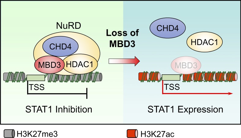
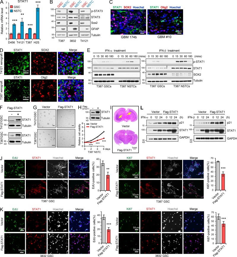
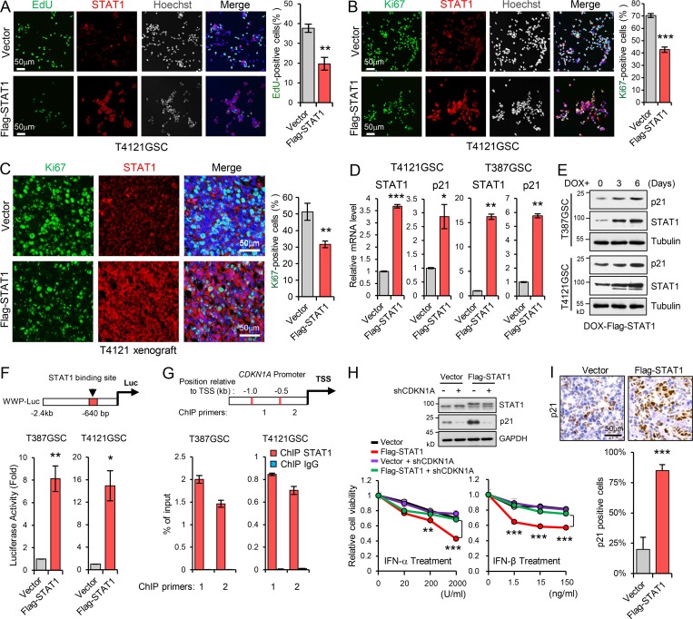
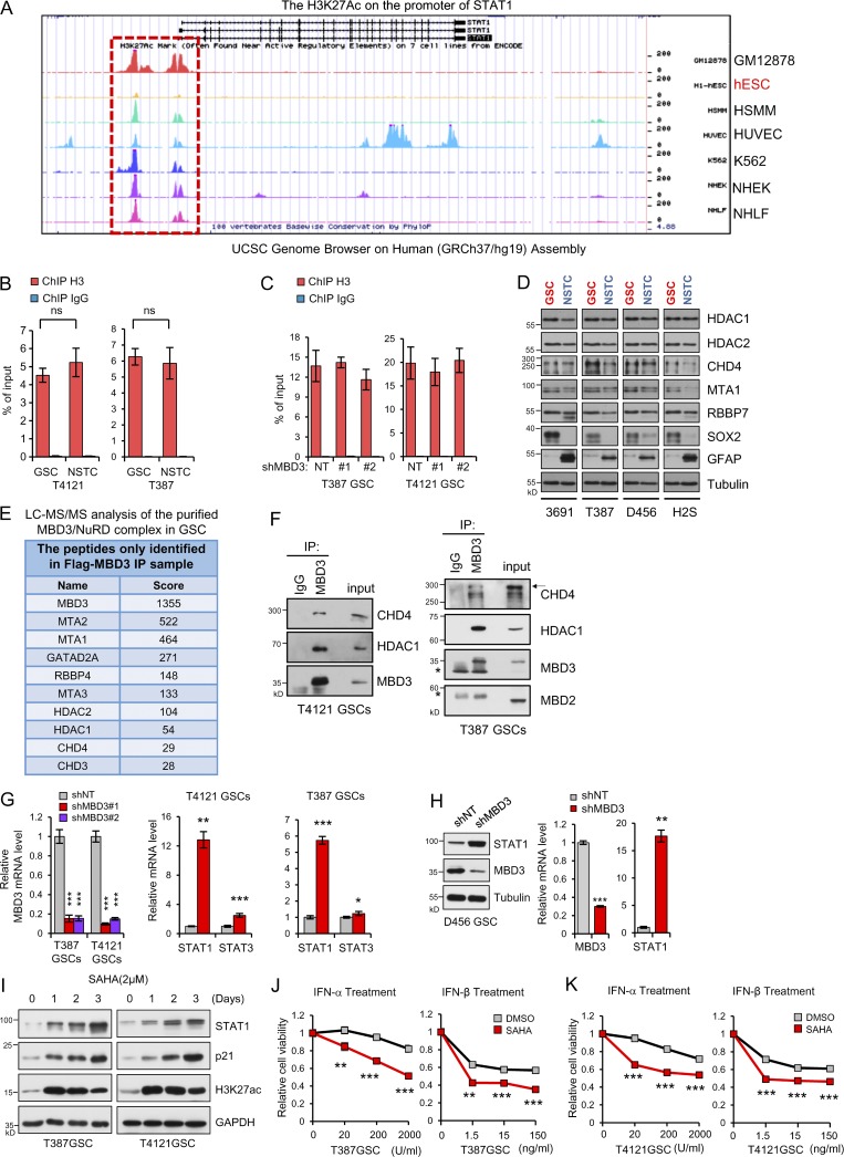
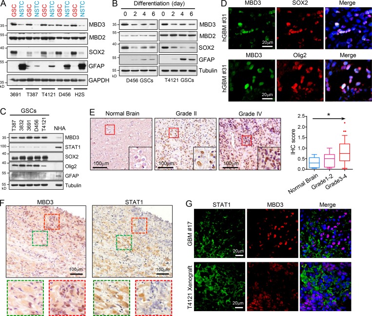
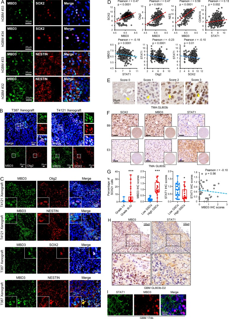
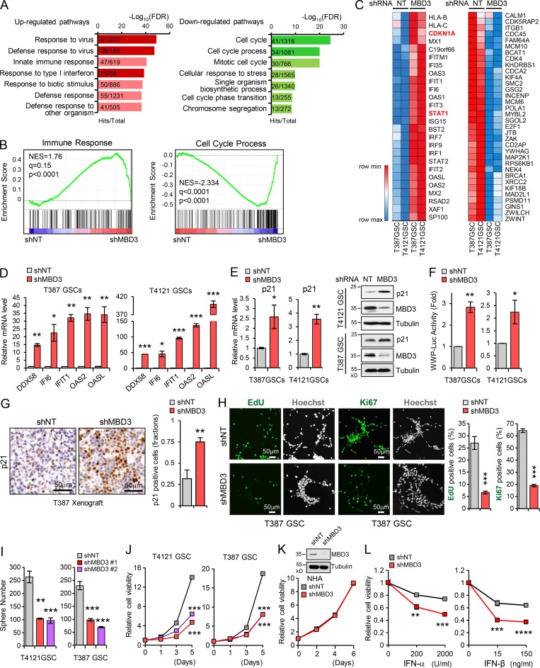
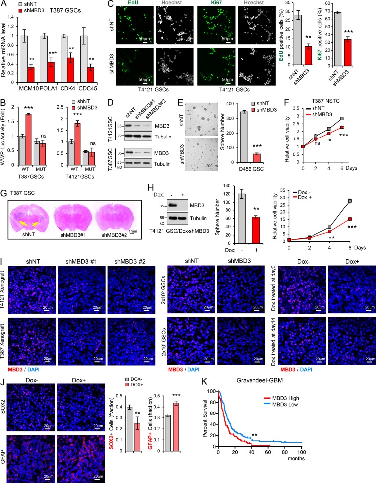
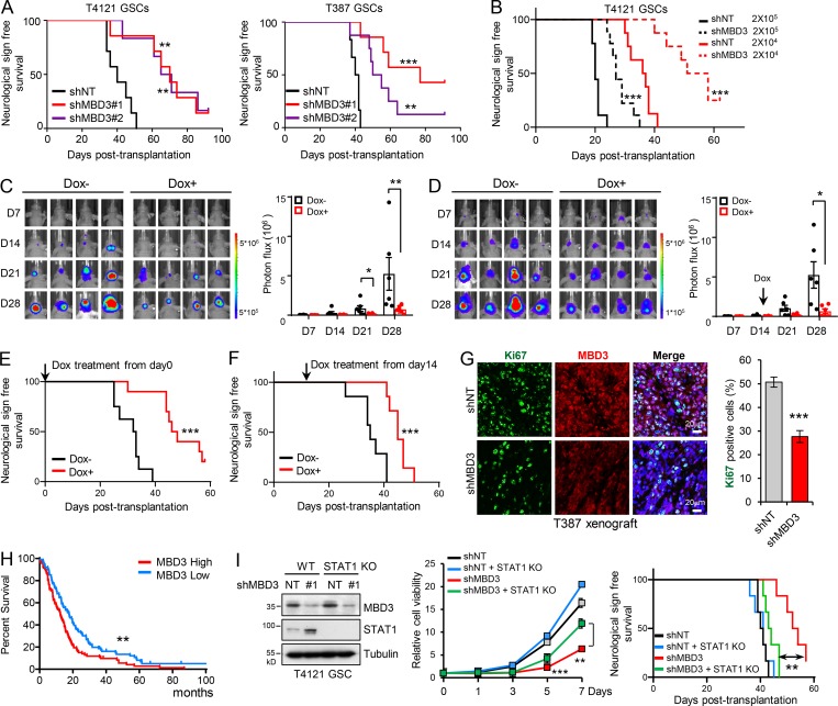
Type I interferons (IFNs) are known to mediate antineoplastic effects during tumor progression. Type I IFNs can be produced by multiple cell types in the tumor microenvironment; however, the molecular mechanisms by which tumor cells evade the inhibition of immune microenvironment remain unknown. Here we demonstrate that glioma stem-like cells (GSCs) evade type I IFN suppression through downregulation of STAT1 to initiate tumor growth under inhospitable conditions. The downregulation of STAT1 is mediated by MBD3, an epigenetic regulator. MBD3 is preferentially expressed in GSCs and recruits NuRD complex to STAT1 promoter to suppress STAT1 expression by histone deacetylation. Importantly, STAT1 overexpression or MBD3 depletion induces p21 transcription, resensitizes GSCs to IFN suppression, attenuates GSC tumor growth, and prolongs animal survival. Our findings demonstrate that inactivation of STAT1 signaling by MBD3/NuRD provides GSCs with a survival advantage to escape type I IFN suppression, suggesting that targeting MBD3 may represent a promising therapeutic opportunity to compromise GSC tumorigenic potential.


Conflict of interest statement

Disclosures: The authors declare no competing interests exist.

Figures

None
Graphical abstract
Figure S1.
Figure S1.
GSCs display less sensitivity to type I IFNs suppression. (A) IHC staining of IFN-α and IFN-β in human GBM specimens. Section was counterstained with hematoxylin. (B) Co-IF staining of IFN-α or IFN-β (green) with macrophage marker CD68, T cell marker CD3, natural killer cell marker CD56, and dendritic cell marker CD11c (red) in human GBM specimens. Nuclei were counterstained with Hoechst (blue). (C) GSCs tumor initiation in the in vivo serial transplantation. 5 × 10³ GSCs (T4121 or T387) were intracranially injected into the brains of nude mice (nu/nu) for the primary xenograft. All recipients developed tumor and died within 50 d, and these tumor cells (5 × 10³) could be serially transplanted at the second round, suggesting the long-term self-renewal and tumorigenicity of GSCs. Kaplan–Meier survival curves of mice implanted with GSCs are shown (top). Summary of the serial transplantation experiment is shown (bottom). (D) In vivo limiting dilution assays were performed with T387 GSCs and matched NSTCs. Kaplan–Meier survival plots are shown. n = 8 for each group, log-rank test. (E and F) Co-IF staining of OASL or CXCL10 (green) with SOX2 (red; E) or with GFAP or b3-tubulin (red; F) in human GBM specimens. Nuclei were counterstained with Hoechst (blue). Quantification are shown (percentage of CXCL10+/− or OASL+/− cells in GFAP+ cells, n = 10; percentage of CXCL10+/− cells in Tubb3+ cells, n = 10; percentage of OASL+/− cells in Tubb3+ cells, n = 15; unpaired Student’s t test). Data are represented as mean ± SD. (G) Limiting dilution analysis of the effect of IFN-α or IFN-β on T387 GSCs. Cells were plated in a limiting dilution manner (1–200 cells per well, 10 wells per condition). 10 d later, each well was evaluated for the presence or absence of tumorsphere. ***, P < 0.001.
Figure 1.
Figure 1.
GSCs display less sensitivity to type I IFN suppression. (A and B) GSCs and matched NSTCs (T387 and T4121) were treated with IFN-α (200 U/ml; A, n = 3) or IFN-β (15 ng/ml; B, n = 3) for 12 h. The mRNA levels of IRGs CXCL10, OASL, IFI27, and IFI6 were analyzed by real-time qPCR. (C and D) CD133high and CD133low glioma cells isolated from GBM PDX were treated with IFN-α (200 U/ml; C, n = 3–4) or IFN-β (15 ng/ml; D, n = 3–4) for 12 h. The mRNA levels of IRGs IFI27, IFI6 and OAS2 were analyzed by real-time qPCR. Data were normalized to untreated GSC group that was set to 1. The y axis represents fold changes (A–D). (E) Co-IF staining of CXCL10 or OASL (green) and putative stem cell markers (SOX2 or Olig2, red) in human GBM specimens. Nuclei were counterstained with Hoechst (blue). Representative images are shown (left). Quantifications are shown (right, n = 3). (F–H) T387 GSCs (F, n = 3) or T4121 GSCs (G, n = 3) and the matched NSTCs, or the CD133high/CD133low glioma cells isolated from GBM PDX (H, n = 4), were treated with indicated dose of IFN-α or IFN-β for 3 d. Cell viability was assessed at day 3 and normalized to the untreated control. Data are represented as mean ± SD (A, B, and E–G) or mean ± SEM (C, D, and H). *, P < 0.05; **, P < 0.01; ***, P < 0.001, as assayed by unpaired Student’s t test or Welch’s t test.
Figure 2.
Figure 2.
Low STAT1 expression is associated with GSC proliferation and tumorigenicity. (A) Heatmap representation of the 40 upregulated proteins (average change more than twofold) screened by MS in four NSTCs compared with matched GSCs derived from human GBM tumors. Raw data were log2 transformed. A relative color scheme uses the minimum and maximum values in each row to convert values to colors. Red is high expression and blue is low. (B) Immunoblot (IB) analysis of STAT1, STAT3, SOX2, Olig2, and GFAP (astrocyte marker) in four GSCs and matched NSTCs. (C) IB analysis of STAT1, SOX2, and GFAP during GSC differentiation induced by serum (10% FBS). (D) Co-IF staining of STAT1 (green) and SOX2, Olig2, and CD15 (red) in human GBM specimens and mouse GBM orthotopic xenografts. Nuclei were counterstained with Hoechst (blue). (E) GSCs and matched NSTCs (T4121) were treated with IFN-α (200 U/ml) or IFN-β (15 ng/ml) for indicated times. Phosphorylated-STAT1, STAT1, SOX2, and tubulin were examined by IB analysis. (F and G) Flag-vector or Flag-STAT1–overexpressing GSCs (T4121) were untreated (F, n = 3) or treated with IFN-α (200 U/ml) or IFN-β (15 ng/ml; G, n = 3) for 12 h. mRNA levels of indicated genes were analyzed by real-time qPCR. Data were normalized to untreated GSC group that was set to 1. The y axis represents fold changes. Unpaired Student’s t test or Welch’s t test. (H and I) Overexpression of Flag-STAT1 impaired sphere formation of GSCs (H) and inhibited cell viability of GSCs (I). Quantification of sphere number (per 2,000 cells) formed by GSCs are shown (H, n = 3). Cell viability of GSCs was assessed at indicated time and normalized to day 0 in each group (I, n = 3). Unpaired Student’s t test. (J) T4121 GSCs expressing vector or Flag-STAT1 were treated with indicated dose of IFN-α (n = 3) or IFN-β (n = 5) for 3 d. Cell viability was assessed and normalized to the untreated control. Unpaired Student’s t test. (K) GSCs expressing vector or Flag-STAT1 were transplanted into brains of nude mice (2 × 10⁴ cells/mouse, nu/nu nude mice). Kaplan–Meier survival curves of mice implanted with T4121 GSCs (vector, n = 11; Flag-STAT1, n = 10; left) or T387 GSCs (vector, n = 9; Flag-STAT1, n = 6; right) are shown. Log-rank test. Data are represented as mean ± SD. *, P < 0.05; **, P < 0.01; ***, P < 0.001.
Figure S2.
Figure S2.
Low STAT1 expression is associated with GSC proliferation and tumorigenicity. (A) mRNA levels of STAT1 in 4 GSCs and matched NSTCs were analyzed by real-time qPCR (n = 3). (B) IB analysis of p-STAT3 (Tyr705), STAT3, SOX2, Olig2, and GFAP in GSCs and matched NSTCs. (C and D) Co-IF staining of STAT1 (green) and SOX2 or Olig2 (red) in human GBM specimens (C) and mouse xenografts (D). Nuclei were counterstained with Hoechst (blue). (E) GSCs and matched NSTCs (T387) were treated with IFN-α (200 U/ml) or IFN-β (15 ng/ml) for indicated times. Phosphorylated-STAT1, STAT1, SOX2, and tubulin were examined by IB analysis. (F) IB analysis of Flag-STAT1 overexpression in GSCs. (G) Tumorsphere formation assay of GSCs expressing vector or Flag-STAT1. Tumorsphere images were assessed by bright-field microscopy. Scale bar represents 100 μm. (H) Cell viability of NSTCs (T387) expressing vector or Flag-STAT1 (n = 3). (I) Representative images of cross sections (H&E stain) of mouse brains (nu/nu) 31 d after transplantation with T4121 GSC expressing vector or Flag-STAT1. (J and K) Overexpression of Flag-STAT1 inhibited GSC proliferation. EdU incorporation assay (J and K, left) and Ki67 staining (J and K, right) in T387 GSCs (J, n = 3) and 3832 GSCs (K, n = 6) expressing vector or Flag-STAT1. Representative images are shown. The percentage of EdU+ or Ki67+ cells was quantified. (L) Flag-vector- or Flag-STAT1–overexpressing GSCs (T4121) were treated with IFN-α (200 U/ml) or IFN-β (15 ng/ml) for the indicated times. The indicated protein levels were analyzed by IB. Data are represented as mean ± SD. **, P < 0.01; ***, P < 0.001, as assayed by unpaired Student’s t test.
Figure 3.
Figure 3.
Low STAT1 expression is essential for GSC proliferation through p21 regulation. (A and B) Overexpression of Flag-STAT1 inhibited GSC proliferation. EdU incorporation assay (A, n = 3) and Ki67 staining (B, n = 3) in T4121 GSCs expressing vector or Flag-STAT1. Representative images are shown (left). The percentage of EdU+ or Ki67+ cells was quantified (right). (C) Co-IF staining of Ki67 (green) and STAT1 (red) in GBM xenografts derived from T4121 GSCs expressing vector or Flag-STAT1 (left). The percentage of Ki67+ cells was quantified (n = 3, right). (D) Real-time qPCR analysis of STAT1 and p21 expression in T4121 and T387 GSC expressing vector or Flag-STAT1 (n = 3). (E) IB analysis of STAT1 and p21 expression in T4121 and T387 GSCs expressing Dox-inducible-Flag-STAT1. GSCs were treated with 100 ng/ml Dox for 0, 3, and 6 d. (F) The p21 promoter-reporter construct (WWP-Luc) is shown on top. Transcriptional activation of p21 was measured using a p21 promoter luciferase reporter assay (bottom). Luciferase activity was measured 60 h after transfection, and activity was normalized to the level of control vector expression (n = 3). (G) ChIP analyses on p21 (CDKN1A) promoter. Assays were performed with STAT1 antibody, and immunoprecipitates were subjected to qPCR analyses. Schematic showing the ChIP primer location with respect to the TSS of the p21 promoter (n = 3). (H) Relative cell viability of GSCs transduced with shCDKN1A in the setting of Flag-STAT1 overexpression. Cells were treated with indicated dose of IFN-α or IFN-β for 3 d. IB analyses of STAT1 and p21 expression in GSCs are shown (top). Data were normalized to untreated GSC group (n = 3). (I) IHC staining of p21 in GBM xenografts derived from T4121 GSCs expressing vector or Flag-STAT1 (top). The percentage of p21+ cells was quantified (bottom, n = 3). Data are represented as mean ± SD. *, P < 0.05; **, P < 0.01; ***, P < 0.001, as assayed by unpaired Student’s t test or Welch’s t test.
Figure S3.
Figure S3.
The MBD3/NuRD complex promotes H3K27 deacetylation on STAT1 promoter to inhibit STAT1 expression in GSCs. (A) The UCSC Genome Browser shows the acetylation of H3K27 on the promoter of STAT1 in GM12878 (B-lymphocyte), hESC (human embryonic stem cells), HSMM (skeletal muscle myoblasts), HUVEC (human umbilical vein endothelial cell), K562 (leukemia), NHEK (epidermal keratinocytes), and NHLF (lung fibroblasts) cells. (B and C) ChIP analyses on STAT1 promoter in GSCs/NSTCs or GSCs expressing shNT/shMBD3s. Assays were performed with the H3 antibody, and immunoprecipitates were subjected to qPCR analyses (n = 3). (D) IB analysis of the indicated genes in 4 GSCs and matched NSTCs derived from 4 human GBM tumors. (E) Liquid chromatography-tandem MS (LC MS/MS) analysis of the purified MBD3/NuRD complex in GSC. Flag IP was performed in T387 GSC expressing Flag-MBD3. The components of NuRD complex were identified with MS. (F) IP of MBD3 was performed in T4121GSCs (left) and T387GSCs (right). The IB for CDH4, HDAC1, MBD3, and MBD2 are shown. IgG was used as an antibody control for IPs. Asterisks indicate nonspecific bands. (G) Real-time qPCR analysis of mRNA levels of MBD3, STAT1, and STAT3 in T4121GSCs or T387GSCs expressing shNT or shMBD3s (n = 3). (H) Knockdown of MBD3 increased the expression of STAT1 in both mRNA and protein in D456 GSCs (n = 3). (I) IB analysis of STAT1, p21, and H3K27ac in GSCs treated with SAHA for the indicated times. (J and K) T387 GSCs (J, n = 3) and T4121 GSCs (K, n = 3) were treated with the indicated dose of IFN-α/IFN-β in the absence or presence of SAHA (2 mM) for 3 d. Cell viability was assessed and normalized to the untreated control. Data are represented as mean ± SD. *, P < 0.05; **, P < 0.01; ***, P < 0.001, as assayed by unpaired Student’s t test or Welch’s t test. ns, not significant.
Figure 4.
Figure 4.
The MBD3/NuRD complex promotes H3K27 deacetylation on STAT1 promoter to inhibit STAT1 expression in GSCs. (A) ChIP analyses on STAT1 promoter. Assays were performed with the H3K27ac (left, n = 3) and H3K27me3 (right, n = 3) antibodies, and immunoprecipitates were subjected to qPCR analyses. (B) ChIP analysis with MBD3 antibody showing the enrichment of MBD3 at the promoter of STAT1 (around primer 6) in T4121GSCs and T387GSCs. Schematic showing the ChIP primer location with respect to the TSS of the STAT1 promoter (top). (C and D) ChIP analysis on the promoter of STAT1 in T4121GSCs (n = 3) and T387GSCs (n = 3) expressing shNT or two independent shMBD3s. Assays were performed with the indicated antibodies, and immunoprecipitates were subjected to qPCR analyses (primer 6). (E) IB analysis of STAT1, STAT3, and MBD3 in T387GSCs and T4121GSCs expressing shNT or two independent shMBD3s. (F) ChIP analyses on STAT1 promoter in GSCs and matched NSTCs. Assays were performed with the HDAC1 (left, n = 3) and CHD4 (right, n = 3) antibodies, and immunoprecipitates were subjected to qPCR analyses (primer 6). (G) Proposed model for MBD3/NuRD-mediated regulation of STAT1 transcription. In GSCs, MBD3 is highly expressed and binds to STAT1 promoter, recruits the NuRD complex (including CHD4 and HDAC1) to suppress STAT1 expression by H3K27 deacetylation. Loss of MBD3 disassembles the NuRD complex, increases H3K27 acetylation, and promotes STAT1 transcription. Data are represented as mean ± SD. *, P < 0.05; **, P < 0.01; ***, P < 0.001, as assayed by unpaired Student’s t test or Welch’s t test.
Figure 5.
Figure 5.
MBD3 is preferentially expressed in GSCs. (A) IB analysis of MBD3, MBD2, SOX2, and GFAP in GSCs and matched NSTCs derived from five human GBM tumors. (B) IB analysis of MBD3, MBD2, SOX2, and GFAP during GSC differentiation. (C) IB analysis of MBD3, STAT1, SOX2, and Olig2 in GSCs and NHAs. (D) Co-IF staining of MBD3 (green) and SOX2/Olig2 (red) in human GBM specimens. Nuclei were counterstained with Hoechst (blue). (E) IHC staining of MBD3 in brain tumor tissue microarray. Section was counterstained with hematoxylin (left). Box plot of histoscore of MBD3 (right). Normal brain tissue (n = 5), low-grade gliomas (I–II, n = 15), and high-grade gliomas (III–IV, n = 39). One-way ANOVA; *, P < 0.05. (F) IHC staining of MBD3 (left) and STAT1 (right) in serial sections of human GBM specimens. Sections were counterstained with hematoxylin. (G) Co-IF staining of STAT1 (green) and MBD3 (red) in human GBM specimen and mouse GBM orthotopic xenograft. Nuclei were counterstained with Hoechst (blue).
Figure S4.
Figure S4.
MBD3 is preferentially expressed in GSCs. (A) Co-IF staining of MBD3 (green) and SOX2, NESTIN (red) in human GBM specimens. Nuclei were counterstained with Hoechst (blue). (B and C) Co-IF staining of MBD3 (green) and SOX2, Olig2, NESTIN (red) in mouse GBM xenografts. Nuclei were counterstained with Hoechst (blue). (D) Pairwise correlation analysis of the indicated genes in TCGA GBM database. Pearson correlation coefficient (r) value and P value are shown (n = 538). (E–H) IHC staining of SOX2, MBD3, and STAT1 in the serial sections of human glioma tissue microarrays. Sections were counterstained with hematoxylin (E, F, and H). IHC score of MBD3 in brain tumor tissue microarray (E). Boxplot (G, left) and correlation analysis (G, right; n = 35) of histoscores of the tissue microarray stained for indicated proteins are shown. Low-grade gliomas (I–II, n = 13) and high-grade gliomas (III–IV, n = 42). SOX2+ cells were quantified to imply the fraction of GSCs in tumor (G; low GSCs, n = 21; high GSCs, n = 19). The scale bar represents 50 μm (F). *, P < 0.05; ***, P < 0.001, as assayed by unpaired Student’s t test. (I) Co-IF staining of STAT1 (green) and MBD3 (red) in human GBM specimens. Nuclei were counterstained with Hoechst (blue).
Figure 6.
Figure 6.
Depletion of MBD3 leads to upregulation of IFN signaling and growth inhibition in GSCs. (A) Overrepresented gene ontology terms among upregulated gene sets (left) and downregulated gene sets (right) in shMBD3-GSCs compared with the shNT-GSCs. (B) Gene set enrichment analysis shows the enrichment of gene sets positive related to immune response (left) and negative related to cell cycle process (right) in shMBD3-GSCs compared with the shNT-GSCs. (C) Heatmap representation of upregulated genes involved in IFN response (left) and downregulated genes involved in cell cycle process (right) in shMBD3-GSCs compared with the shNT-GSCs. (D) Real-time qPCR analysis of mRNA level of IRGs in T387 or T4121 GSCs expressing shNT or shMBD3 (n = 3). (E) Real-time qPCR (left) and IB (right) analysis of p21 expression in T387 or T4121 GSCs expressing shNT or shMBD3 (n = 3). (F) p21 promoter (WWP-Luc) luciferase reporter assay showed that knockdown of MBD3 induced the transcription activation of p21 in GSCs (n = 3). (G) IHC staining of p21 in GBM xenografts derived from T387 GSCs expressing shNT or shMBD3 (left). The percentage of p21+ cells was quantified (right; n = 3). (H) Knockdown of MBD3 impaired GSC proliferation assessed by EdU incorporation assay and Ki67 staining in T387 GSCs. Representative images are shown (left). The percentage of EdU+ or Ki67+ cells was quantified (right; n = 3). (I and J) Knockdown of MBD3 with two shRNA sequences inhibited GSC sphere formation (I) and cell viability (J; n = 3). (K) Knockdown of MBD3 had no obvious effect on cell viability of NHA (n = 3). (L) T4121 GSCs expressing shNT or shMBD3 were treated with indicated dose of IFN-α or IFN-β for 3 d, and cell viability was assessed and normalized to the untreated control (n = 3). Data are represented as mean ± SD. *, P < 0.05; **, P < 0.01; ***, P < 0.001, as assayed by unpaired Student’s t test or Welch’s t test.
Figure S5.
Figure S5.
Depletion of MBD3 upregulates IFN signaling and inhibits GSC growth. (A) Real-time qPCR analysis of mRNA levels of MCM10, POLA1, CDK4, and CDC45 in T387GSCs expressing shNT or shMBD3 (n = 3). (B) CDKN1A promoter (WWP-Luc) luciferase reporter assay showed that MBD3 depletion had no effect on the reporter with STAT1 binding site mutation. Binding sites of STAT1 on CDKN1A promoter was mutated from 5′-TTCCCGGAA-3′ to 5′-AAGCTTGAA-3′ (n = 3). (C) Knockdown of MBD3 inhibited GSC proliferation assessed by EdU incorporation assay and Ki67 staining in T4121 GSCs expressing shNT or shMBD3. Representative images are shown (left). The percentage of EdU+ or Ki67+ cells was quantified (right; n = 3). (D) IB analysis showed the knockdown of MBD3 with two different shRNAs in T4121GSCs and T387GSCs. (E) Knockdown of MBD3 inhibited D456 GSC sphere formation. (F) Cell viability of T387 NSTCs expressing shNT or shMBD3 (n = 3). (G) Representative images of cross sections (H&E stain) of mouse brains (nu/nu) 38 d after transplantation with T387 GSC expressing shNT, shMBD3#1, or shMBD3#2. (H) T4121 GSCs transduced with Tet-on-shMBD3 were treated with Dox (100 ng/ml) or vehicle control. IB analysis showed the knockdown of MBD3 in T4121 GSCs (left). Inducible knockdown of MBD3 inhibited T4121 GSCs tumorsphere formation (middle) and cell viability (right; n = 3). (I) IF staining of MBD3 (red) in xenograft tissues to assess the efficiency of MBD3 knockdown in vivo in Fig. 7 (A–D), respectively. (J) IF staining of SOX2 or GFAP (red) in xenografts of T4121 GSCs (Dox-shMBD3) implanting mice (nu/nu) treated with or without Dox. Quantification of SOX2 or GFAP percentage are shown (right, n = 5). (K) Kaplan–Meier survival analysis of patients with high (n = 76) and low (n = 79) expression of MBD3 in Gravendeel GBM dataset. Log-rank test. For A–J, data are represented as mean ± SD. *, P < 0.05; **, P < 0.01; ***, P < 0.001, as assayed by unpaired Student’s t test or Welch’s t test.
Figure 7.
Figure 7.
Highly expressed MBD3 promotes GSC malignant progression. (A) GSCs expressing shNT or shMBD3s were transplanted into brains of nude mice (5 × 10⁴ cells/mouse). Kaplan–Meier survival curves of mice implanted with T4121 GSCs (shNT, n = 7; shMBD3#1, n = 6; shMBD3#2, n = 7) or T387 GSCs (shNT, n = 6; shMBD3#1, n = 8; shMBD3#2, n = 7) are shown. Log-rank test. (B) T4121 GSCs expressing shNT or shMBD3 were transplanted into brains of nude mice in a limiting dilution manner (2 × 10⁵ or 2 × 10⁴ cells/mouse, n = 9 or n = 8, respectively). Kaplan–Meier survival plots are shown. Log-rank test. (C–F) Luciferase-labeled T4121GSCs were transduced with the Tet-on-inducible shMBD3 and then transplanted into the brains of nude mice (2 × 10⁴ cells/mouse). Mice were treated with vehicle control or Dox (2 mg/ml in drinking water) to induce expression of shMBD3 from day 0 (C and E) or day 14 (D and F). GBM xenografts were tracked by bioluminescence, and the representative images from animals at the indicated time are shown (C and D, left). Bioluminescent quantification indicated that induced knockdown of MBD3 inhibited GSC tumor initiation and growth (C and D, right). Kaplan–Meier survival plots of mice are shown (E, shNT, n = 8; shMBD3, n = 10; F, n = 7 for each group). Unpaired Student’s t test for C and D. Log-rank test for E and F. (G) Co-IF staining of Ki67 and MBD3 in GBM xenografts derived from T387 GSCs expressing shNT or shMBD3 (left). The percentage of Ki67+ cells was quantified (right, n = 3). Data are represented as mean ± SD (unpaired Student’s t test). (H) Kaplan–Meier survival analysis of patients with high (n = 93) and low expression (n = 88) of MBD3 in REMBRANDT GBM dataset. Log-rank test. (I) Knockout of STAT1 rescued the inhibition of MBD3 depletion on GSC viability and tumor initiation. IB of WT and STAT1 KO GSCs transduced with shNT or shMBD3 (left). Cell viability was assessed with GSCs as indicated (middle, n = 3, unpaired Student’s t test). The indicated GSCs were transplanted into brains of nude mice (2 × 10⁴ cells/mouse). Kaplan–Meier survival curves of mice are shown (n = 6 for each group). Log-rank test. Data are represented as mean ± SD (G and I) or mean ± SEM (C and D). *, P < 0.05; **, P < 0.01; ***, P < 0.001. nu/nu nude mice were used in the animal experiments.

Similar articles

Cited by

References

    1. Abbas T., and Dutta A.. 2009. p21 in cancer: intricate networks and multiple activities. Nat. Rev. Cancer. 9:400–414. 10.1038/nrc2657 - DOI - PMC - PubMed
    1. Aguilera C., Nakagawa K., Sancho R., Chakraborty A., Hendrich B., and Behrens A.. 2011. c-Jun N-terminal phosphorylation antagonises recruitment of the Mbd3/NuRD repressor complex. Nature. 469:231–235. 10.1038/nature09607 - DOI - PubMed
    1. Alvarado A.G., Thiagarajan P.S., Mulkearns-Hubert E.E., Silver D.J., Hale J.S., Alban T.J., Turaga S.M., Jarrar A., Reizes O., Longworth M.S., et al. . 2017. Glioblastoma Cancer Stem Cells Evade Innate Immune Suppression of Self-Renewal through Reduced TLR4 Expression. Cell Stem Cell. 20:450–461.e4. 10.1016/j.stem.2016.12.001 - DOI - PMC - PubMed
    1. Badgwell B., Lesinski G.B., Magro C., Abood G., Skaf A., and Carson W. III. 2004. The antitumor effects of interferon-alpha are maintained in mice challenged with a STAT1-deficient murine melanoma cell line. J. Surg. Res. 116:129–136. 10.1016/j.jss.2003.09.005 - DOI - PubMed
    1. Bao S., Wu Q., Sathornsumetee S., Hao Y., Li Z., Hjelmeland A.B., Shi Q., McLendon R.E., Bigner D.D., and Rich J.N.. 2006. Stem cell-like glioma cells promote tumor angiogenesis through vascular endothelial growth factor. Cancer Res. 66:7843–7848. 10.1158/0008-5472.CAN-06-1010 - DOI - PubMed

Publication types

MeSH terms