Review
. 2020 Oct;75(10):2477-2490.
doi: 10.1111/all.14279. Epub 2020 Apr 13.

Hypersensitivities following allergen antigen recognition by unconventional T cells

Affiliations
Review

Hypersensitivities following allergen antigen recognition by unconventional T cells

Marcela de Lima Moreira et al. Allergy. 2020 Oct.

Abstract

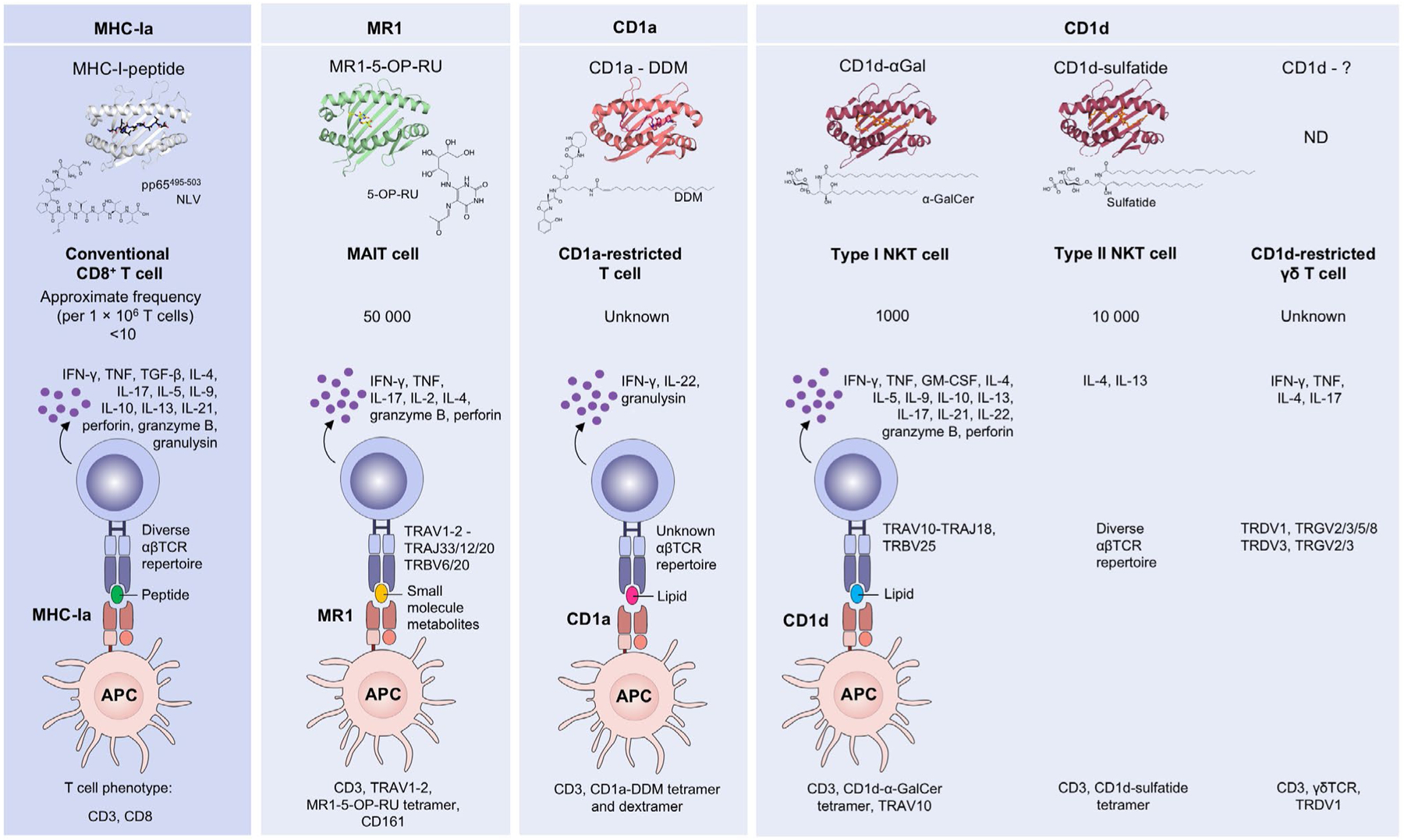
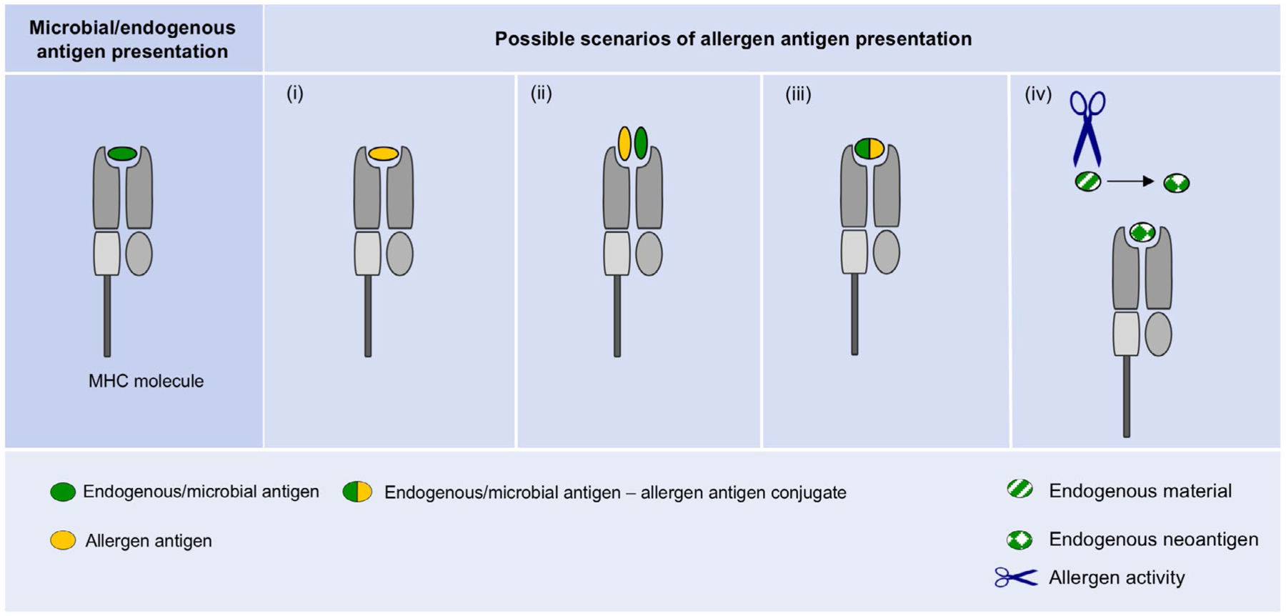
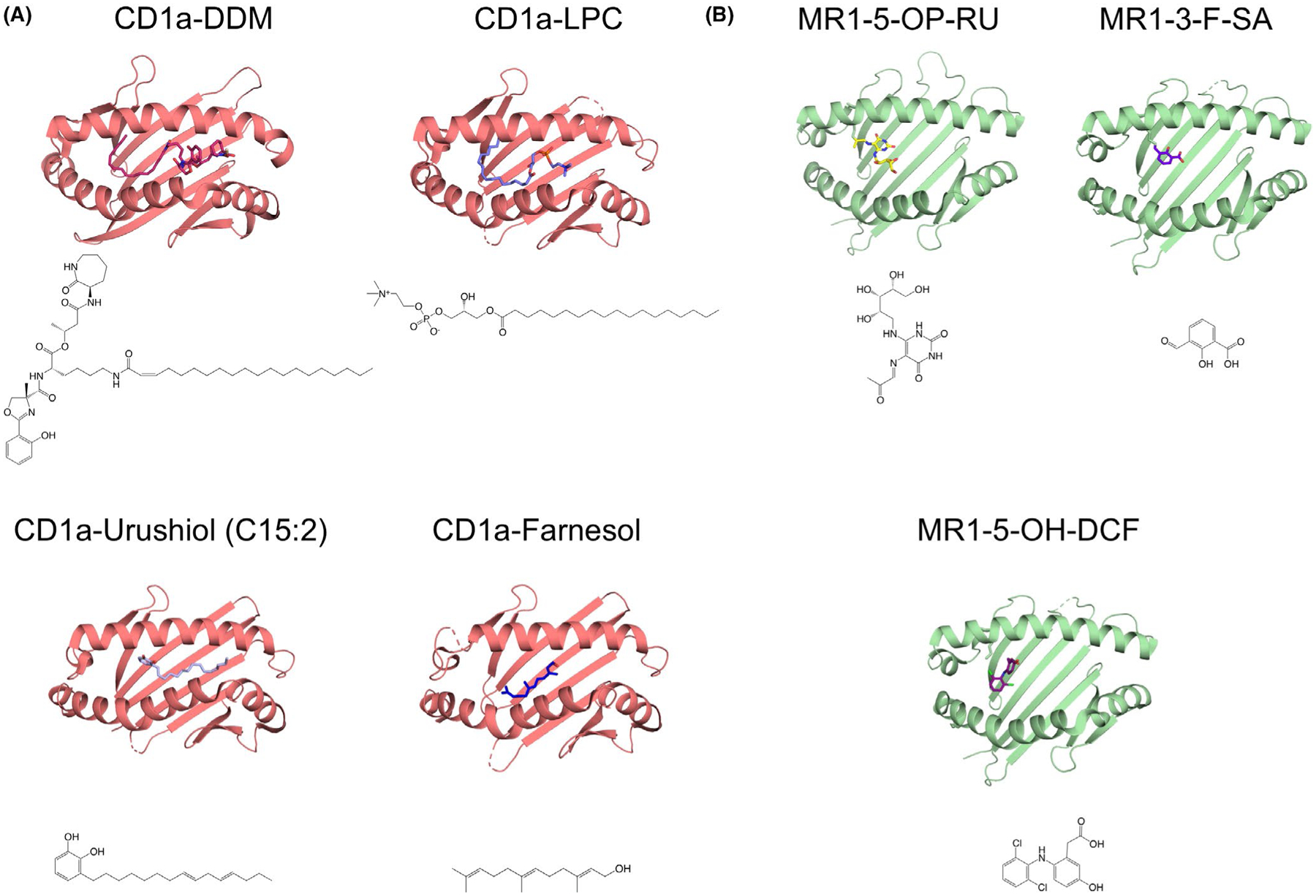
Conventional T cells recognise protein-derived antigens in the context of major histocompatibility complex (MHC) class Ia and class II molecules and provide anti-microbial and anti-tumour immunity. Conventional T cells have also been implicated in type IV (also termed delayed-type or T cell-mediated) hypersensitivity reactions in response to protein-derived allergen antigens. In addition to conventional T cells, subsets of unconventional T cells exist, which recognise non-protein antigens in the context of monomorphic MHC class I-like molecules. These include T cells that are restricted to the cluster of differentiation 1 (CD1) family members, known as CD1-restricted T cells, and mucosal-associated invariant T cells (MAIT cells) that are restricted to the MHC-related protein 1 (MR1). Compared with conventional T cells, much less is known about the immune functions of unconventional T cells and their role in hypersensitivities. Here, we review allergen antigen presentation by MHC-I-like molecules, their recognition by unconventional T cells, and the potential role of unconventional T cells in hypersensitivities. We also speculate on possible scenarios of allergen antigen presentation by MHC-I-like molecules to unconventional T cells, the hallmarks of such responses, and the expected frequencies of hypersensitivities within the human population.

Keywords: CD1; MAIT cells; MR1; NKT cells; antigen.


Conflict of interest statement

CONFLICT OF INTEREST

James McCluskey, Zhenjun Chen and Sidonia BG Eckle are inventors on patents describing MR1 antigens and MR1 tetramers. The other authors have no conflict of interest in relation to this work.

Figures

FIGURE 1
FIGURE 1
Overview of antigen presentation by MHC-I and MHC-I-like molecules and their recognition by T cells. Row 1 displays top-views onto the antigen cleft based on crystal structures of HLA-A*02:01 in complex with the cytomegalovirus pp65-derived peptide antigen NLV495−503 (PDB ID: 2X4R), as a representative of an MHC-Ia molecule; MR1 in complex with the bacterial/fungal small molecule metabolite antigen 5-(2-oxopropylideneamino)-6-d-ribitylaminouracil (5-OP-RU) derived from a biosynthetic precursor to riboflavin (PDB ID: 4NQC); CD1a in complex with the Mycobacterium tuberculosis lipid antigen Dideoxymycobactin (DDM) (PDB ID: IXZ0); and CD1d in complex with the marine sponge–derived lipid antigen α-galactosylceramide (α-GalCer) (PDB ID: 2PO6) or the endogenous lipid antigen sulfatide (PDB ID: 4MQ7). Row 2 shows chemical structures of the relevant antigens, followed by the name and approximate frequency of the relevant MHC-restricted T-cell type in row 3. The frequency of CD1d-restricted γδ T cells, most of which recognise endogenous lipid antigens and sulfatides, is estimated to be 0.05%−3.5% of CD1d-α-GalCer reactive cells. Row 4 shows schematics of the antigen presentation by antigen-presenting cells (APCs) and their recognition by T-cell receptors (TCRs) expressed by T cells. In each case, the antigen type, TCR usage and effector function molecules are highlighted.,,,,,,,– Row 5 includes phenotypic markers commonly used to identify these cells by flow cytometry,–
FIGURE 2
FIGURE 2
Possible scenarios of allergen antigen presentation by MHC-I-like molecules. A schematic of the four possible scenarios of allergen antigen display by MHC-I-like molecules in comparison with microbial/endogenous antigen presentation (left column). (i) The allergen antigen replaces the microbial/endogenous antigen. (ii) The allergen antigen and a microbial or endogenous antigen are simultaneously presented, but as distinct entities. The repertoire of microbial or endogenous antigens may be identical to that presented in the absence of the allergen antigen or distinct. (iii) The allergen antigen is directly conjugated to an antigen (iii), selected from the microbial or endogenous Ag repertoire that is either identical or distinct to that presented in the absence of the allergen antigen. (iv) The allergen might “act on” endogenous material, eliciting presentation of endogenous antigens (in the absence of allergen antigen and referred to as neoantigens), that would not be presented in steady state or that are normally presented at very low levels
FIGURE 3
FIGURE 3
Allergen antigen presentation by CD1a and MR1. (A) Chemical structures of the Mycobacterium tuberculosis–derived antigen Dideoxymycobactin (DDM), the bee/wasp venom allergen-derived antigen Lysophosphatidylcholine (LPC), the poison ivy allergen-derived antigen urushiol (C15:2) and the allergen antigen farnesol contained in cosmetics/perfumes and their presentation by CD1a (PDB IDs: IXZ0, 4X6E, 5JIA, 6NUX), displaying top-views onto the CD1a-antigen complexes. (B) Chemical structures of the bacterial/fungal riboflavin biosynthesis derived antigen 5-(2-oxopropylideneamino)-6-d-ribitylaminouracil (5-OP-RU), the drug-like small molecule antigen 3-formyl-salicylic acid (3-F-SA) and the diclofenac drug metabolite antigen 5-hydroxy-diclofenac (5-OH-DCF) and their presentation by MR1 (PDB IDs: 4NQC, 5U6Q, 5U72), displaying top-views onto the MR1-antigen complexes

Similar articles

Cited by

References

    1. Gell PGH, Coombs RRA. Clinical Aspects of Immunology. 24–25, Broad Street. Oxford: Blackwell Scientific Publications Ltd.; 1963.
    1. Uzzaman A, Cho SH. Chapter 28: Classification of hypersensitivity reactions. Allergy asthma Proc. 2012;33:96–99. - PubMed
    1. Woodfolk JA. T-cell responses to allergens. J Allergy Clin Immunol. 2007;119(2):280–294. - PubMed
    1. Imai K, Kanno M, Kimoto H, Shigemoto K, Yamamoto S, Taniguchi M. Sequence and expression of transcripts of the T-cell antigen receptor α-chain gene in a functional, antigen-specific suppressor-T-cell hybridoma. Proc Natl Acad Sci USA. 1986;83(22):8708–8712. - PMC - PubMed
    1. Fowlkes BJ, Kruisbeek AM, Ton-That H, et al. A novel population of T-cell receptor αβ-bearing thymocytes which predominantly expresses a single Vβ gene family. Nature. 1987;329 (6136):251–254. - PubMed

Publication types

Substances