Review
. 2020 Apr;9(8):e1901682.
doi: 10.1002/adhm.201901682. Epub 2020 Mar 17.

Engineered Biomaterial Platforms to Study Fibrosis

Affiliations
Review

Engineered Biomaterial Platforms to Study Fibrosis

Matthew D Davidson et al. Adv Healthc Mater. 2020 Apr.

Abstract

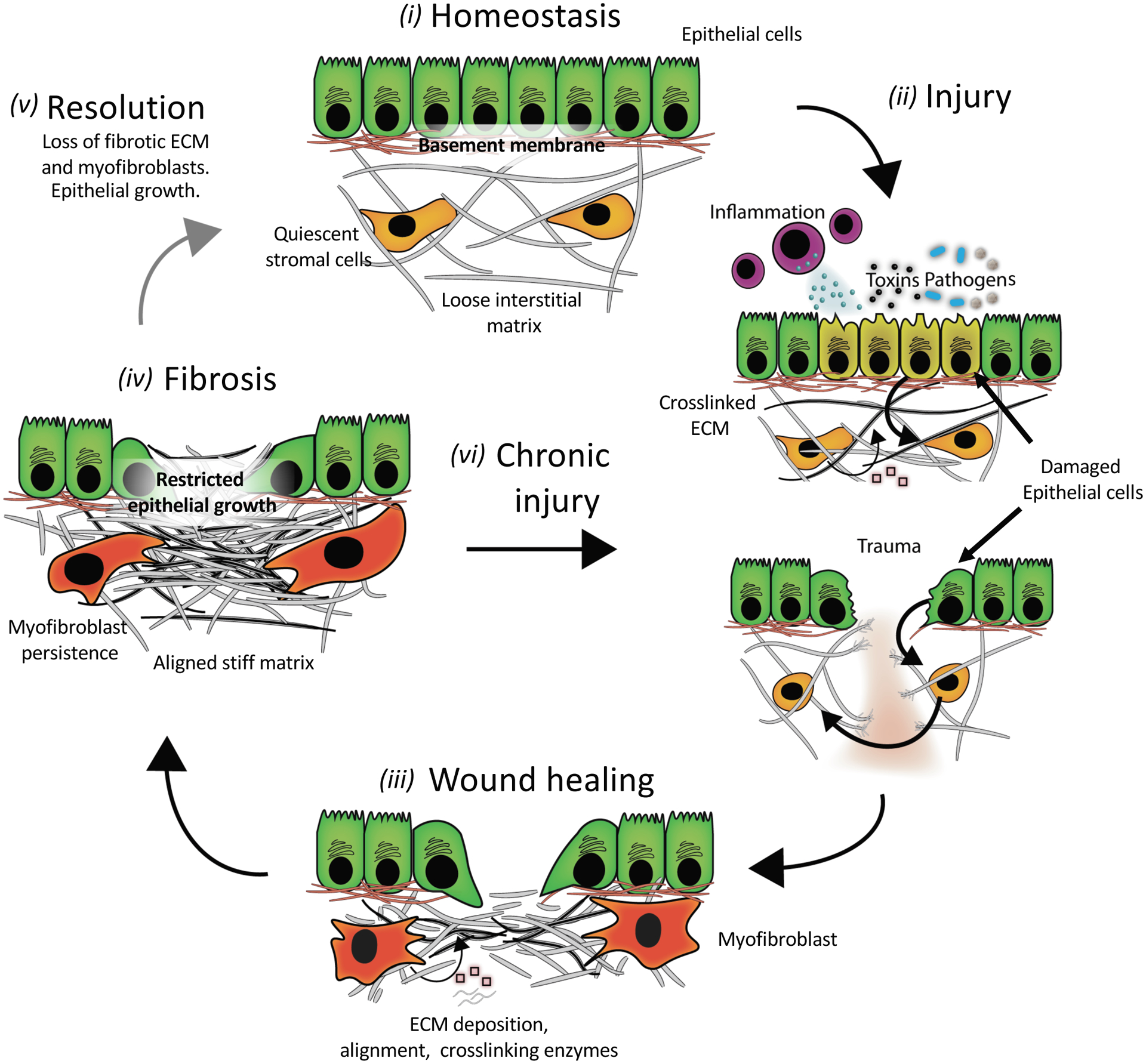
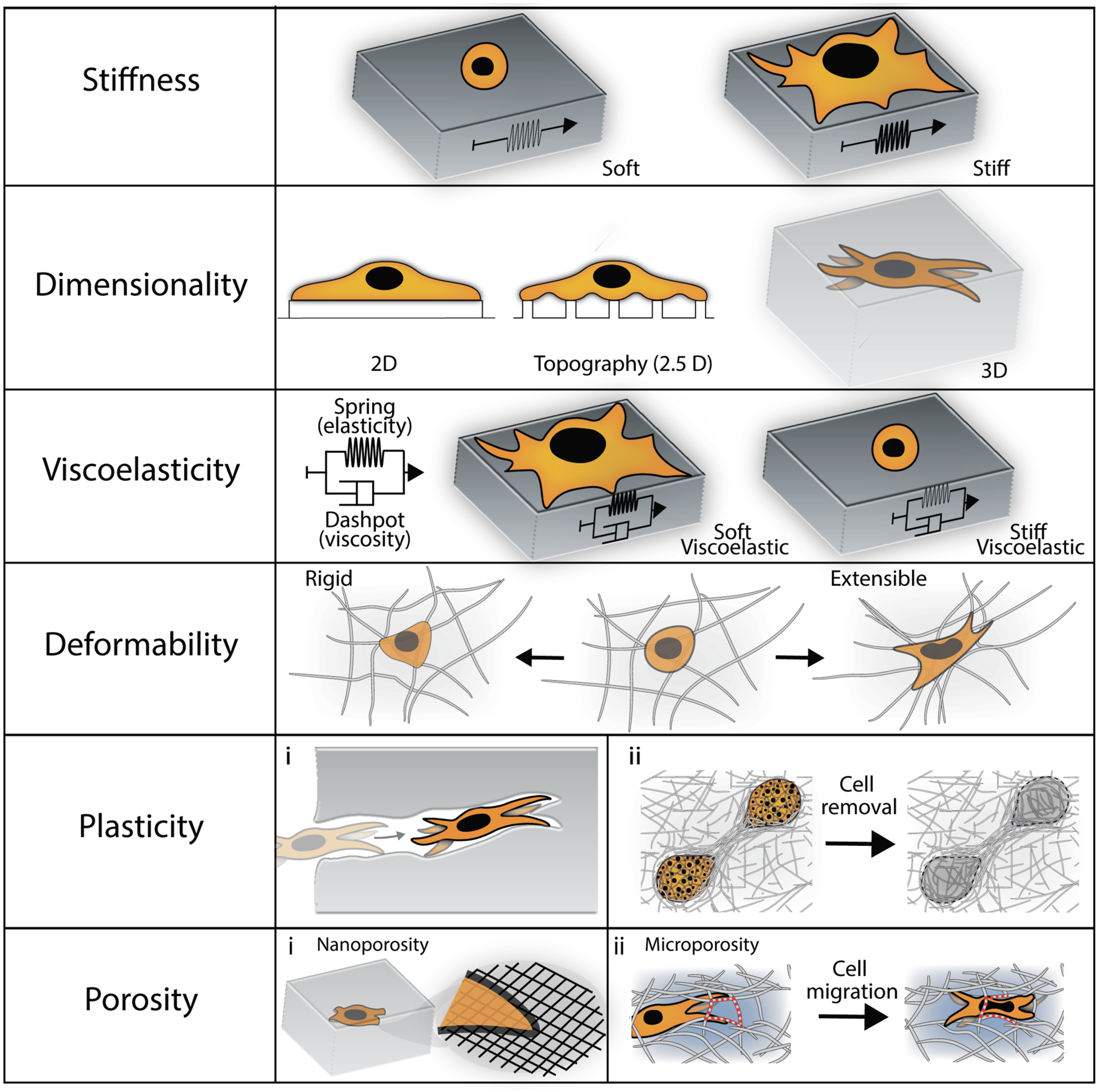
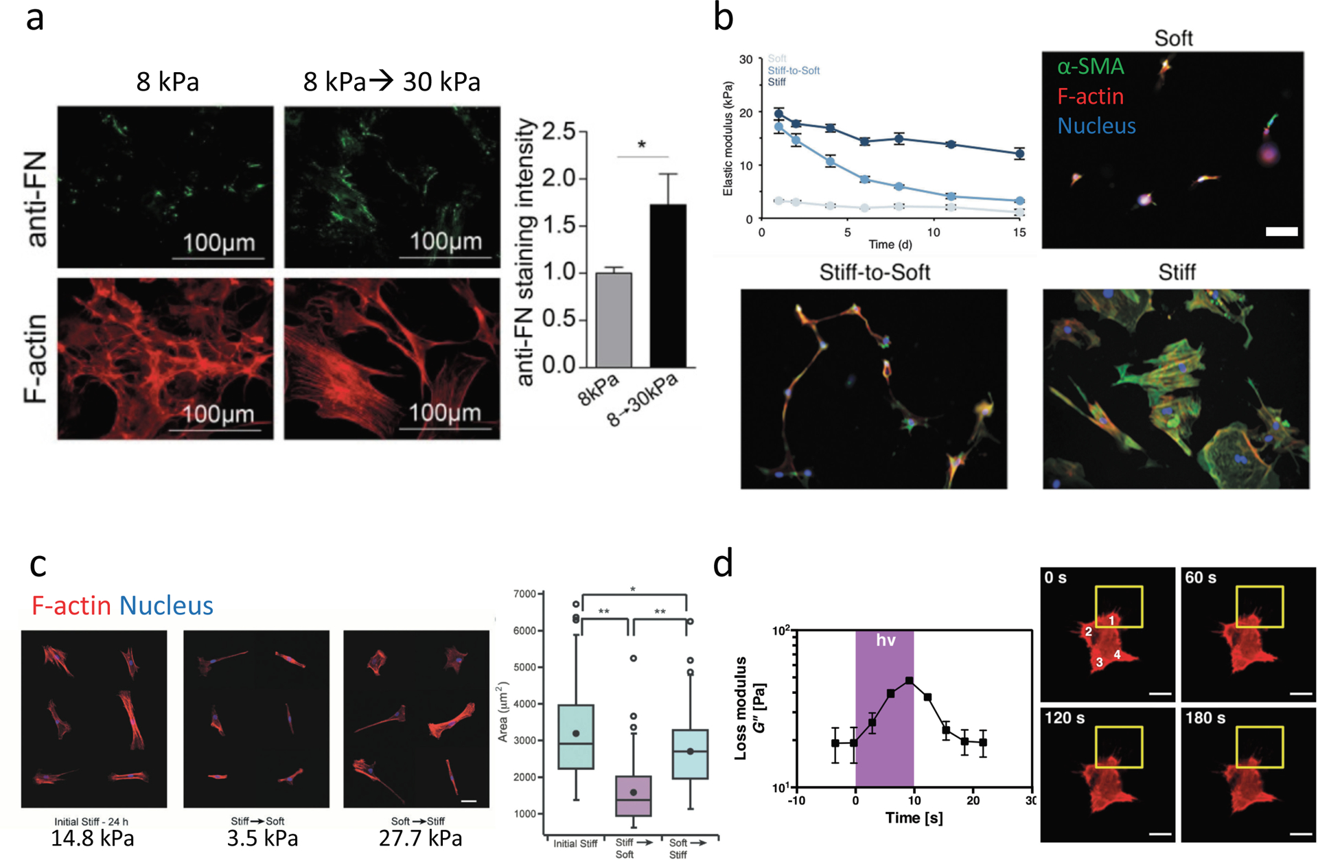
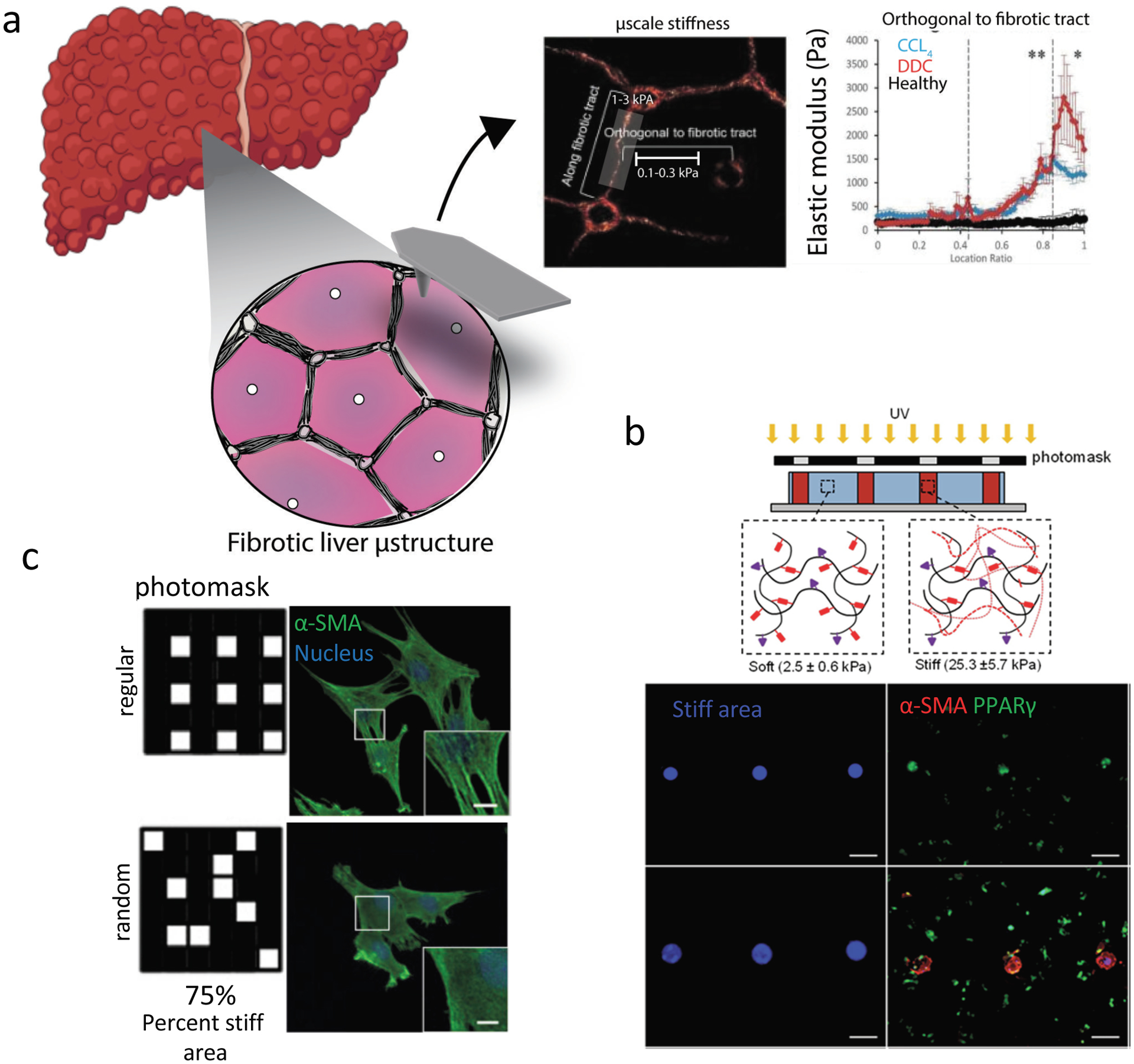
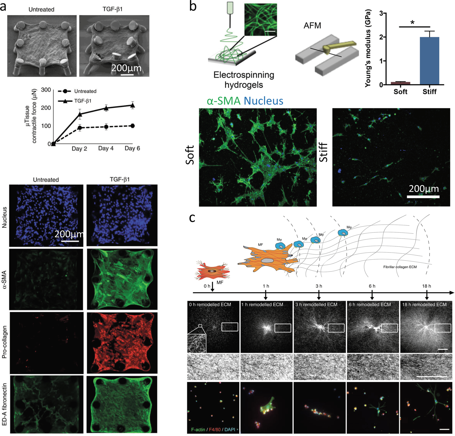
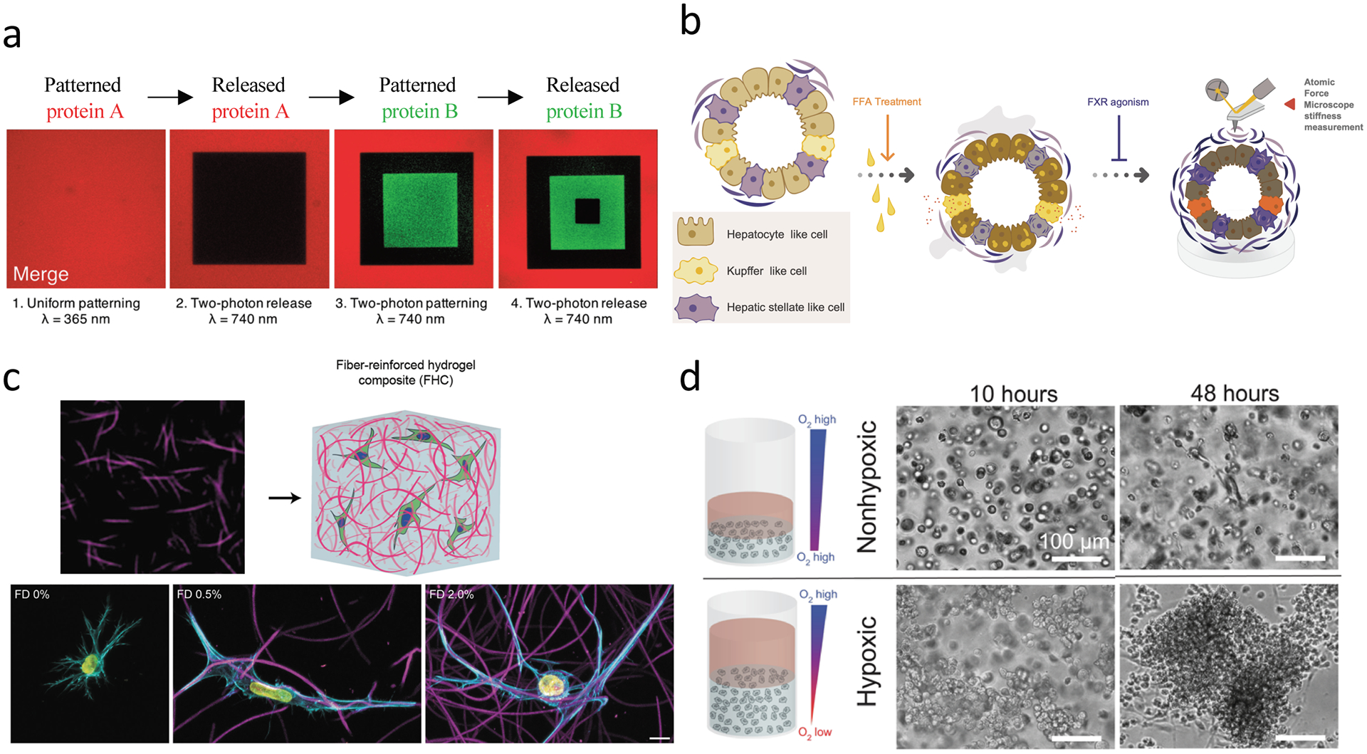
Many pathologic conditions lead to the development of tissue scarring and fibrosis, which are characterized by the accumulation of abnormal extracellular matrix (ECM) and changes in tissue mechanical properties. Cells within fibrotic tissues are exposed to dynamic microenvironments that may promote or prolong fibrosis, which makes it difficult to treat. Biomaterials have proved indispensable to better understand how cells sense their extracellular environment and are now being employed to study fibrosis in many tissues. As mechanical testing of tissues becomes more routine and biomaterial tools become more advanced, the impact of biophysical factors in fibrosis are beginning to be understood. Herein, fibrosis from a materials perspective is reviewed, including the role and mechanical properties of ECM components, the spatiotemporal mechanical changes that occur during fibrosis, current biomaterial systems to study fibrosis, and emerging biomaterial systems and tools that can further the understanding of fibrosis initiation and progression. This review concludes by highlighting considerations in promoting wide-spread use of biomaterials for fibrosis investigations and by suggesting future in vivo studies that it is hoped will inspire the development of even more advanced biomaterial systems.

Keywords: biomaterial systems; biomaterials; extracellular matrices; fibrosis studies; mechanobiology.


Figures

Figure 1.
Figure 1.. A simplified schematic of fibrosis pathogenesis.
(i) An ECM network in a healthy tissue has a stiffness that supports epithelial cell function and stromal cells in a quiescent state. (ii) Common injuries to tissues include trauma, autoimmune reactions (inflammation), chemicals (toxins), and pathogens (bacteria and viral infections). Acute signaling between injured epithelial and stromal cells triggers ECM remodeling (degradation and/or stiffening (pink squares are ECM modifying proteins)). (iii) During the wound-healing response, myofibroblasts secrete ECM (grey lines) and ECM-modifying proteins while also contracting the matrix to re-establish tensional homeostasis. This provisional matrix can provide a substrate for re-epithelialization; however, excessive matrix deposition and stiffening can lead to (iv) scarring or fibrosis that alters epithelial growth and differentiation. Fibrosis is marked by high levels of stiff ECM such as crosslinked collagen, with resulting myofibroblast persistence. In some tissues, fibrosis resolves (v) if the underlying stimulus for injury is removed, but often (vi) chronic injury leads to a progressive cycle of further wound healing and fibrosis without removal of excess ECM.
Figure 2.
Figure 2.. Schematic of important structural and mechanical features to incorporate into biomaterial platforms to investigate fibrosis.
These include various features of the extracellular matrix, as well as outcomes of cellular behavior, such as spreading, morphology and migration.
Figure 3.
Figure 3.. Dynamic hydrogels mimic changes in mechanical properties observed in fibrosis.
(a) Stiffening methacrylated-HA hydrogels can be used to mimic the changes in stiffness that occur during cardiac fibrosis though through photoinitiated (UV) crosslinking of methacrylate groups to stiffen gels from 8 kPa to 30 kPa in the presence of cells. Cardiac fibroblasts seeded on 8 kPa hydrogels and either stiffened to 30 kPa or maintained at 8 kPa showed increased fibrosis markers with increased fibronectin deposition (top and quantified on right) and large f-actin stress fibers (bottom). (b) Degradable HA hydrogels that gradually soften over time due to crosslinker hydrolysis can be used to mimic regression of liver fibrosis. Activated hepatic stellate cells (myofibroblasts) seeded on softening hydrogels (stiff-to-soft) develop an intermediate phenotype (with regards to α-SMA expression and cell spreading) between quiescent stellate cells continuously cultured on soft hydrogels (soft), and activated stellate cells continuously cultured on stiff hydrogels (stiff). Scale bar 100 μm. (c) Dynamic softening and subsequent stiffening hydrogels can be designed to mimic repeated changes in ECM mechanics using HA hydrogels synthesized with photodegradable o-nitrobenzyl and methacrylate crosslinking groups that undergo softening (14.8 kPa→3.5 kPa) through photodegradation of the o-nitrobenzyl crosslinker, and subsequent stiffening (3.5 kPa--> 27.7 kPa) through photoinitiated methacrylate crosslinking. MSCs seeded on hydrogels and then softened showed reduced spread area, while subsequent stiffening increased spread area, as assessed by staining cells for f-actin after each change in stiffness. (d) Photoinitiator (Irgacure 2959)-modified PEG hydrogels polymerized with crosslinkers that participate in additional fragmentation chain transfer reactions (e.g., allyl sulfide bis(azide)) undergo acute changes in loss modulus (G”) upon exposure to light through crosslinker rearrangement, which subsides after cessation of light (left). Live imaging of MSC f-actin shows responses to local changes in viscoelasticity (yellow square) over time by retracting cell projections and locally reducing cell area. Scale bar 20μm. *,**, represent p≤0.05, p≤0.01. Part (a) is adapted with permission from ref.[70] CC-BY-3.0 (https://creativecommons.org/licenses/by-nc-sa/3.0/) 2017 ASCB®, part (b) is adapted with permission from ref. [79] 2016 Oxford University Press, part (c) is adapted with permission from ref. [63] 2017 John Wiley and Sons, part (d) is adapted with permission from ref. [93] 2019 ] CC-BY-3.0 (https://creativecommons.org/licenses/by-nc-sa/3.0/) IOP Publishing.
Figure 4.
Figure 4.. Mechanical heterogeneity in fibrosis and biomaterial systems to mimic this
. (a) Fibrosis in the liver is characterized by fibrotic tracts which ultimately (in cirrhosis) form nodules. When characterized by atomic force microscopy (AFM), fibrotic tracts of the diseased liver have higher elastic moduli than non-fibrotic regions. (b) Patterned stiffness, similar to fibrotic regions in tissue, can be created within soft hydrogels to probe how stiff area influences myofibroblast activation. Stiff patterns can be created within methacrylated-HA hydrogels using a photomask and photoinitiated crosslinking (top, bottom left (blue)). Quiescent hepatic stellate cell activation, assessed by loss of PPARγ and increased α-SMA expression (bottom, right), is promoted by large (multicellular sized) stiff patterns, while cell-sized patterns to not lead to activation. Scale bar is 200 μm. (c) To mimic subcellular heterogeneity in ECM mechanical properties observed in tissues, high resolution photomasks can be used to create small 4 μm2 patterns of softened (photodegraded) gel within photodegradable PEG-o-nitrobenzyl hydrogels. Regular and random patterns of soft matrix can be patterned on stiff matrix, and the response of valvular interstitial cells (VICs) can be probed. VICs seeded on regular patterns have increased myofibroblast features (including α-SMA-containing stress fibers), when compared to VICs seeded on random patterns. Scale bar is 2 μm. Part (a) is adapted with permission from ref. [43] 2016 John Wiley and Sons, part (b) is adapted with permission from ref. [81] 2014 Elsevier, part (c) is adapted with permission from ref. [84] 2017 Elsevier.
Figure 5.
Figure 5.. Fibrous biomaterials to investigate fibrosis.
(a) Lung fibrosis models are fabricated with lung fibroblasts embedded in fibrillar type I collagen hydrogels. Lung fibroblasts contract collagen matrix around microfabricated elastomeric (PDMS) posts to create microtissues, which bend soft microposts in response to microtissue contractile forces (observed through SEM). Lung fibroblast microtissues treated with TGF-ß1 display fibrosis-relevant phenotypes with increased contraction of the matrix (higher levels of micro-post bending in SEM image, (top)), and fibrosis relevant markers (bottom) such as α-SMA labeling and collagen/ED-A fibronectin deposition. (b) To mimic fibrotic fibrous ECM, HA can be modified with different levels of methacrylate groups to give soft (low modification) and stiff (high modification) electrospun fibrous hydrogel networks (top). Fibrous networks are created atop microfabricated wells to isolate cell-fiber interactions. Quiescent hepatic stellate cells undergo myofibroblast activation and form large α-SMA positive multicellular clusters over 7 days on soft fibers (bottom, left), while stiff fibers prevent myofibroblast activation and highly spread cluster formation (bottom, right). (c) To investigate heterotypic cell-cell interactions in fibrous environments, macrophages and fibroblasts can be co-seeded on top of type I collagen gels and their interactions tracked over time with f-actin (myofibroblast) and F4–80 (macrophages) (bottom) labeling, while observing collagen remodeling (center panels). Macrophages are seeded at various times after myofibroblast seeding, and are attracted to myofibroblasts during initial ECM contraction due to dynamic tugging of the ECM which activates macrophage integrin signaling and directs migration (top). Myofibroblast-induced ECM alignment does not appear to direct macrophage migration. Scale bar 100 μm. Part (a) is adapted with permission from ref.[97] CC-BY-4.0 (https://creativecommons.org/licenses/by/4.0/) 2018 Springer Nature, Part (b) is adapted with permission from ref.[99] 2019 American Chemical Society, Part (c) is adapted with permission from ref.[102] CC-BY-4.0 (https://creativecommons.org/licenses/by/4.0/) 2019 Springer Nature.
Figure 6.
Figure 6.. Emerging technologies to study fibrosis.
(a) Sequential tethering and release of signaling factors (e.g., ECM ligands or fibrogenic factors such as TGF-ß1) can be achieved using allyl sulfide-modified PEG hydrogels, and could be used to understand spatial heterogeneity in ECM factors/properties and their contribution to fibrosis development. (b) Multicellular organoids have been developed from iPSCs, which enable stromal cell-epithelial cell interactions using personalized cell cultures. These methods could be expanded to use in biomaterial systems where the influence of ECM properties on fibrosis outcomes could be studied. (c) Heterogeneity in ECM topography and mechanical properties can be recapitulated with composite hydrogel systems composed of Dextran-vinyl sulfone (VS) electrospun fibers embedded in tunable bulk photocrosslinked hydrogels (gelatin-methacrylate hydrogel shown here). Fibroblasts display increased spreading and protrusions in 3D in response to local fiber density. Scale bar is 10 μm. (d) Hypoxia-inducible hydrogels can be fabricated with ferulic acid-modified polymers, where oxygen is consumed in a laccase-mediated reaction that crosslinks ferulic acid groups and simultaneously creates a hypoxic environment. Hypoxia-inducible gels promote multi-cellular vascular network formation with endothelial-colony-forming cells (ECFCs, bottom), while non-hypoxic gels show low levels of single cell network formation (top). Scale bar is 100 μm. Hypoxia-inducible gels have tunable mechanical properties, and thus could be explored to probe the impact of hypoxia and mechanics on fibrosis outcomes. Part (a) is adapted with permission from ref.[117] https://pubs.acs.org/doi/full/10.1021/acscentsci.8b00325 2019 American Chemical Society, part (b) is adapted with permission from ref.[121] 2019 Elsevier, part (c) is adapted with permission from ref.[86] 2019 American Chemical Society, part (d) is adapted with permission from ref.[131] CC BY-NC 4.0 https://creativecommons.org/licenses/by-nc/4.0/legalcode 2019 AAAS.

References

    1. Lampi MC, Reinhart-King CA, Sci. Transl. Med 2018, 10, 475. - PubMed
    1. Yang L, Van Der Werf KO, Koopman BFJM, Subramaniam V, Bennink ML, Dijkstra PJ, Feijen J, J. Biomed. Mater. Res. - Part A 2007, 82, 160. - PubMed
    1. Ban E, Franklin JM, Nam S, Smith LR, Wang H, Wells RG, Chaudhuri O, Liphardt JT, Shenoy VB, Biophys. J 2018, 114, 450. - PMC - PubMed
    1. Tomasek JJ, Gabbiani G, Hinz B, Chaponnier C, Brown RA, Nat. Rev. Mol. Cell Biol 2002, 3, 349. - PubMed
    1. Wang H, Abhilash AS, Chen CS, Wells RG, Shenoy VB, Biophys. J 2014, 107, 2592. - PMC - PubMed

Publication types

Substances