. 2020 Apr 8;142(14):6661-6674.
doi: 10.1021/jacs.0c00188. Epub 2020 Mar 27.

Design of a Functionalized Metal-Organic Framework System for Enhanced Targeted Delivery to Mitochondria

Affiliations

Design of a Functionalized Metal-Organic Framework System for Enhanced Targeted Delivery to Mitochondria

Salame Haddad et al. J Am Chem Soc. .

Abstract

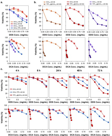
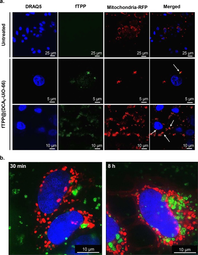
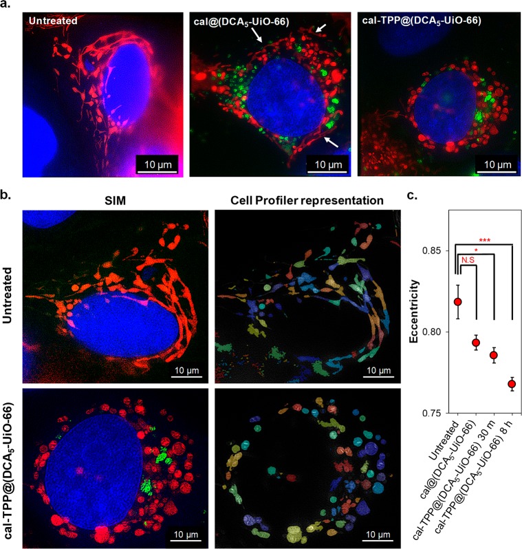
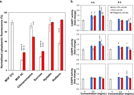
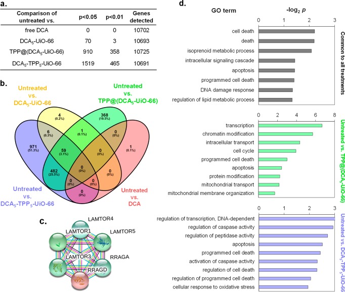
Mitochondria play a key role in oncogenesis and constitute one of the most important targets for cancer treatments. Although the most effective way to deliver drugs to mitochondria is by covalently linking them to a lipophilic cation, the in vivo delivery of free drugs still constitutes a critical bottleneck. Herein, we report the design of a mitochondria-targeted metal-organic framework (MOF) that greatly increases the efficacy of a model cancer drug, reducing the required dose to less than 1% compared to the free drug and ca. 10% compared to the nontargeted MOF. The performance of the system is evaluated using a holistic approach ranging from microscopy to transcriptomics. Super-resolution microscopy of MCF-7 cells treated with the targeted MOF system reveals important mitochondrial morphology changes that are clearly associated with cell death as soon as 30 min after incubation. Whole transcriptome analysis of cells indicates widespread changes in gene expression when treated with the MOF system, specifically in biological processes that have a profound effect on cell physiology and that are related to cell death. We show how targeting MOFs toward mitochondria represents a valuable strategy for the development of new drug delivery systems.


Conflict of interest statement

The authors declare no competing financial interest.

Figures

Figure 1
Figure 1
MTS viability assay of MCF-7 cells after incubation with different systems. (a) Cell viability as a function of DDS (top) and equivalent DCA concentration (bottom) after incubation for 72 h with different DCA-loaded DDSs. (b) Cell viability as a function of DDS (top) and equivalent DCA concentration (bottom) after incubation for 72 h with DCAx-UiO-66 and TPP@(DCAx-UiO-66). (c) Cell viability as a function of DDS (top) and equivalent DCA concentration (bottom) after incubation for 4–72 h with DCA5-UiO-66, TPP@(DCA5-UiO-66), and DCA5-TPP5-UiO-66. Error bars—sometimes smaller than the symbol sizes—represent the standard error of the mean of five replicate measurements.
Figure 2
Figure 2
Microscopy imaging of MCF-7 cells. (a) Confocal microscopy images of cells incubated for 2 h with fTPP@(DCA5-UiO-66). (b) SIM images of cells incubated for 30 min (left) and 8 h (right) with cal-TPP@(DCA5-UiO-66). fTPP and calcein are shown in green, mitochondria stained with RFP are shown in red, and nuclei stained with DRAQ-5 are shown in blue. White arrows in confocal microscopy show overlap between red and green signals.
Figure 3
Figure 3
SIM imaging of MCF-7 cells. (a) Images of untreated cells and cells treated with cal@(DCA5-UiO-66) and cal-TPP@(DCA5-UiO-66) for 8 h; mitochondria are colored in red, MOFs in green, and nuclei in blue; white arrows indicate stringy mitochondria. (b) Images showing a shape analysis of mitochondria using Cell Profiler software. Top row, untreated cell; bottom row, cell after 8 h of incubation with cal-TPP@(DCA5-UiO-66). (c) The effects of different treatments on the eccentricity of mitochondria. Results show the average eccentricity of at least 200 mitochondria. Error bars represent the standard error of the mean. Statistical significance was assessed using one-way ANOVA followed by Tukey’s multiple comparisons test.
Figure 4
Figure 4
Final fate of MOF nanoparticles in MCF-7 cells. (a) Effects of pharmacological endocytosis inhibitors on the uptake of cal@(DCA5-UiO-66) (white bars) and cal-TPP@(DCA5-UiO-66) (red bars) by MCF-7 cells, measured by flow cytometry. (b) Caspase 7, 8, and 9 activities after incubation of MCF-7 cells for 4 and 8 h with DCA5-UiO-66, DCA5-TPP5-UiO-66, and TPP@(DCA5-UiO-66). Samples were run in triplicate; error bars represent the standard error of the mean. Statistical significance was assessed using one-way ANOVA followed by Dunnett’s multiple comparisons test.
Figure 5
Figure 5
Biochemical effects of different treatments on MCF-7 cells. (a) Number of differentially expressed genes between cells treated with different conditions. (b) Venn diagram analysis of differentially expressed genes in microarrays of MCF-7 cells treated with DCA5-UiO-66, DCA5-TPP5-UiO-66, and TPP@(DCA5-UiO-66) compared to untreated control. (c) Gene network displaying interconnected genetic targets in common for all three treatments. (d) Significant gene ontology (GO) terms of associated biological processes from 59 differentially expressed genes (p < 0.05) in MCF-7 cells in common for all three treatments (top), treated with TPP@(DCA5-UiO-66) (middle), and treated with DCA5-TPP5-UiO-66 (bottom).

References

    1. Porporato P. E.; Filigheddu N.; Pedro J. M. B.-S.; Kroemer G.; Galluzzi L. Mitochondrial Metabolism and Cancer. Cell Res. 2018, 28 (3), 265–280. 10.1038/cr.2017.155. - DOI - PMC - PubMed
    1. Voet D.; Voet J. G.; Pratt C. W.. Fundamentals of Biochemistry, 2nd ed.; John Wiley and Sons: 2006.
    1. Alkarakooly Z.; Kilaparty S. P.; Al-Anbaky Q. A.; Khan M. S.; Ali N. Dichloroacetic Acid (DCA)-Induced Cytotoxicity in Human Breast Cancer Cells Accompanies Changes in Mitochondrial Membrane Permeability and Production of Reactive Oxygen Species. J. Cancer Ther. 2014, 5, 1234–1248. 10.4236/jct.2014.513125. - DOI
    1. Fulda S. Tumor Resistance to Apoptosis. Int. J. Cancer 2009, 124 (3), 511–515. 10.1002/ijc.24064. - DOI - PubMed
    1. Hanahan D.; Weinberg R. A. The Hallmarks of Cancer. Cell 2000, 100 (1), 57–70. 10.1016/S0092-8674(00)81683-9. - DOI - PubMed

Publication types

MeSH terms

Substances