. 2020 Aug;63(2):198-208.
doi: 10.1165/rcmb.2019-0170OC.

The Transient Receptor Potential Channel Vanilloid 1 Is Critical in Innate Airway Epithelial Responses to Protease Allergens

Affiliations

The Transient Receptor Potential Channel Vanilloid 1 Is Critical in Innate Airway Epithelial Responses to Protease Allergens

Caspar Schiffers et al. Am J Respir Cell Mol Biol. 2020 Aug.

Abstract

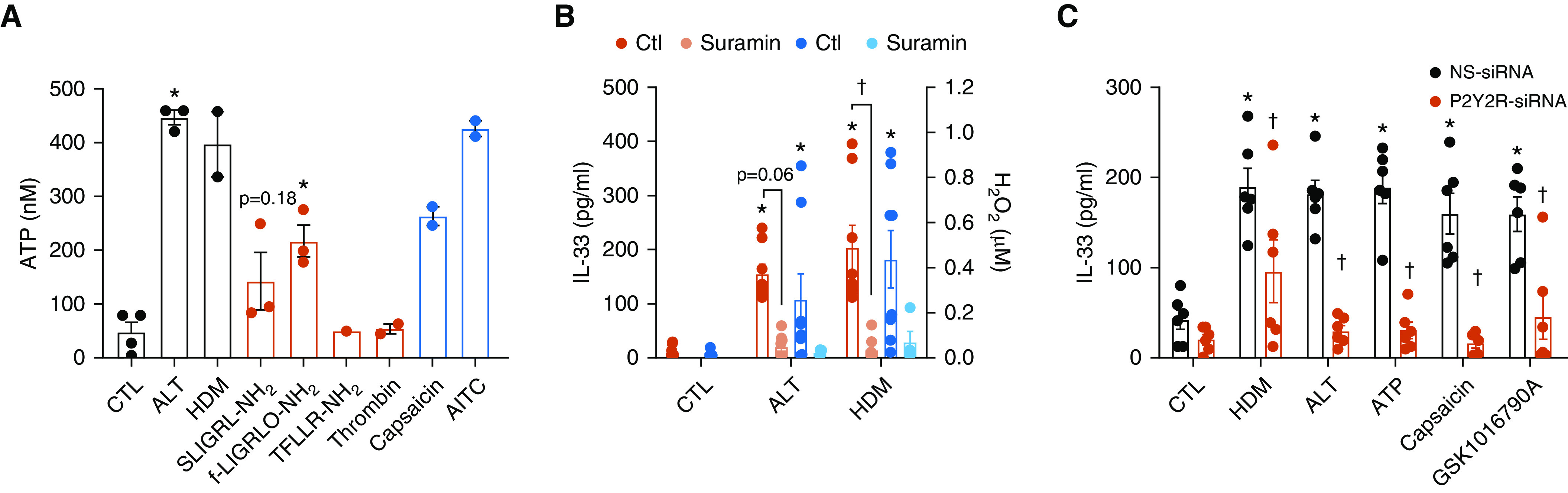
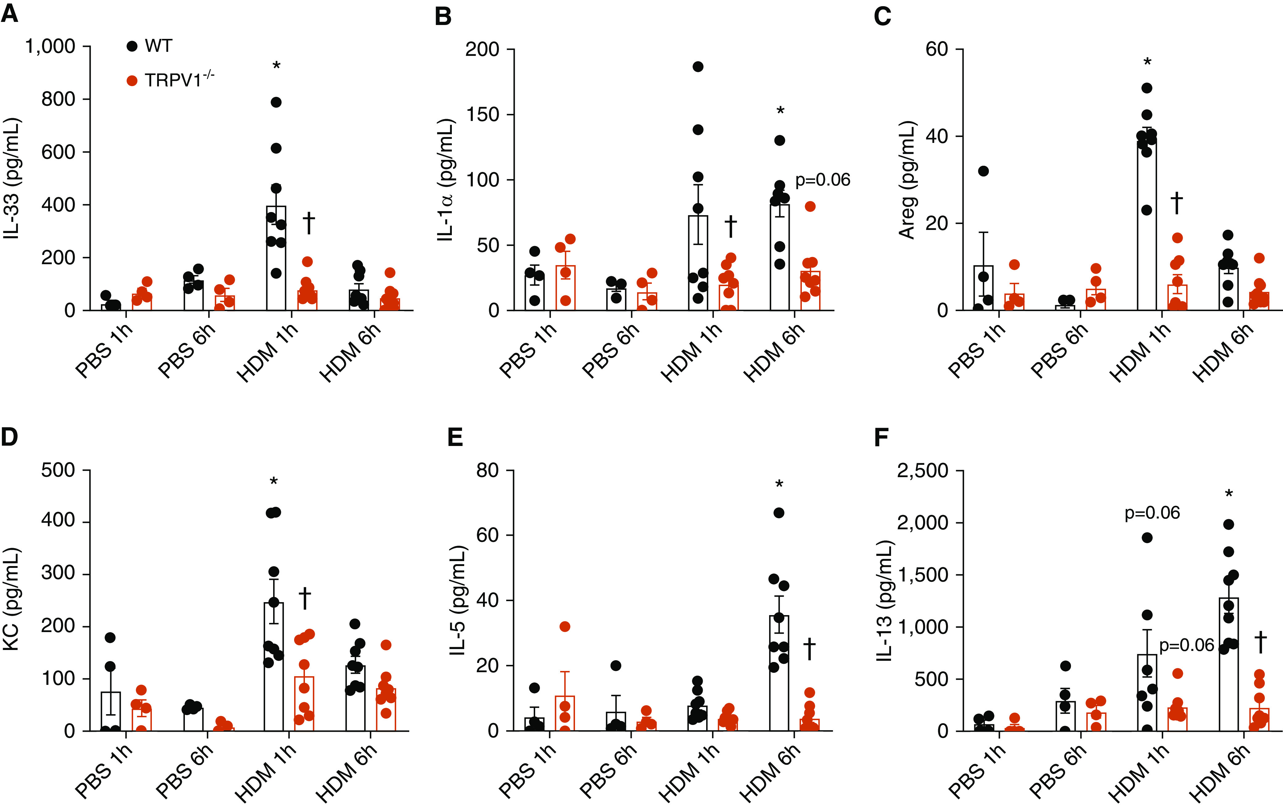
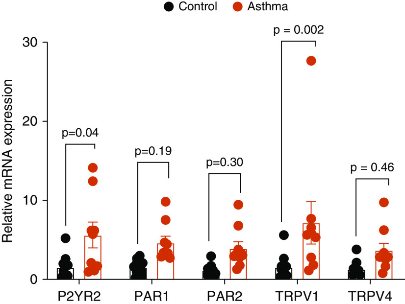
The airway epithelium plays a critical role in innate responses to airborne allergens by secreting IL-1 family cytokines such as IL-1α and IL-33 as alarmins that subsequently orchestrate appropriate immune responses. Previous studies revealed that epithelial IL-33 secretion by allergens such as Alternaria alternata or house dust mite involves Ca2+-dependent signaling, via initial activation of ATP-stimulated P2YR2 (type 2 purinoceptor) and subsequent activation of the nicotinamide adenine dinucleotide phosphate (NADPH) oxidase DUOX1. We sought to identify proximal mechanisms by which epithelial cells sense these allergens and here highlight the importance of PAR2 (protease-activated receptor 2) and TRP (transient receptor potential) Ca2+ channels such as TRPV1 (TRP vanilloid 1) in these responses. Combined studies of primary human nasal and mouse tracheal epithelial cells, as well as immortalized human bronchial epithelial cells, indicated the importance of both PAR2 and TRPV1 in IL-33 secretion by both Alternaria alternata and house dust mite, based on both pharmacological and genetic approaches. TRPV1 was also critically involved in allergen-induced ATP release, activation of DUOX1, and redox-dependent activation of EGFR (epidermal growth factor receptor). Moreover, genetic deletion of TRPV1 dramatically attenuated allergen-induced IL-33 secretion and subsequent type 2 responses in mice in vivo. TRPV1 not only contributed to ATP release and P2YR2 signaling but also was critical in downstream innate responses to ATP, indicating potentiating effects of P2YR2 on TRPV1 activation. In aggregate, our studies illustrate a complex relationship between various receptor types, including PAR2 and P2YR2, in epithelial responses to asthma-relevant airborne allergens and highlight the central importance of TRPV1 in such responses.

Keywords: ATP; DUOX1; IL-33; lung; type 2 immune responses.


Figures

Figure 1.
Figure 1.
Protease allergens induce IL-33 release from nasal epithelial (HNE) cells via PAR2 (protease-activated receptor 2) and TRPV1 (transient receptor potential vanilloid 1). (A) Assessment of protease activity in allergen extracts, relative to papain (μg/ml; mean ± SE; n = 2–6) after exposure to 0, 3, 10, and 30 μg/ml allergen (Alternaria alternata [ALT] or house dust mite [HDM]). *P < 0.05 versus no allergen by one-way ANOVA. (B) Inhibition of allergen-induced IL-33 release from HNE cells by the serine protease inhibitors PMSF (100 μM) or antipain (50 μg/ml) or by the PAR2 inhibitor FSLLRY-NH2 (10 μM). (C) IL-33 release and extracellular H2O2 production induced by agonists of PAR2 (SLIGRL-NH2, 50 μM; f-LIGRLO-NH2, 5 μM), but not by agonists of PAR1 (thrombin, 30 nM; TFLLR-NH2, 50 μM). *P < 0.05 versus PBS control by one-way ANOVA. (D) Inhibition of allergen-induced IL-33 release by the TRPV1 inhibitors JNJ17203212 (1 μM) or capsazepine (10 μM), but not by the TRPV4 inhibitor RN1734 (10 μM). (E) Stimulation of IL-33 release or H2O2 production by TRP channel agonists, capsaicin (10 μM), allyl isothiocyanate (AITC, 100 μM), GSK1016790A (10 nM), or acrolein (ACR; 30 μM). *P < 0.05 versus control by one-way ANOVA. Data represent mean ± SE from independent studies from two to four different donors. *P < 0.05 versus control treatment. #H2O2 production by capsaicin could not be assessed because of its interference with the H2O2 assay. CTL = control; GSK = GSK1016790A; PMSF = phenylmethyl sulfonyl fluoride.
Figure 2.
Figure 2.
Importance of type 2 purinoceptor (P2YR2) and DUOX1 in innate allergen responses. (A) Effects of protease allergens (black), PAR agonists (red), or TRPV1 agonists (blue) on extracellular ATP release from HNE cells after 15 minutes of stimulation. *P < 0.05 versus control by one-way ANOVA. (B) Inhibition of allergen-induced H2O2 production and IL-33 secretion from HNE cells by the P2YR2 inhibitor suramin (100 μM). Data represent mean ± SE from independent studies from three to four different donors. *P < 0.05 versus control by one-way ANOVA. P < 0.05 versus corresponding treatment without inhibitor by one-way ANOVA. (C) Measurement of IL-33 release from HBE1 cells (mean ± SE; n = 5–6) after 2 hours of stimulation with HDM (50 μg/ml), ALT (50 μg/ml), ATP (100 μM), the TRPV1 agonist capsaicin (10 μM), or the TRPV4 agonist GSK1016790 (10 nM), after siRNA silencing of P2YR2. *P < 0.05 versus control treatment. P < 0.05 versus corresponding non-specific (NS)-siRNA treatment. DUOX1 = dual oxidase 1.
Figure 3.
Figure 3.
Involvement of TRPV1 in allergen-induced innate epithelial responses. (A) Human bronchial epithelial (HBE1) cells were pretreated with either the TRPV1 inhibitor JNJ17203212 (1 μM) or capsazepine (10 μM) and stimulated for 2 hours with HDM (50 μg/ml) or ATP (100 μM) for analysis of IL-33 secretion in the medium (mean ± SE; n = 4). (B) Activation of IL-33 release from HBE1 cells by 2-hour stimulation with TRPV1 agonists capsaicin (10 μM) or AITC (100 μM) or the TRPV4 agonist GSK1016790 (10 nM) (mean ± SE; n = 4). (C) Representative Western blot analysis of HBE1 cell lysates for EGFR (epidermal growth factor receptor) phosphorylation after 10-minute stimulation with HDM (50 μg/ml) or ATP (100 μM), in the absence or presence of TRPV1 inhibitors JNJ17203212 (1 μM) or capsazepine (10 μM), or after stimulation with the TRPV1 agonist capsaicin (10 μM) or TRPV4 agonist GSK1016790 (10 nM). (D and E) Semi-quantitative analysis (mean ± SE; n = 4) of pEGFR/tEGFR ratios, calculated using ImageJ software and normalized to either HDM or AITC groups. Effect of TRPV1-siRNA on (F) IL-33 secretion (mean ± SE; n = 6) or (G) extracellular H2O2 production (mean ± SE; n = 2) on stimulation with HDM (50 μg/ml), ALT (50 μg/ml), ATP (100 μM), the TRPV1 agonist capsaicin (10 μM), or the TRPV4 agonist GSK1016790 (10 nM). Note that H2O2 production by capsaicin stimulation could not be assessed because of its interference with H2O2 assay. *P < 0.05 versus control treatment. P < 0.05 versus corresponding treatment without inhibitor or NS-siRNA treatment. capz = capsazepine; JNJ = JNJ17203212; pEGFR = phosphorylated EGFR on Y1068; tEGFR = total EGFR.
Figure 4.
Figure 4.
TRPV1 deficiency attenuates innate HDM responses. Mouse tracheal epithelial cells (MTECs) isolated from wild-type (WT) or TRPV1−/− mice were stimulated with HDM (50 μg/ml) or ATP (100 μM) for 2 hours, and production of (A) IL-33, (B) IL-1⍺, or (C) KC was measured in the medium by ELISA (mean ± SE; n = 8). *P < 0.05 compared with control treatment. P < 0.05 compared with corresponding treatment in WT MTECs. KC = keratinocyte chemoattractant (also known as neutrophil chemokine CXCL1).
Figure 5.
Figure 5.
TRPV1 deficiency attenuates innate HDM responses and type 2 inflammation in vivo. WT C57Bl6/J and TRPV1−/− mice were challenged with HDM extract or vehicle control, and lung lavage was collected after 1 or 6 hours, for ELISA analysis of (A) IL-33, (B) IL-1⍺, (C) Areg (amphiregulin), (D) KC, (E) IL-5, or (F) IL-13 (mean ± SE; n = 4–8). *P < 0.05 compared with corresponding PBS control. P < 0.05 compared with corresponding treatment in WT mice.
Figure 6.
Figure 6.
DUOX1 deficiency attenuates innate cytokine responses in response to capsaicin. WT C57BL6/NJ and DUOX1−/− mice were challenged with capsaicin (30 μg/kg), and BAL was collected after 1 hour for ELISA analysis of (A) IL-33, (B) IL-1⍺, or (C) KC (mean ± SE; n = 5–6). *P < 0.05 compared with corresponding PBS control. P < 0.05 compared with corresponding treatment in WT mice.
Figure 7.
Figure 7.
Enhanced mRNA expression of P2YR2 and TRPV1 in HNE cells isolated from patients with asthma compared with control subjects. Relative mRNA expression of P2YR2, PAR1, PAR2, TRPV1, and TRPV4 in HNEs isolated from control subjects (black) or patients with asthma (red). Data points represent the averages of duplicate analysis (mean ± SE) in cells isolated from 10 control donors and 9 donors with asthma.

References

    1. Holtzman MJ, Byers DE, Alexander-Brett J, Wang X. The role of airway epithelial cells and innate immune cells in chronic respiratory disease. Nat Rev Immunol. 2014;14:686–698. - PMC - PubMed
    1. Lambrecht BN, Hammad H. Allergens and the airway epithelium response: gateway to allergic sensitization. J Allergy Clin Immunol. 2014;134:499–507. - PubMed
    1. Kouzaki H, Iijima K, Kobayashi T, O’Grady SM, Kita H. The danger signal, extracellular ATP, is a sensor for an airborne allergen and triggers IL-33 release and innate Th2-type responses. J Immunol. 2011;186:4375–4387. - PMC - PubMed
    1. Cayrol C, Girard JP. Interleukin-33 (IL-33): a nuclear cytokine from the IL-1 family. Immunol Rev. 2018;281:154–168. - PubMed
    1. Roan F, Obata-Ninomiya K, Ziegler SF. Epithelial cell-derived cytokines: more than just signaling the alarm. J Clin Invest. 2019;129:1441–1451. - PMC - PubMed

Publication types