. 2020 Mar 17;12(6):5244-5258.
doi: 10.18632/aging.102944. Epub 2020 Mar 17.

Increased expression of the mitochondrial derived peptide, MOTS-c, in skeletal muscle of healthy aging men is associated with myofiber composition

Affiliations

Increased expression of the mitochondrial derived peptide, MOTS-c, in skeletal muscle of healthy aging men is associated with myofiber composition

Randall F D'Souza et al. Aging (Albany NY). .

Abstract

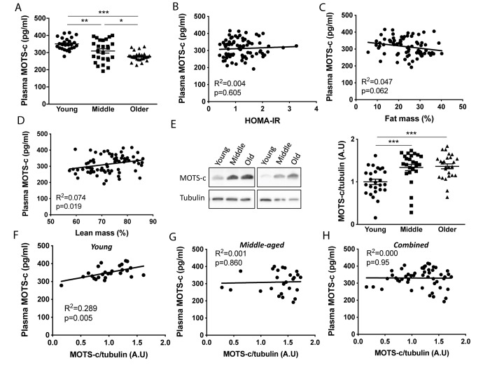
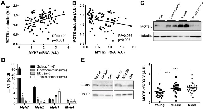
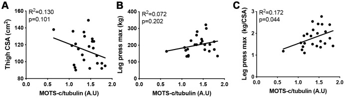
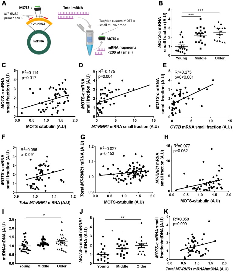
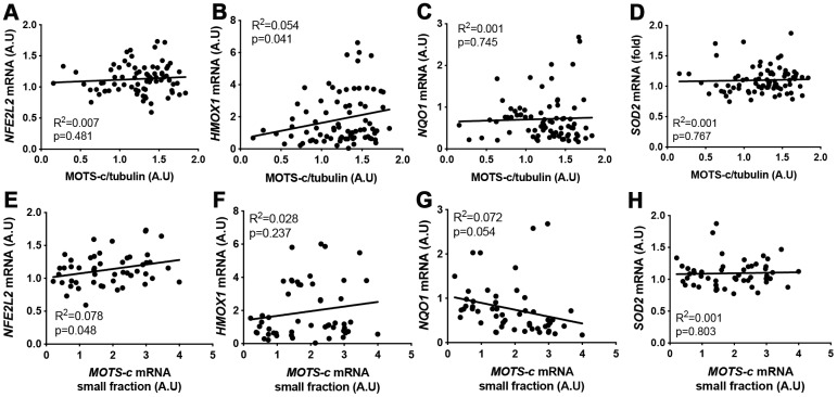
Mitochondria putatively regulate the aging process, in part, through the small regulatory peptide, mitochondrial open reading frame of the 12S rRNA-c (MOTS-c) that is encoded by the mitochondrial genome. Here we investigated the regulation of MOTS-c in the plasma and skeletal muscle of healthy aging men. Circulating MOTS-c reduced with age, but older (70-81 y) and middle-aged (45-55 y) men had ~1.5-fold higher skeletal muscle MOTS-c expression than young (18-30 y). Plasma MOTS-c levels only correlated with plasma in young men, was associated with markers of slow-type muscle, and associated with improved muscle quality in the older group (maximal leg-press load relative to thigh cross-sectional area). Using small mRNA assays we provide evidence that MOTS-c transcription may be regulated independently of the full length 12S rRNA gene in which it is encoded, and expression is not associated with antioxidant response element (ARE)-related genes as previously seen in culture. Our results suggest that plasma and muscle MOTS-c are differentially regulated with aging, and the increase in muscle MOTS-c expression with age is consistent with fast-to-slow type muscle fiber transition. Further research is required to determine the molecular targets of endogenous MOTS-c in human muscle but they may relate to factors that maintain muscle quality.

Keywords: MOTS-c; aging; mitochondria; mitochondrial derived peptides; muscle.


Conflict of interest statement

CONFLICTS OF INTEREST: PC and CL are consultants and shareholders of CohBar, Inc. All other authors declare no conflicts of interest.

Figures

Figure 1
Figure 1
Plasma MOTS-c levels decrease and skeletal muscle levels increase with aging. Plasma MOTS-c (A), correlated with HOMA-IR (B), fat mass (C), lean mass (D), and muscle MOTS-c expression (E) in young, middle-aged and older males. Representative blots are independent and from different participants. Correlation between plasma MOTS-c and muscle MOTS-c expression in young (F), middle (G), and combined (H). Significance was determined using linear regression or one-way ANOVA. Data is presented as means ± SE, n=26 per group except for body composition measurements where data was not available for a young (n=25) and middle-aged (n=25) participant. *p<0.05, **p<0.01, ***p<0.001.
Figure 2
Figure 2
MOTS-c expression is higher in slow-type muscle. Correlations between muscle MOTS-c expression and MYH7 (A) and MYH2 (B) mRNA levels in young, middle-aged and older men. Mouse extensor digitorum longus (EDL), gastrocnemius, tibialis anterior (TA) and soleus (SOL) muscle MOTS-c expression (C), and mRNA levels of fiber type markers (D). Two independent COXIV representative blots with different participants and quantification of MOTS-c relative to COXIV expression (E) in muscle samples from young, middle-aged and older males. Significance was determined using linear regression or one-way ANOVA. Data is presented as means ± SE for n=26 per group. ***p<0.001; #p<0.0001 vs soleus muscle.
Figure 3
Figure 3
The association between MOTS-c and muscle area and function. Thigh cross-sectional area (CSA) (A), maximal leg press load (B) and maximal leg press load relative to CSA (C) was correlated with muscle MOTS-c expression in older men. Significance was determined using linear regression or one-way ANOVA. Due to missing pQCT/leg press data n=24.
Figure 4
Figure 4
Muscle MOTS-c and 12S rRNA transcription with aging. 12S rRNA mRNA (MT-RNR1) and MOTS-c mRNA analysis technique (A). Young, middle-aged and older male muscle MOTS-c mRNA levels in the small RNA fraction were determined (B) and correlated with muscle MOTS-c protein expression (C), MT-RNR1 (D) and CYTB (E) mRNA levels in the small RNA fraction and MT-RNR1 mRNA levels in the total RNA fraction (F). MT-RNR1 mRNA levels in the total (G) and small (H) RNA fraction were correlated with MOTS-c protein expression. Muscle mitochondrial to nuclear DNA (mtDNA/nDNA) (I), and MOTS-c mRNA levels relative to mtDNA (J), and correlation of MOTS-c mRNA and MT-RNR1 mRNA in the total RNA relative to mtDNA (K). Significance was determined using linear regression or one-way ANOVA. Results are shown as means ± SE, due to limited sample availability and assay failure, for small RNA fraction assays n=14-18 per group, and for mtDNA n=26 for young, 24 for middle and 25 for old. *p<0.05, **p<0.01, ***p<0.001.
Figure 5
Figure 5
MOTS-c muscle protein or small mRNA expression does not correlate well with antioxidant-response element (ARE) related genes. Correlation between MOTS-c protein (AD) or small mRNA (EH) expression and mRNA levels of NFE2L2, HMOX1, NQO1 and SOD2 in muscle samples from young, middle-aged and older men. Significance was determined using linear regression for n=78 (protein correlations) or n=52 (MOTS-c mRNA correlations).

References

    1. Flatt T. A new definition of aging? Front Genet. 2012; 3:148. 10.3389/fgene.2012.00148 - DOI - PMC - PubMed
    1. Martin GM, Austad SN, Johnson TE. Genetic analysis of ageing: role of oxidative damage and environmental stresses. Nat Genet. 1996; 13:25–34. 10.1038/ng0596-25 - DOI - PubMed
    1. Jang JY, Blum A, Liu J, Finkel T. The role of mitochondria in aging. J Clin Invest. 2018; 128:3662–70. 10.1172/JCI120842 - DOI - PMC - PubMed
    1. Petersen KF, Befroy D, Dufour S, Dziura J, Ariyan C, Rothman DL, DiPietro L, Cline GW, Shulman GI. Mitochondrial dysfunction in the elderly: possible role in insulin resistance. Science. 2003; 300:1140–42. 10.1126/science.1082889 - DOI - PMC - PubMed
    1. López-Otín C, Blasco MA, Partridge L, Serrano M, Kroemer G. The hallmarks of aging. Cell. 2013; 153:1194–217. 10.1016/j.cell.2013.05.039 - DOI - PMC - PubMed

Publication types