. 2020 Mar 9;61(3):25.
doi: 10.1167/iovs.61.3.25.

Connexin 50-R205G Mutation Perturbs Lens Epithelial Cell Proliferation and Differentiation

Affiliations

Connexin 50-R205G Mutation Perturbs Lens Epithelial Cell Proliferation and Differentiation

Nikki Tjahjono et al. Invest Ophthalmol Vis Sci. .

Abstract

Purpose: To investigate the underlying mechanisms for how the mouse Cx50-R205G point mutation, a homologue of the human Cx50-R198W mutation that is linked to cataract-microcornea syndrome, affects proper lens growth and fiber cell differentiation to lead to severe lens phenotypes.

Methods: EdU labeling, immunostaining, confocal imaging analysis, and primary lens epithelial cell culture were performed to characterize the lens epithelial cell (LEC) proliferation and fiber cell differentiation in wild-type and Cx50-R205G mutant lenses in vivo and in vitro.

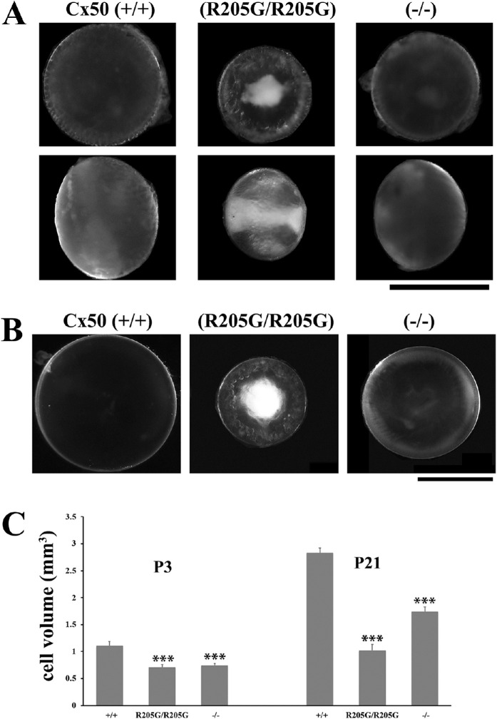
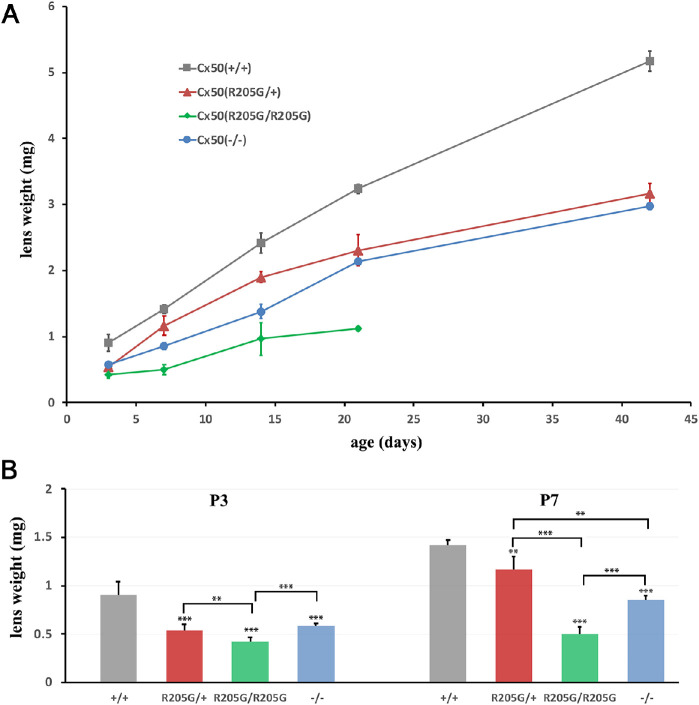
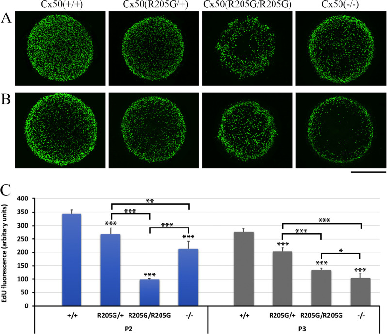
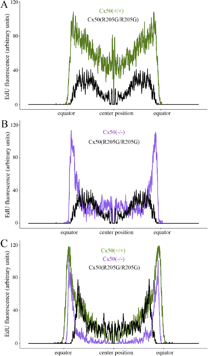
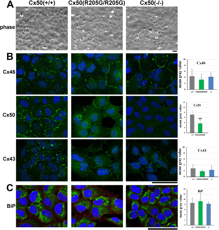
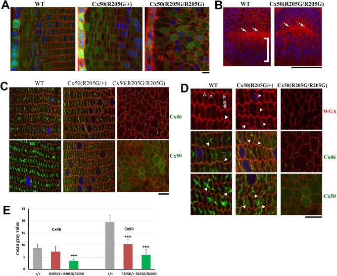
Results: The Cx50-R205G mutation severely disrupts the lens size and transparency. Heterozygous and homozygous Cx50-R205G mutant and Cx50 knockout lenses all show decreased central epithelium proliferation while only the homozygous Cx50-R205G mutant lenses display obviously decreased proliferating LECs in the germinative zone of neonatal lenses. Cultured Cx50-R205G lens epithelial cells reveal predominantly reduced Cx50 gap junction staining but no change of the endoplasmic reticulum stress marker BiP. The heterozygous Cx50-R205G lens fibers show moderately disrupted Cx50 and Cx46 gap junctions while the homozygous Cx50-R205G lens fibers have drastically reduced Cx50 and Cx46 gap junctions with severely altered fiber cell shape in vivo.

Conclusions: The Cx50-R205G mutation inhibits both central and equatorial lens epithelial cell proliferation to cause small lenses. This mutation also disrupts the assembly and functions of both Cx50 and Cx46 gap junctions in lens fibers to alter fiber cell differentiation and shape to lead to severe lens phenotypes.


Conflict of interest statement

Disclosure: N. Tjahjono, None; C.-hong Xia, None; R. Li, None; S. Chu, None; J. Wang, None; X. Gong, None

Figures

Figure 1.
Figure 1.
The homozygous Cx50(R205G/R205G) mutant lenses show more severe phenotype than the Cx50(–/–) knockout lenses. (A) Lens images of postnatal day 3 (P3) mice reveal early-onset growth defect and severe cataract in the homozygous Cx50(R205G/R205G) mutant lens, in comparison to the Cx50(+/+) wild-type and the Cx50(–/–) knockout lenses. The upper panels show lenses viewed from the anterior surface, while the lower panels display lenses viewed from the equator, and the anterior-posterior axis is from left to right. Scale bar: 1 mm. (B) Anterior view of fresh lenses of 3-week-old Cx50(+/+) wild-type, homozygous Cx50(R205G/R205G) and Cx50(–/–) knockout mice. Scale bar: 1 mm. (C) Lens volume comparison of P3 and P21 wild-type, homozygous Cx50(R205G/R205G), and Cx50(–/–) knockout mice. The P3 homozygous Cx50(R205G/R205G) lenses show approximately 40% reduction (P < 0.001) and the Cx50(–/–) knockout lenses have approximately 33% reduction (P < 0.001) when compared with the wild-type control. At P21, homozygous Cx50(R205G/R205G) lenses are approximately 64% smaller (P < 0.001) and the Cx50(–/–) knockout lenses are approximately 39% smaller (P < 0.001) than the wild-type lenses. Data are mean ± SD, n = 6–8 lenses of each genotype, with the Student's t-test for statistical analysis, ***P < 0.001, indicating statistically significant when compared with the wild-type.
Figure 2.
Figure 2.
The lens growth curves of Cx50(+/+) wild-type, Cx50(–/–) knockout, heterozygous Cx50(R205G/+), and homozygous Cx50(R205G/R205G) lenses based on the lens wet weight at postnatal ages. (A) Individual wet lens weight was measured, and the average lens wet weight was obtained from mice of each genotype (n = 3–7 mice). The average lens weight for each genotype was plotted at each age point from P3 to P42 days. The Cx50-R205G mutation displays a semidominant inheritance pattern, in which the heterozygous Cx50(R205G/+) lenses are smaller than the wild-type Cx50(+/+) lenses, while the homozygous Cx50(R205G/R205G) lenses are the smallest. (B) Statistical analysis and the wet weight bar graphs of different lenses at P3 and P7. Compared to the wild-type lenses, the P3 Cx50(–/–) lenses have approximately 36% reduction (P < 0.001), heterozygous Cx50(R205G/+) lenses show approximately 41% reduction (P < 0.001), and homozygous Cx50(R205G/R205G) lenses display approximately 54% reduction (P < 0.001); at P7, the Cx50(–/–) knockout lenses show approximately 40% reduced weight (P < 0.001), the heterozygous Cx50(R205G/+) lenses have approximately 18% reduction (P < 0.01), and homozygous Cx50(R205G/R205G) lenses have approximately 65% reduction (P < 0.001). The mean values per data point are presented as ± SD (n = 3–10 mice). Student’s t-test was used for statistical analysis, **P < 0.01, ***P < 0.001, indicating statistically significant for the comparison.
Figure 3.
Figure 3.
The Cx50-R205G mutation disrupts lens epithelial cell proliferation based on EdU labeling examination of P2 and P3 lenses in vivo. (A) Flattened Z-stack images of EdU-labeled lens epithelium of P2 mice. Compared with the wild-type Cx50(+/+), the heterozygous Cx50(R205G/+), the homozygous Cx50(R205G/R205G), and the Cx50(–/–) knockout lenses show reduced EdU labeling in central epithelium, while homozygous Cx50(R205G/R205G) lens also displays severely abnormal EdU labeling in peripheral epithelium (equator/germinative zone or GZ). (B) Flattened images of EdU-labeled lens epithelium of P3 mice. The homozygous Cx50(R205G/R205G) mutant has aberrant GZ proliferating cells with increased EdU labeling in the central epithelium (in comparison to P2 homozygous Cx50-R205G), the heterozygous Cx50(R205G/+) mutant lenses show increased EdU labeling in all epithelia, and the Cx50(–/–) knockout lens shows low EdU labeling in central epithelium. Scale bar (A, B), 500 µm. (C) Statistical comparison and bar graphs of EdU fluorescence in the Cx50-R205 mutant, Cx50-knockout, and wild-type lenses. Average EdU fluorescence, which correlates to proliferating epithelial cells, is obtained by determining the area of the fluorescent region of each flattened image and normalized by lens diameter. Compared with the wild-type control, among the homozygous Cx50(R205G/R205G), the heterozygous Cx50(R205G/+), and the Cx50(–/–) knockout, the homozygous Cx50(R205G/R205G) mutant mice have the most reduced total EdU fluorescence at P2 (approximately 71% reduction, P < 0.001), and the Cx50(–/–) knockout lenses display the most reduced total EdU fluorescence at P3 (approximately 63%, P < 0.001). Data presented as mean ± SD, n = 3–5 samples per data point, Student's t-test for statistical analysis. *P < 0.05, **P < 0.01, ***P < 0.001, indicating statistically significant when compared with the wild-type or compared as indicated.
Figure 4.
Figure 4.
Quantification of EdU labeling from the peripheral (equator/germinative zone) to the central epithelium of P2 and P3 lenses. (A) Line scans along the flattened EdU fluorescent images of P2 lenses, crossing through the diameter of the lens (a position of approximately 1500 µm is the center of the lens, and the position at which the fluorescence jumps from zero is the lens equator). The P2 Cx50(R205G/R205G) lens has lower epithelial cell proliferation by EdU labeling at all locations along the lens diameter compared with the P2 wild-type Cx50(+/+) (P < 0.01). (B) A comparison of line scans of EdU-labeled P2 Cx50(–/–) knockout and P2 Cx50(R205G/R205G) lenses. The P2 Cx50(R205G/R205G) lens has significantly lower proliferation in both central and peripheral (germinative zone) epithelium compared with the P2 Cx50(–/–) (P < 0.01). (C) Line scan comparison of EdU-labeled P3 wild-type Cx50(+/+), Cx50(–/–), and Cx50(R205G/R205G) lenses. The P3 Cx50(R205G/R205G) lens has a surge in proliferation but still shows decreased proliferation in peripheral epithelium when compared with that of both wild-type and Cx50(–/–) (P < 0.01). For all line scan charts, about 154 to 166 line scans per sample were averaged for each age and genotype (n = 3–5); SEM ribbon not shown due to large sample size and small SE. Student’s t-tests for the central epithelium performed at positions 1250 to 1750 µm.
Figure 5.
Figure 5.
The Cx50 gap junctions are significantly reduced in cultured Cx50(R205G/R205G) primary lens epithelial cells in vitro. (A) No apparent morphologic difference is observed in cultured primary lens epithelial cells isolated from lenses of wild-type Cx50(+/+), Cx50(R205G/R205G), and Cx50(–/–) knockout mice. Scale bar: 50 µm. (B) Immunostaining of cultured primary lens epithelial cells with anti-Cx46, anti-Cx50, and anti-Cx43 antibodies (green signals, costained with DAPI in blue). Compared with the typical Cx46 (green) gap junctions at the cell-cell boundary of wild-type cells, reduced Cx46 gap junctions occurred in some Cx50(R205G/R205G) lens epithelial cells, while normal Cx46 junctions appeared in the Cx50(–/–) lens epithelial cells; however, quantitation of Cx46 staining by ImageJ reveals no statistically significant difference between Cx50(R205G/R205G) and wild-type cells (P > 0.05, Student’s t-test). Compared with robust Cx50 gap junctions at the cell-cell boundary of wild-type lens epithelial cells, faint and thin Cx50 gap junctions are detected in the Cx50(R205G/R205G) lens epithelial cells and no Cx50 gap junction in Cx50(–/–) lens epithelial cells. Statistical analysis reveals significantly reduced Cx50 gap junction staining in Cx50(R205G/R205G) cells (**P < 0.01, mean ± SD, Student’s t-test). Typical punctate Cx43 gap junctions are detected in wild-type epithelial cells, and Cx43 staining signal appears to be slightly reduced in the Cx50(R205G/R205G) and Cx50(–/–) lens epithelial cells; however, the Cx43 gap junction staining difference between wild-type and Cx50(R205G/R205G) cells is not statistically significant (P = 0.16). The bar graphs show quantitation of the membrane-stained connexin signal intensity by ImageJ. Three 63× images of staining were converted to grayscale and staining intensity was measured on five to six cells of each image; average mean gray intensity of each cell subtracted from background was plotted. Cell culture and staining were repeated at least three times, and similar results were observed. (C) Immunostaining of cultured primary lens epithelium cells with an anti-BiP antibody (BiP signal, green; DAPI, blue). BiP expression was detected in cytosols of all cells, enriched in endoplasmic reticulum. The bar graphs display quantitation of BiP signal intensity by ImageJ. Compared with the wild-type cells, the Cx50(R205G/R205G) and Cx50 knockout cells show comparable BiP expression; the differences are not statistically significant (P > 0.05, mean ± SD, Student’s t-test). Scale bar: (B, C) 50 µm.
Figure 6.
Figure 6.
The Cx50-R205G mutation disrupts meridional rows and fulcrum at lens equator and alters lens fiber cell shape and gap junctions. (A) Lens fiber cell morphology revealed by wheat germ agglutinin (WGA; red) and DAPI (blue) stained cross sections of GFP-positive (green) Cx50(+/+) wild-type, Cx50(R205G/+), and Cx50(R205G/R205G) lenses from 3-week-old mice. Images collected from the lens periphery to interior 100-µm fibers are displayed. The heterozygous Cx50-R205G lens fiber cells show hexagonal cell shape with normal fiber-to-fiber overlay organization while homozygous Cx50-R205G lens fiber cells display rounded cell shape and irregular organization. Scale bar: 10 µm. (B) Equatorial images of P13 wild-type and Cx50(R205G/R205G) lenses stained with WGA (red) and DAPI (blue). In the wild-type lens, meridional rows (indicated by ]) are aligned and straight, but the meridional rows of Cx50(R205G/R205G) lenses are misaligned. The Cx50(R205G/R205G) lens fulcrum (indicated by white arrows) is disrupted, unlike the straight line of the wild-type lens fulcrum (white arrows). Scale bar: 100 µm. (C) Cx46 and Cx50 gap junction expression in lens sections costained with either anti-Cx46 or anti-Cx50 antibodies (green), WGA (red), and DAPI (blue). In the wild-type lens, Cx46 and Cx50 gap junction plaques are enriched in the ball-and-sockets and also expressed on the long and narrow sides of fibers. Heterozygous Cx50-R205G fiber cells display reduced and short or small gap junction plaques along the broad and short sides, while the homozygous Cx50(R205G/R205G) fibers only have sparse, very short, or dot-like Cx46 and Cx50 plaques. All lens sections were from 3-week-old mice. Scale bar: 10 µm. (D) Enlarged images of lens fibers costained with anti-Cx46 or anti-Cx50 (green), WGA (red), and DAPI (blue). Typical Cx46 and Cx50 gap junctions are detected in wild-type ball-and-sockets (indicated by white arrowheads) while short or dot-like Cx46 and Cx50 signals are observed in Cx50(R205G/+) fiber cell boundaries with aberrant membrane structures (indicated by arrowheads). The caret indicates the short side of the hexagonal shaped fibers in the wild-type, while the asterisk marks the long side. All lens sections were from 3-week-old mice. Scale bar: 10 µm. (E) Bar graphs of Cx46 and Cx50 staining intensity quantitation. Compared with the wild-type lens section staining, the Cx50(R205G/R205G) section shows significantly reduced Cx46 (P < 0.001) and Cx50 (P < 0.001) staining intensity; the heterozygous Cx50(R205G/+) displays significantly reduced Cx50 staining intensity (P < 0.001) compared with the wild-type, but the difference of Cx46 staining intensity between the heterozygous and wild-type is not statistically significant (P = 0.27). Data are shown as mean ± SD, Student’s t-test, ***P < 0.001, indicating statistically significant when compared with the wild-type.

References

    1. Piatigorsky J. Lens differentiation in vertebrates: a review of cellular and molecular features. Differentiation. 1981; 19: 134–153. - PubMed
    1. Bassnett S, Sikic H. The lens growth process. Prog Retin Eye Res. 2017; 60: 181–200. - PMC - PubMed
    1. Mathias RT, Rae JL. The lens: local transport and global transparency. Exp Eye Res. 2004; 78: 689–698. - PubMed
    1. Mathias RT, White TW, Gong X. Lens gap junctions in growth, differentiation, and homeostasis. Physiol Rev. 2010; 90: 179–206. - PMC - PubMed
    1. Gong X, Cheng C, Xia CH. Connexins in lens development and cataractogenesis. J Membr Biol. 2007; 218: 9–12. - PubMed

Publication types

MeSH terms

Supplementary concepts