. 2020 May 7;48(8):4371-4381.
doi: 10.1093/nar/gkaa153.

Loop-closure kinetics reveal a stable, right-handed DNA intermediate in Cre recombination

Affiliations

Loop-closure kinetics reveal a stable, right-handed DNA intermediate in Cre recombination

Massa J Shoura et al. Nucleic Acids Res. .

Abstract

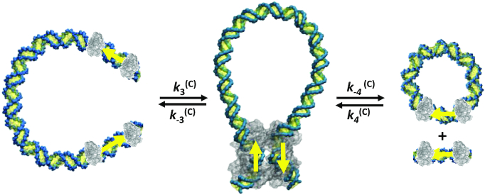
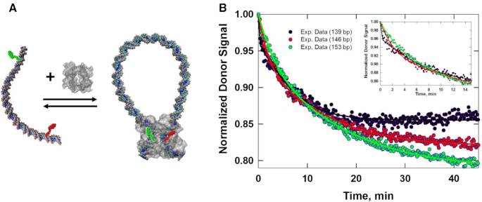
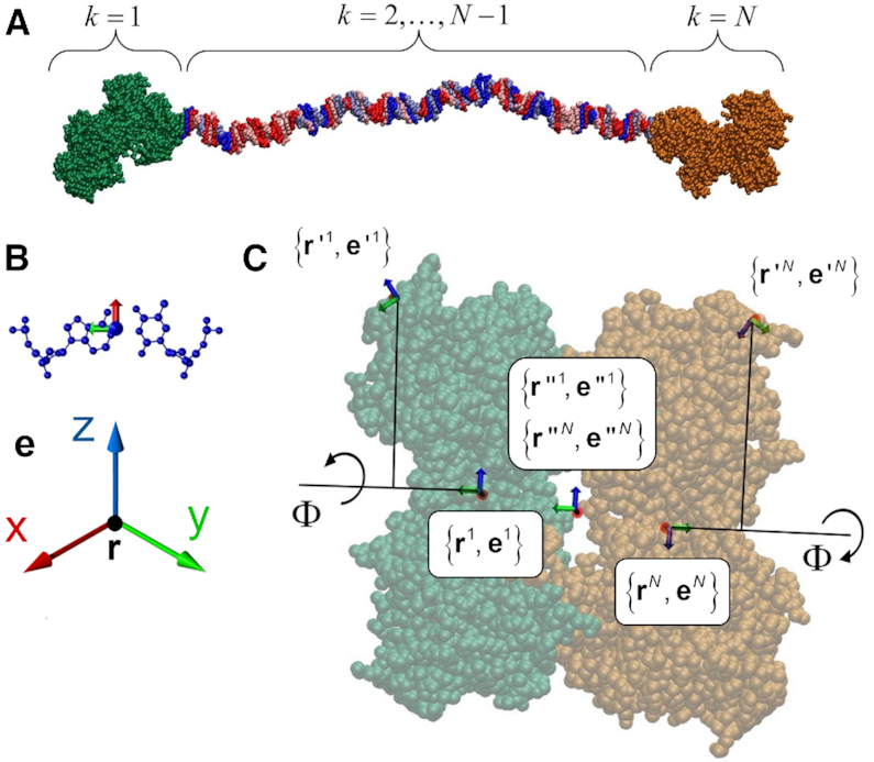
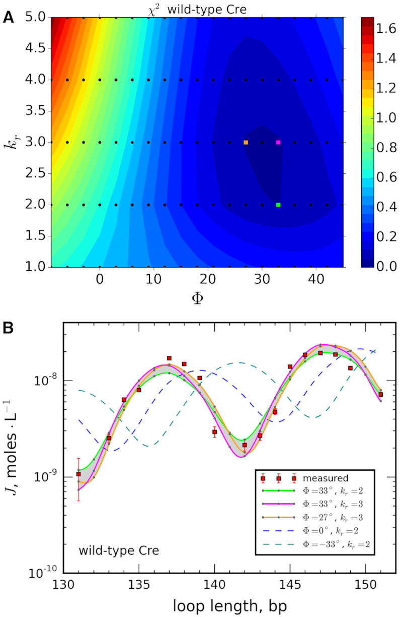
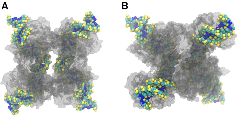
In Cre site-specific recombination, the synaptic intermediate is a recombinase homotetramer containing a pair of loxP DNA target sites. The enzyme system's strand-exchange mechanism proceeds via a Holliday-junction (HJ) intermediate; however, the geometry of DNA segments in the synapse has remained highly controversial. In particular, all crystallographic structures are consistent with an achiral, planar Holliday-junction (HJ) structure, whereas topological assays based on Cre-mediated knotting of plasmid DNAs are consistent with a right-handed chiral junction. We use the kinetics of loop closure involving closely spaced (131-151 bp) loxP sites to investigate the in-aqueo ensemble of conformations for the longest-lived looped DNA intermediate. Fitting the experimental site-spacing dependence of the loop-closure probability, J, to a statistical-mechanical theory of DNA looping provides evidence for substantial out-of-plane HJ distortion, which unequivocally stands in contrast to the square-planar intermediate geometry from Cre-loxP crystal structures and those of other int-superfamily recombinases. J measurements for an HJ-isomerization-deficient Cre mutant suggest that the apparent geometry of the wild-type complex is consistent with temporal averaging of right-handed and achiral structures. Our approach connects the static pictures provided by crystal structures and the natural dynamics of macromolecules in solution, thus advancing a more comprehensive dynamic analysis of large nucleoprotein structures and their mechanisms.


Figures

Figure 1.
Figure 1.
Products generated by Cre recombinase acting on the DNA molecules used in this study. In the first step DNA fragments flanked by directly repeated loxP sites (indicated by yellow arrows) form a DNA loop via site synapsis, which is mediated by a Cre-protein tetramer. Two sequential cleavage/strand-exchange events generate a short linear molecule and a DNA circle as recombinant products in the second step.
Figure 2.
Figure 2.
Intramolecular synapsis and recombination kinetics obtained from time-dependent FRET measurements. (A) Schematic of the intramolecular reaction carried out on a DNA fragment bearing donor- and acceptor-labeled loxP sites. (B) Donor-fluorescence signal, which monitors donor quenching via FRET during site synapsis and recombination. Fluorescence decays are shown for molecules having 139-, 146- and 153-bp DNA loops. Rate constants were obtained by fitting the fluorescence decay to a system of ordinary differential equations that describe the time-dependent concentrations of reactants, intermediates and products along the intramolecular recombination pathway (40). The best-fit numerical solution is given by the solid curve. The fluorescence decay and fit to the data over the first 15 minutes of the recombination reaction are shown in the inset.
Figure 3.
Figure 3.
Nucleoprotein model. (A) DNA molecule with a pair of Cre-bound loxP sites at each end. The molecule shown is modeled as a chain of formula image rigid bodies. Each rigid body in the model is colored as follows: Cre-loxP protein-DNA complexes, green and orange; DNA base pairs: adenine (blue), thymine (light blue), cytosine (red), guanine (pink). (B) Definition of reference frames embedded within DNA base pairs: formula image axes (red) point toward the major groove, formula image axes (green) point toward the primary strand, and formula image axes (blue) point in the 5′ to 3′ direction of the sense strand. (C) Definition of the dihedral angle between Cre half tetramers in the synapse. The geometry of the Cre-loxP synaptic complex was modified from the nearly planar crystal structure geometry by applying rotations of the Cre-bound loxP sites as shown. Frame formula image is rotated into the page by an angle formula image about the axis formula image and frame formula image is rotated out of the page by the same angle formula image about the axis formula image, increasing the dihedral angle of the loop ends in the protein synapse by 2formula image. For a definition of the body-fixed reference frames formula image see Materials and Methods, and an extended discussion of the synapse model in Supplementary Information.
Figure 4.
Figure 4.
Contour map of solutions and optimal fits of calculated J-factor curves to measured J-factor values for wild-type Cre-mediated looping. (A) Contour plots of the mean-square error, MSE, for fitting of experimental J factors to NMA-computed values for the wild-type Cre synapse as a function of the dihedral angle formula image and rotational flexibility constant formula imagefor the pair of Cre half-tetramers (see Methods). (B) Measured values of J for wild-type Cre along with an envelope of optimally fitted J-factor curves, corresponding to values formula image indicated by the three highlighted points in the contour map shown in (A). Each data point is the average of at least three independent measurements and error bars indicate ±1 standard error. The envelope of optimal formula image values spans the range 27° (orange)formula image33° (green) with 2 ≤ formula image≤ 3 kBrad−2. A particular solution in this range for formula image = 33° with formula image 3 kBrad−2 is shown in magenta. Calculated J-factor curves for formula image0° (blue dashed curve) and formula image−33° (cyan dashed curve) with formula image 2 kBrad−2, provided for comparison, do not provide satisfactory fits to the experimental data.
Figure 5.
Figure 5.
3D models of Cre synaptic complexes according to (A) crystal structure 5CRX and (B) minimum-elastic energy structure corresponding to the optimum fit of the NMA model to our experimental J-factor data.
Figure 6.
Figure 6.
Contour map of solutions and optimal fits of calculated J-factor curves to measured J-factor values for Cre-mediated looping by the R101A mutant Cre protein. The R101A mutation strongly reduces the protein's Holliday-junction resolution activity, promoting the accumulation of junction-containing synaptic complexes. Contour plots (A) and optimally fitted J-factor curves (B) for mutant Cre were obtained as described in Figure 4. Optimal fits were obtained for 42° (green)formula image48° (orange) and formula image2 kBrad−2. A particular solution for formula image = 48° with formula image 3 kBrad−2 is also shown in magenta. The J-factor dependence for formula image33° and formula image2 kBrad−2, which lies within the best-fit envelope of the wild-type protein, is shown for comparison (blue dashed curve).

Similar articles

Cited by

References

    1. Castán A., Hernández P., Krimer D.B., Schvartzman J.B.. Drolet M. DNA catenation reveals the dynamics of DNA topology during replication. DNA Topoisomerases: Methods and Protocols. 2018; NY: Springer New York; 75–86. - PubMed
    1. Keszthelyi A., Minchell N.E., Baxter J.. The causes and consequences of topological stress during DNA replication. Genes (Basel). 2016; 7:134. - PMC - PubMed
    1. Dorman C.J., Dorman M.J.. DNA supercoiling is a fundamental regulatory principle in the control of bacterial gene expression. Biophys. Rev. 2016; 8:209–220. - PMC - PubMed
    1. Racko D., Benedetti F., Dorier J., Burnier Y., Stasiak A.. Generation of supercoils in nicked and gapped DNA drives DNA unknotting and postreplicative decatenation. Nucleic Acids Res. 2015; 43:7229–7236. - PMC - PubMed
    1. Rice P.A. Serine resolvases. Microbiol Spectr. 2015; 3:doi:10.1128/microbiolspec.MDNA3-0045-2014. - PMC - PubMed

Publication types