. 2020 Mar 8;12(3):627.
doi: 10.3390/cancers12030627.

Impaired Anti-Tumor T cell Response in Hepatocellular Carcinoma

Affiliations

Impaired Anti-Tumor T cell Response in Hepatocellular Carcinoma

Nada Chaoul et al. Cancers (Basel). .

Abstract

Different subsets of lymphocytes have the capacity to promote or counteract the progression of solid cancers, including hepatocellular carcinoma (HCC). Therefore, to determine the infiltrative ability and functional status of major immune cell subtypes into tumor may lead to novel insights from the perspective of immunotherapy. After obtaining single cell suspensions from freshly collected specimens of HCC tumor, along with paired peritumor tissues and peripheral blood mononuclear cells (PBMCs) from 14 patients, we flow-cytometrically identified and quantified the relative frequencies of lymphocyte subsets within the tissues of origin. We found that the recruitment in the tumor of cytotoxic cells, namely the terminally differentiated CD4+ and CD8+ T cells (TEFF), is impaired, whereas the effector memory CD4+ T cells (TEM) are more attracted in this site. Concerning the other subsets, the frequency of NK CD56hi and NKT CD56hi cells infiltration in the tumor is increased, whereas that of NKT CD56low is reduced. Although CD4+ and CD8+ T cells settled in the tumor show a higher degree of activation than the circulating counterpart, they occur with a more exhausted phenotype. Overall, these data demonstrate the prevalently immunosuppressive nature of HCC microenvironment, and prompt us to search for strategies to enhance the activity of anti-tumor immune cell subsets.

Keywords: HCC; T cell response; antitumor immunity; immunotherapy.


Conflict of interest statement

The authors declare no conflict of interest.

Figures
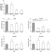
Figure 1
Figure 1
Hepatocellular carcinoma (HCC) patients have imbalanced circulating lymphocyte proportions and an impaired immune infiltration to the tissues. Cell suspensions were prepared from peripheral blood mononuclear cells (PBMC), tumor and peritumor taken during surgery and were analyzed by flow cytometry. The PBMC from healthy donors were used as control. (a) Percentage of immune infiltration was identified by the total CD45+ cell portion in each tissue. (be) Percentage of lymphoid subsets within total CD45+ cells. Grey histograms represent cells obtained from healthy donors, while white histograms represent cells from HCC patients. Results are expressed as Mean ± SEM from cumulative results (n = 9–14 patients or controls). * p < 0.05 and ** p < 0.01, as determined by Mann–Whitney’s test between each lymphoid subset, in each tissue.
Figure 2
Figure 2
Decreased frequencies of CD56low NK and NKT cells in tumors. Cell suspensions were prepared from PBMC, tumor and peritumor taken during surgery and were analyzed by flow cytometry. The PBMC from healthy donors were used as control. The intensity of CD56 expression in NK and NKT cells was analyzed in all tissues and the expression of CD56hi (hatched histograms) and CD56lo (white histograms) NK and NKT cells are shown in (a) and (b), respectively. Results are expressed as Mean ± SEM from cumulative results (n = 9–14 patients or controls). * p < 0.05, as determined by Mann–Whitney’s test between each lymphoid subset, in each tissue.
Figure 3
Figure 3
Tregs are increased in HCC patients while Effector T cells are reduced. Cell suspensions were prepared from PBMC, tumor and peritumor taken during surgery and were analyzed by flow cytometry. The PBMC from healthy donors were used as control. The frequencies of T cell subsets were analyzed in detail. (a) Frequencies of total CD4+ T cells, and CD8+ T cells within total CD3+ T cells are shown; (b) Frequencies of Tregs (CD4+ Foxp3+ T-bet T cells), Th1 (CD4+ Foxp3 T-bet+ cells) and T-bet+ Tregs (CD4+ Foxp3+ T-bet+ cells) within CD4+ T cells. (c,d) Naïve (TN), central memory (TCM), effector memory (TEM) and effector cell (TEFF) subsets within CD4+ T cells and CD8+ T cells. Results are expressed as Mean ± SEM from cumulative results (with n = 9–14 patients or controls). * p < 0.05 and ** p < 0.01 as determined by Mann–Whitney’s test between each lymphoid subset, in each tissue.
Figure 4
Figure 4
The percentage of PD-1+ and CD69+ T cells are increased in the tumor. Cell suspensions were prepared from PBMC, tumor and peritumor taken during surgery and were analyzed by flow cytometry. PBMC from healthy donors were used as control. The expression of PD-1 and CD69 on T cells from PBMC, tumor and peritumor of each patient and on T cells from PBMC of healthy controls was quantified. (a) Percentage of CD69+ CD4+ T cells and CD-69+ CD8+ T cells; (b) gMFI of CD69; (c) Percentage of PD-1+ CD4+ T cells and PD-1+ CD8+ T cells; (d) Geometric mean fluorescence intensity (gMFI) of PD-1. Results are expressed as Mean ± SEM from cumulative results (with n = 9–14 patients or controls). See Table 5 for statistical analysis.

References

    1. Nordenstedt H., White D.L. El-Serag HB: The changing pattern of epidemiology in hepatocellular carcinoma. Dig. Liver Dis. 2010;42:S206–S214. doi: 10.1016/S1590-8658(10)60507-5. - DOI - PMC - PubMed
    1. Raza A., Sood G.K. Hepatocellular carcinoma review: Current treatment, and evidence-based medicine. World J. Gastroenterol. 2014;20:4115–4127. doi: 10.3748/wjg.v20.i15.4115. - DOI - PMC - PubMed
    1. Lang L. FDA approves sorafenib for patients with inoperable liver cancer. Gastroenterology. 2008;134:379. doi: 10.1053/j.gastro.2007.12.037. - DOI - PubMed
    1. Llovet J.M., Ricci S., Mazzaferro V., Hilgard P., Gane E., Blanc J.F., de Oliveira A.C., Santoro A., Raoul J.L., Forner A., et al. Sorafenib in advanced hepatocellular carcinoma. N. Engl. J. Med. 2008;359:378–390. doi: 10.1056/NEJMoa0708857. - DOI - PubMed
    1. Granito A., Marinelli S., Negrini G., Menetti S., Benevento F., Bolondi L. Prognostic significance of adverse events in patients with hepatocellular carcinoma treated with sorafenib. Ther. Adv. Gastroenterol. 2016;9:240–249. doi: 10.1177/1756283X15618129. - DOI - PMC - PubMed