Review
. 2020 Mar 9;12(3):620.
doi: 10.3390/polym12030620.

Polymer- and Hybrid-Based Biomaterials for Interstitial, Connective, Vascular, Nerve, Visceral and Musculoskeletal Tissue Engineering

Affiliations
Review

Polymer- and Hybrid-Based Biomaterials for Interstitial, Connective, Vascular, Nerve, Visceral and Musculoskeletal Tissue Engineering

Anatolii Abalymov et al. Polymers (Basel). .

Abstract

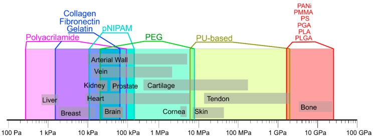
In this review, materials based on polymers and hybrids possessing both organic and inorganic contents for repairing or facilitating cell growth in tissue engineering are discussed. Pure polymer based biomaterials are predominantly used to target soft tissues. Stipulated by possibilities of tuning the composition and concentration of their inorganic content, hybrid materials allow to mimic properties of various types of harder tissues. That leads to the concept of "one-matches-all" referring to materials possessing the same polymeric base, but different inorganic content to enable tissue growth and repair, proliferation of cells, and the formation of the ECM (extra cellular matrix). Furthermore, adding drug delivery carriers to coatings and scaffolds designed with such materials brings additional functionality by encapsulating active molecules, antibacterial agents, and growth factors. We discuss here materials and methods of their assembly from a general perspective together with their applications in various tissue engineering sub-areas: interstitial, connective, vascular, nervous, visceral and musculoskeletal tissues. The overall aims of this review are two-fold: (a) to describe the needs and opportunities in the field of bio-medicine, which should be useful for material scientists, and (b) to present capabilities and resources available in the area of materials, which should be of interest for biologists and medical doctors.

Keywords: cells; connective; hybrid; hydrogels; interstitial; layer-by-layer; musculoskeletal; nervous; polymers; tissue engineering; vascular; visceral.


Conflict of interest statement

The authors declare no conflict of interest.

Figures

Figure 1
Figure 1
Mechanical properties of natural tissues and polymers. Data are composed based on data from the following publications [25,131,132].
Figure 2
Figure 2
(A) Scheme of ultrasonic mineralization of polycaprolactone fibers and recrystallization of vaterite to calcite. (B) Microphotographs show pristine PCL fibers. (C) the mineralized scaffold based on PCL material. (D) process of recrystallization vaterite to calcite. (E) Micrograph of histological sections, 21 days after implantation in the withers of the mouse. Yellow dashed lines indicate the location of the matrix. Reprinted with permission from Elsevier, 2018 [138].
Figure 3
Figure 3
(A) Synergistic triggering of the integrin-VEGF signal, which is connected to the Poly (ethylene adipate) network. (B) In vitro and in vivo fluorescence microphotographs of cells. (C) Scheme of the effect of VEGF associated with nanofibrils on cell signaling. Reprinted with permission from Elsevier, 2017 [159].
Figure 4
Figure 4
(A) Image of a tracheal implant (appearance and section). SEM micrograph of the outer wall and slice. Scalebar 2 mm. Histological Section 8 weeks after implantation. Reprinted with permission from Elsevier, 2015 [8]. (B) Schematics showing lungs, where 3D printed scaffolds shown in (A) can be incorporated, and which can be modeled by microfluidic devices depicted in (C). (C) Schematics of the co-culture system and images of assembled fluidic devices with cells including confocal laser scanning images of epithelial and endothelial cells co-cultured on a 75/25 (PCL/gelatin) fiber mesh. Reprinted from [173] with permission from Nature Publishing Group, © 2017.
Figure 5
Figure 5
(a) Scheme depicting the creation of a composite material based on titanium and alginate gel. (b) Atomic force microscopy topography images of functionalized surfaces. (c) Fluorescence photographs showing osteoblast cells on the surface of a composite material. (d) Analysis of the amount of active enzyme by a different combination of components of the composite material. Reprinted with permission of Wiley, 2018 [228].

References

    1. Blanpain C., Fuchs E. Plasticity of epithelial stem cells in tissue regeneration. Science (80-) 2014;344:1242281. doi: 10.1126/science.1242281. - DOI - PMC - PubMed
    1. Mobaraki M., Abbasi R., Vandchali S.O., Ghaffari M., Moztarzadeh F., Mozafari M. Corneal repair and regeneration: Current concepts and future directions. Front. Bioeng. Biotechnol. 2019;7 doi: 10.3389/fbioe.2019.00135. - DOI - PMC - PubMed
    1. Takeo M., Lee W., Ito M. Wound healing and skin regeneration. Cold Spring Harb. Perspect. Med. 2015;5:a023267. doi: 10.1101/cshperspect.a023267. - DOI - PMC - PubMed
    1. Michalopoulos G.K. Liver regeneration. J. Cell. Physiol. 2007;213:286–300. doi: 10.1002/jcp.21172. - DOI - PMC - PubMed
    1. Daly A.C., Riley L., Segura T., Burdick J.A. Hydrogel microparticles for biomedical applications. Nat. Rev. Mater. 2020;5:20–43. doi: 10.1038/s41578-019-0148-6. - DOI - PMC - PubMed

LinkOut - more resources