. 2020 Mar 9;9(3):658.
doi: 10.3390/cells9030658.

Conducting Polymer Mediated Electrical Stimulation Induces Multilineage Differentiation with Robust Neuronal Fate Determination of Human Induced Pluripotent Stem Cells

Affiliations

Conducting Polymer Mediated Electrical Stimulation Induces Multilineage Differentiation with Robust Neuronal Fate Determination of Human Induced Pluripotent Stem Cells

Eva Tomaskovic-Crook et al. Cells. .

Abstract

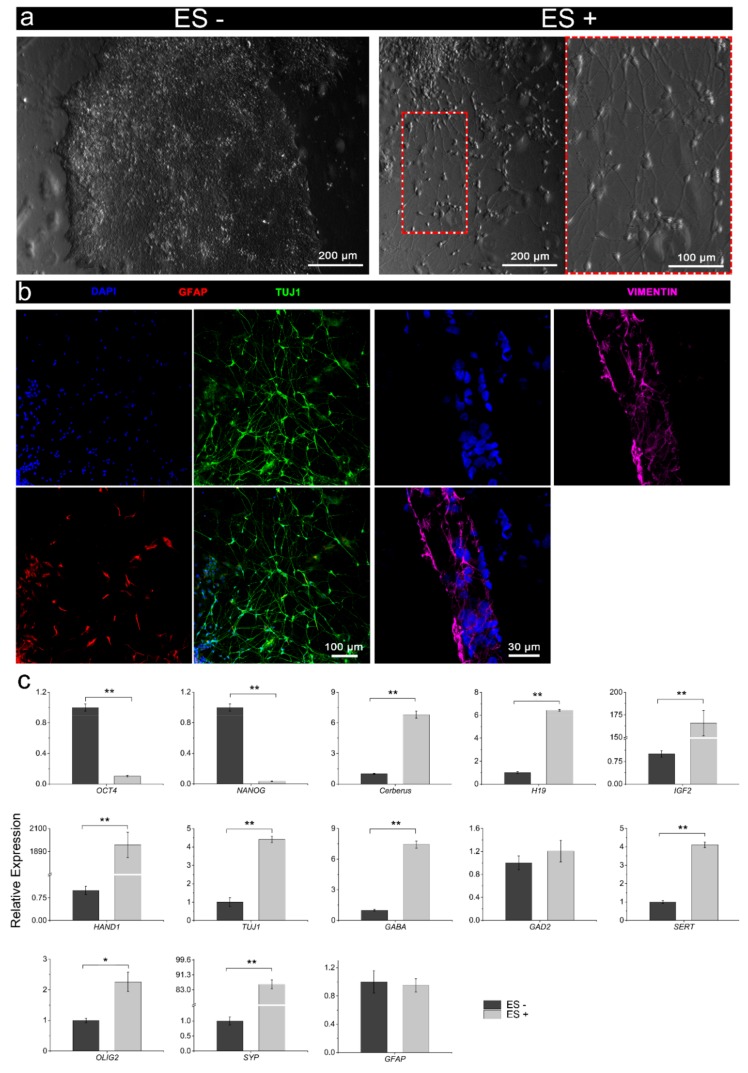
Electrical stimulation is increasingly being used to modulate human cell behaviour for biotechnological research and therapeutics. Electrically conductive polymers (CPs) such as polypyrrole (PPy) are amenable to in vitro and in vivo cell stimulation, being easy to synthesise with different counter ions (dopants) to augment biocompatibility and cell-effects. Extending our earlier work, which showed that CP-mediated electrical stimulation promotes human neural stem cell differentiation, here we report using electroactive PPy containing the anionic dopant dodecylbenzenesulfonate (DBS) to modulate the fate determination of human induced pluripotent stem cells (iPSCs). Remarkably, the stimulation without conventional chemical inducers resulted in the iPSCs differentiating to cells of the three germ lineages-endoderm, ectoderm, and mesoderm. The unstimulated iPSC controls remained undifferentiated. Phenotypic characterisation further showed a robust induction to neuronal fate with electrical stimulation, again without customary chemical inducers. Our findings add to the growing body of evidence supporting the use of electrical stimulation to augment stem cell differentiation, more specifically, pluripotent stem cell differentiation, and especially neuronal induction. Moreover, we have shown the versatility of electroactive PPy as a cell-compatible platform for advanced stem cell research and translation, including identifying novel mechanisms of fate regulation, tissue development, electroceuticals, and regenerative medicine.

Keywords: conductive polymers; differentiation; ectodermal; electrical stimulation; endodermal; induced pluripotent stem cells; mesodermal; neuronal; polypyrrole.


Conflict of interest statement

The authors declare no conflict of interest. The funders had no role in the design of the study; in the collection, analyses, or interpretation of data; in the writing of the manuscript, or in the decision to publish the results”.

Figures

Figure 1
Figure 1
Schematic of the human iPSC stimulation setup and regimen using a PPy:DBS-based stimulation module: (a) Human iPSCs were harvested and transferred onto Matrigel®-coated PPy:DBS film within a stimulation module. Cells were stimulated in iPSC culture medium resulting in robust induction of neuroectoderm favouring neuronal fate determination. (b) iPSCs were initially maintained for 24 h (day 0–day 1) in culture medium before stimulating at a current density of ± 0.1 mA/cm2 for 8 h per day for 3 days (day 1–day 4), followed by ± 0.25 mA/cm2 for 8 h every 2 days for 6 days (day 4–day 10).
Figure 2
Figure 2
Electrochemical activity and stability of PPy:DBS film employed for iPSC stimulation: (a) CV voltammogram for PPy:DBS substrate in PBS electrolyte, with clear oxidation and reduction peaks indicating the presence of a redox active polymer. Arrows indicate the direction of potential scan (scan rate: 0.1 V/s, over a potential range of −0.7 to +0.7 V). (b) An example of the biphasic current (i) waveform applied, overlayed with the output voltage waveform used to calculate impedances, with Vt, Va and Vp denoting peak voltage output, initial voltage drop, and remaining voltage drop, respectively.
Figure 3
Figure 3
Immunophenotypic characterisation of the iPSCs: (ac) iPSCs cultured on PPy:DBS substrate and stained with nuclear DAPI colocalised with pluripotency markers (a) Oct4, (b) SOX2, and (c) SSEA4. (d) Flow cytometry of the iPSCs confirming high level expresson OCT4, SSEA4, TRA-1-60, and TRA-1-8 (black histograms). Red histograms indicate the isotype controls. Scale bars as indicated.
Figure 4
Figure 4
Immunophenotypic characterisation of iPSC differentiability: expression of neuroectodermal progenitor cell markers Pax6, nestin, and vimentin, early neuronal marker Tuj, and glial cell marker GFAP at (a) day 6, (b) day 12, or (c) day 18 of iPSC differentiation. Day 18 cell cultures were distinguished by more prominent neurite outgrowths emanating from Tuj1-expressing neurons. GFAP-expressing cells frequently appeared contigous with neurons, signified by two adjacent DAPI-labelled nuclei (arrows) and converged fluorescent labelling of adjoining glia and neurons. Scale bars as indicated.
Figure 5
Figure 5
Immunophenotypic characterisation of iPSC differentiation following the conventional chemical induction with (ES+) and without (ES-) electrical stimulation. Expression of (a) early neuronal marker Tuj and neuroectodermal progenitor cell markers, (b) nestin, and (c) Pax6. Electrically stimulated cell cultures were distinguished by a higher antigen expression and diffuse nestin-labelled neurites outgrowths. Scale bars as indicated. (d) Quantitative assessment of Tuj1, nestin, and Pax6 immunocytochemistry (integrated density per cell; IntDen/Cell), confirming qualitative assessment. Mean ± S.D.; n = 3–5. One-way analysis of variance (ANOVA) with a Bonferroni post-hoc test. *p < 0.001.
Figure 6
Figure 6
Characterisation of the iPSCs following electrical stimulation (ES+) versus no stimulation (ES-) without chemical inducers. (a) Bright-field micrographs of ES- (left panel) and ES+ (right panel) iPSC cultures, with ES- cultures consisting of classical iPSC-colonies with high-density undifferentiated cells, while ES+ cultures consisted of more dispersed and differentiated cells with clear neuronal morphology. ES+ cell cultures were distinguished by polarised neurons possessing dendritic arborizations and elongated axonal-like projections. (b) Immunophenotyping confirmed the neural induction of ES+ cultures, showing high-density Tuj1-expressing neurons with diffuse intersecting neurites, and to a lesser extent GFAP-expressing cells, as well as cells expressing the neuroectodermal progenitor cell marker vimentin. Scale bars as indicated. (c) Comparative gene expression (pluripotency: OCT4, NANOG; endodermal: Cerberus, H19; mesodermal: IGF2, HAND1; neuroectodermal: TUJ1, GABA, GAD2, SERT, OLIG2, SYP, GFAP) of the iPSCs following electrical stimulation (ES+) or without electrical stimulation (ES-). Relative gene expression represents data normalized to β-actin and expressed relative to the non-stimulated iPSCs. Mean ± S.D.; n = 3. One-way ANOVA with a Bonferroni post hoc test. *p < 0.01; **p < 0.001.

References

    1. Balint R., Cassidy N.J., Cartmell S.H. Electrical stimulation: A novel tool for tissue engineering. Tissue Eng. Part. B. Rev. 2013;19:48–57. doi: 10.1089/ten.teb.2012.0183. - DOI - PubMed
    1. Kringelbach M.L., Jenkinson N., Owen S.L., Aziz T.Z. Translational principles of deep brain stimulation. Nat. Rev. Neurosci. 2007;8:623–635. doi: 10.1038/nrn2196. - DOI - PubMed
    1. Moffa A.H., Martin D., Alonzo A., Bennabi D., Blumberger D.M., Bensenor I.M., Daskalakis Z., Fregni F., Haffen E., Lisanby S.H., et al. Efficacy and acceptability of transcranial direct current stimulation (tDCS) for major depressive disorder: An individual patient data meta-analysis. Prog. Neuropsychopharmacol. Biol. Psychiatry. 2019 doi: 10.1016/j.pnpbp.2019.109836. - DOI - PubMed
    1. Deyo R.A., Walsh N.E., Martin D.C., Schoenfeld L.S., Ramamurthy S. A controlled trial of transcutaneous electrical nerve stimulation (TENS) and exercise for chronic low back pain. N. Engl. J. Med. 1990;322:1627–1634. doi: 10.1056/NEJM199006073222303. - DOI - PubMed
    1. Tomaskovic-Crook E., Zhang P., Ahtiainen A., Kaisvuo H., Lee C.Y., Beirne S., Aqrawe Z., Svirskis D., Hyttinen J., Wallace G.G., et al. Human Neural Tissues from Neural Stem Cells Using Conductive Biogel and Printed Polymer Microelectrode Arrays for 3D Electrical Stimulation. Adv. Healthc. Mater. 2019;8:e1900425. doi: 10.1002/adhm.201900425. - DOI - PubMed

Publication types

MeSH terms

LinkOut - more resources