Review
. 2020 Mar 9;9(3):736.
doi: 10.3390/jcm9030736.

Glucocerebrosidase: Functions in and Beyond the Lysosome

Affiliations
Review

Glucocerebrosidase: Functions in and Beyond the Lysosome

Daphne E C Boer et al. J Clin Med. .

Abstract

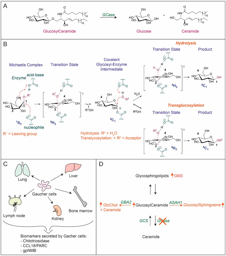
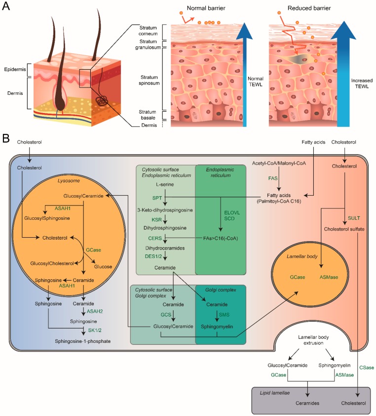
Glucocerebrosidase (GCase) is a retaining β-glucosidase with acid pH optimum metabolizing the glycosphingolipid glucosylceramide (GlcCer) to ceramide and glucose. Inherited deficiency of GCase causes the lysosomal storage disorder named Gaucher disease (GD). In GCase-deficient GD patients the accumulation of GlcCer in lysosomes of tissue macrophages is prominent. Based on the above, the key function of GCase as lysosomal hydrolase is well recognized, however it has become apparent that GCase fulfills in the human body at least one other key function beyond lysosomes. Crucially, GCase generates ceramides from GlcCer molecules in the outer part of the skin, a process essential for optimal skin barrier property and survival. This review covers the functions of GCase in and beyond lysosomes and also pays attention to the increasing insight in hitherto unexpected catalytic versatility of the enzyme.

Keywords: Gaucher disease; glucocerebrosidase; glucosylceramide; lysosome; skin.


Conflict of interest statement

The authors declare no conflicts of interest

Figures

Figure 1
Figure 1
(A) Structure of glucosylceramide (GlcCer) and degradation by GCase to glucose and ceramide. (B) Catalytic activity GCase: Hydrolyzation of β-glucosides and transglucosylation activity. (C) Occurrence of Gaucher cells and the biomarkers they secrete in plasma. (D) Metabolic adaptations to GCase deficiency: increase of GlcCer as a result of lack of degradation by GCase. Accumulated GlcCer is converted by ASAH1 to glucosylsphingosine, Glucosylated cholesterol (GlcChol) formed by GBA2 increases, and GM3 levels rise because increased anabolism by glycosyltransferases to complex GSLs. Enzymes are depicted in green. ASAH1: acid ceramidase, GBA2: cytosolic β-glucosidase, GCase: β-glucocerebrosidase, GCS: glucosylceramide synthase.
Figure 2
Figure 2
Schematic overview of the human skin and the main processes involved around GCase and its related lipids. (A) Schematic overview of a cross section of the skin showing the epidermis, dermis and subcutaneous tissue. The middle illustration shows a more detailed view of the epidermis under healthy conditions. The right illustration depicts a more detailed view of the epidermis with a reduced barrier. Exogenous compounds can get into deeper layers of the epidermis when the barrier is reduced, resulting in an immune response. It also leads to an increased transepidermal water loss (TEWL). (B) Schematic overview of the main processes involved around GCase within the cell. Arrows indicate the transport or conversion of lipids; associated enzymes are listed adjacent to their abbreviations. ASAH1: acid ceramidase, ASAH2: neutral ceramidase, ASMase: acid sphingomyelinase, CERS: ceramide synthase family, CSase: cholesterol sulfatase, DES1/2: dihydroceramide desaturase 1 and 2, ELOVL: elongation of very long chain fatty acids family, FAS: fatty acid synthase, GCase: β-glucocerebrosidase, GCS: glucosylceramide synthase, KSR: 3-ketosphinganine reductase, PLA-2: phospholipase, SCD: stearoyl-CoA desaturase, SMS: sphingomyelin synthase, SPT: serine palmitoyltransferase, SULT: cholesterol sulfotransferase type 2 isoform 1b.
Figure 3
Figure 3
The life cycle of GCase in and beyond the lysosome. AMRF: Action myoclonus renal failure syndrome, ER: endoplasmic reticulum, LB: lamellar body, LIMP2: lysosomal membrane protein 2LRO: lysosome related organelle.

Similar articles

Cited by

References

    1. Weinreb N.J., Brady R.O., Tappel A.L. The lysosomal localization of sphingolipid hydrolases. Biochim. Biophys. Acta. 1968;159:141–146. doi: 10.1016/0005-2744(68)90251-9. - DOI - PubMed
    1. Brady R.O., Kanfer J.N., Bradley R.M., Shapiro D. Demonstration of a deficiency of glucocerebroside-cleaving enzyme in Gaucher’s disease. J. Clin. Investig. 1966;45:1112–1115. doi: 10.1172/JCI105417. - DOI - PMC - PubMed
    1. Gaucher P.C.E. Ph.D. Thesis. Faculté de Médecine; Paris, France: 1882. De L’epithelioma Primitif de la Rate, Hypertrophie Idiopathique de la Rate Sans Leucemie.
    1. Beutler E., Grabowski G.A. Glucosylceramide lipidosis-Gaucher disease. In: Sriver C.R., Beaudet A.L., Sly W.S., Valle D., editors. The Metabolic and Molecular Bases of Inherited Disease. 8th ed. McGraw-Hill; New York, NY, USA: 2001.
    1. Smith L., Mullin S., Schapira A.H.V. Insights into the structural biology of Gaucher disease. Exp. Neurol. 2017;298:180–190. doi: 10.1016/j.expneurol.2017.09.010. - DOI - PubMed

LinkOut - more resources