Review
. 2020 Mar 9;8(1):7.
doi: 10.3390/jdb8010007.

Game of Tissues: How the Epidermis Thrones C. elegans Shape

Affiliations
Review

Game of Tissues: How the Epidermis Thrones C. elegans Shape

Cátia A Carvalho et al. J Dev Biol. .

Abstract

The versatility of epithelial cell structure is universally exploited by organisms in multiple contexts. Epithelial cells can establish diverse polarized axes within their tridimensional structure which enables them to flexibly communicate with their neighbors in a 360° range. Hence, these cells are central to multicellularity, and participate in diverse biological processes such as organismal development, growth or immune response and their misfunction ultimately impacts disease. During the development of an organism, the first task epidermal cells must complete is the formation of a continuous sheet, which initiates its own morphogenic process. In this review, we will focus on the C. elegans embryonic epithelial morphogenesis. We will describe how its formation, maturation, and spatial arrangements set the final shape of the nematode C. elegans. Special importance will be given to the tissue-tissue interactions, regulatory tissue-tissue feedback mechanisms and the players orchestrating the process.

Keywords: C. elegans; epidermal-muscle axis; epidermal-neuroblast axis; epithelial morphogenesis.


Conflict of interest statement

The authors declare no conflict of interest. The funders had no role in the design of the study; in the collection, analyses, or interpretation of data; in the writing of the manuscript, or in the decision to publish the results.

Figures
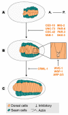
Figure 1
Figure 1
Dorsal intercalation. Epithelial cells are born dorsally as two rows (A), the dorsal cells reorganize their shape to intercalate within themselves (B) covering the dorsal length of the embryo (C). Players in orange are required in the epidermis for dorsal intercalation. All inhibitory signs are representative of the outcome of the absence of a player. Embryos oriented with anterior (A.) on the left and posterior (P.) on the right.
Figure 2
Figure 2
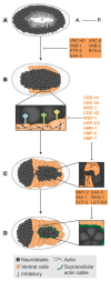
Ventral enclosure. Ventral enclosure encompasses the following “steps”: In the end of gastrulation, the neuroblasts close the ventral cleft (A) allowing the migration of the ventral epidermal cells (B). The “leading cells” initiate the migration (C) followed by the “pocket cells” (D), enclosing the embryo in a continuous epidermal layer. In each ventral enclosure “step”, players in grey and highlighted in orange are required in neuroblasts and epidermis, whereas players in orange are required in the epidermal tissue. All inhibitory signs are indicative of the outcome of the absence of a player. Embryos oriented with anterior (A.) on the left and posterior (P.) on the right.
Figure 3
Figure 3
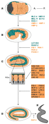
Elongation. The embryo progressively elongates from an initial 50 µm oval shape (A), through 1,5-fold (B), two-fold (C) and three-fold stages (D) until it hatches as a larva (E). Players in blue are required mainly in the seam cells, players in orange are required mainly in the dorsal/ventral cells, players in grey highlighted in orange are required in muscle and epidermal tissues. All inhibitory signs are representative of the outcome of the absence of a player. Embryos oriented with anterior (A.) on the left and posterior (P.) on the right. FOs—Fibrous Organelles, M—muscle.

References

    1. Hardin J. Epidermal morphogenesis. WormBook. 2005:1–22. doi: 10.1895/wormbook.1.35.1. - DOI - PMC - PubMed
    1. Chisholm A.D., Hsiao T.I. The C. elegans epidermis as a model skin. I: development, patterning, and growth. Wiley Interdiscip. Rev. Dev. Biol. 2012 doi: 10.1002/wdev.79. - DOI - PMC - PubMed
    1. Thompson K.W., Joshi P., Dymond J.S., Gorrepati L., Smith H., Krause M., Eisenmann D.M. The Paired-box protein PAX-3 regulates the choice between lateral and ventral epidermal cell fates in C. elegans. Dev. Biol. 2016;41:191–207. doi: 10.1016/j.ydbio.2016.03.002. - DOI - PMC - PubMed
    1. Koh K., Rothman J.H. ELT-5 and ELT-6 are required continuously to regulate epidermal seam cell differentiation and cell fusion in C. elegans. Development. 2001;128:2867–2880. - PubMed
    1. Cassata G., Shemer G., Morandi P., Donhauser R., Podbilewicz B., Baumeister R. ceh-16/engrailed patterns the embryonic epidermis of Caenorhabditis elegans. Development. 2005;132:739–749. doi: 10.1242/dev.01638. - DOI - PubMed

LinkOut - more resources