. 2020 Mar 9;9(3):667.
doi: 10.3390/cells9030667.

Oral Administration of Alpha Linoleic Acid Rescues Aβ-Induced Glia-Mediated Neuroinflammation and Cognitive Dysfunction in C57BL/6N Mice

Affiliations

Oral Administration of Alpha Linoleic Acid Rescues Aβ-Induced Glia-Mediated Neuroinflammation and Cognitive Dysfunction in C57BL/6N Mice

Waqar Ali et al. Cells. .

Abstract

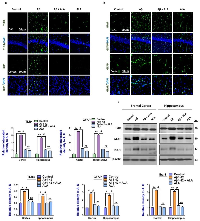
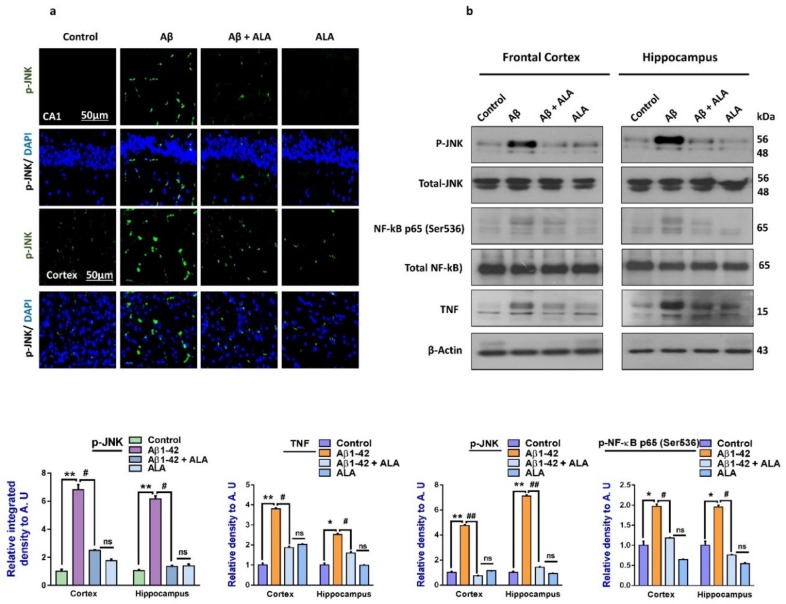
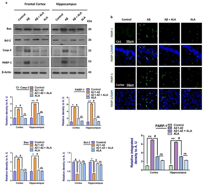
In this work, we evaluated the effects of alpha linoleic acid (ALA), an omega-3 polyunsaturated fatty acid, on amyloid-beta-induced glial-cell-mediated neuroinflammation, amyloidogenesis, and cognitive dysfunction in mice. After an infusion of Aβ1-42 (Aβ1-42, 5 μL/5 min/mouse, intracerebroventricular injection (i.c.v), and respective treatments of ALA (60 mg/kg per oral for six weeks), neuroinflammation, apoptotic markers, and synaptic markers were evaluated by Western blot and immunofluorescence analyses. According to our findings, the infusion of Aβ1-42 activated Toll-like receptor 4 (TLR4), glial fibrillary acidic protein (GFAP), and ionized calcium adaptor molecule 1 (Iba-1) in the frontal cortices and hippocampi of the Aβ1-42-injected mice to a greater extent than the Aβ1-42 + ALA-cotreated mice. Similarly, there was an elevated expression of phospho-c-Jun-N-terminal kinase (p-JNK), phospho-nuclear factor-kB p65 (p-NF-kB p65 (Ser536)), and tissue necrosis factor (TNF) in the Aβ1-42 infused mouse brains; interestingly, these markers were significantly reduced in the Aβ + ALA-cotreated group. The elevated expression of pro-apoptotic markers was observed during apoptotic cell death in the Aβ1-42-treated mouse brains, whereas these markers were markedly reduced in the Aβ + ALA-cotreated group. Moreover, Aβ1-42 infusion significantly increased amyloidogenesis, as assessed by the enhanced expression of the amyloid precursor proteins (APP) beta-amyloid cleaving enzyme-1 (BACE-1) and amyloid-beta (Aβ1-42) in the mouse brains, whereas these proteins were markedly reduced in the Aβ + ALA-cotreated group. We also checked the effects of ALA against Aβ-triggered synaptic dysfunction and memory dysfunction, showing that ALA significantly improved memory and synaptic functions in Aβ-treated mouse brains. These results indicated that ALA could be an applicable intervention in neuroinflammation, apoptotic cell loss, amyloidogenesis, and memory dysfunction via the inhibition of TLR4 and its downstream targets in Aβ + ALA-cotreated mouse brains.

Keywords: Alzheimer’s disease; neurodegeneration; neuroinflammation; omega-3 fatty acids.


Conflict of interest statement

The funders had no role in the design of the study; in the collection, analyses, or interpretation of data; in the writing of the manuscript, or in the decision to publish the results.

Figures

Figure 1
Figure 1
Effects of alpha linoleic acid against amyloid-beta (Aβ)-induced activated Toll-like receptor 4 (TLR4), glial fibrillary acidic protein (GFAP), and ionized calcium adaptor molecule 1 (Iba-1) in mouse brains. (a) and (b): Immunofluorescence results of TLR4 and GFAP in the frontal cortices and hippocampi (CA1) of the treated mice groups, with respective bar graphs. Magnification: 30×; scale bar: 30 and 50 µm. (c) Western blot results of Toll-like receptor 4 (TLR4), glial fibrillary acidic proteins (GFAP), and ionized calcium-binding adaptor molecule 1 (Iba-1), with respective bar graphs. * Significantly different from the Aβ-treated group; # significantly different from the Aβ + ALA-cotreated group. Significance = * p < 0.05; ** p < 0.01; # p < 0.05; and ## p < 0.01. Aβ: amyloid beta; ALA: alpha linoleic acid; DAPI: 4′,6-diamidino-2-phenylindole; ns = non significant.
Figure 2
Figure 2
Effects of alpha linoleic acid against Aβ-induced activated p-JNK, p-NF-kB, and TNF-α in mouse brains. (a) Immunofluorescence results of p-JNK in the frontal cortices and hippocampi (CA1) of the treated mice groups (n = 10 mice per group) with respective bar graphs. Magnification: 30×; scale bar 50 µm. (b) Western blot results of p-JNK, p-NF-kB, and TNF-α in the experimental groups with respective bar graphs. * Significantly different from the Aβ-treated group; # significantly different from the Aβ + ALA-cotreated group. Significance = * p < 0.05; ** p < 0.01; # p < 0.05; and ## p < 0.01. Aβ: amyloid beta; ALA: alpha linoleic acid; DAPI: 4′,6-diamidino-2-phenylindole; ns = non significant.
Figure 3
Figure 3
Effects of alpha linoleic acid against Aβ-induced apoptotic cell death in mouse brains. (a) Immunoblot results of BCL2-associated X protein (Bax), Bcl-2, caspase-3, and poly-ADP-ribosyltransferase (PARP-1) in the frontal cortices and hippocampi (CA1) of the treated mice groups (n = 16 mice per group, 8 for immunofluorescence and 8 for Western blot) with respective bar graphs. (b) Immunofluorescence results of PARP-1 in the frontal cortices and hippocampi of the experimental groups (n = 10 mice per group) with respective bar graphs. Magnification: 30×; scale bar: 50 µm. * Significantly different from the Aβ-treated group; # significantly different from the Aβ + ALA-cotreated group. Significance = * p < 0.05; ** p < 0.01; # p < 0.05; and ## p < 0.01. Aβ: amyloid beta; ALA: alpha linoleic acid; DAPI: 4′,6-diamidino-2-phenylindol; ns = non significant.
Figure 4
Figure 4
Effects of alpha linoleic acid against Aβ-induced AD-like changes in the mouse brains. (a) Immunofluorescence results of Aβ in the frontal cortex and hippocampus of the experimental groups (n = 16 mice per group, 8 for IF and 8 for WB), with their respective bar graphs, magnification 30×, scale bar 50 µm. (b) Immunoblots results of Aβ, BACE-1, and p-Tau in the frontal cortex and hippocampus (CA1) of the treated mice groups (n = 10 mice per group), with respective bar graphs. * Significantly different from the Aβ-treated group; # significantly different from the Aβ + ALA-cotreated group. Significance = * p < 0.05; ** p < 0.01; # p < 0.05; and ## p < 0.01. Aβ: amyloid beta; ALA: alpha linoleic acid; DAPI: 4′,6-diamidino-2-phenylindole; ns = non significant.
Figure 5
Figure 5
Effects of alpha linoleic acid against Aβ-induced synaptic and memory dysfunctions in mice. (a) Immunofluorescence results of PSD-95 in the frontal cortices and hippocampi of the experimental groups (n = 10 mice per group) with respective bar graphs. Magnification: 30×; scale bar: 50 µm. (b) Immunoblot results of PSD-95 and SNAP-23 in the frontal cortices and hippocampi (CA1) of the treated mice groups (n = 16 mice per group, 8 for IF and 8 for WB) with respective bar graphs. (c) Bar graph showing the mean time taken to escape (sec) during the training days. (d) Final escape time during the probe test. (e) Number of platform crossings. (f) Time spent in the target quadrant. (g) Spontaneous alternation behavior. * Significantly different from the Aβ-treated group; # significantly different from the Aβ + ALA-cotreated group. Significance = * p < 0.05; ** p < 0.01; # p < 0.05; and ## p < 0.01. Aβ: amyloid beta; ALA: alpha linoleic acid; DAPI: 4′,6-diamidino-2-phenylindole; ns = non significant.

References

    1. Ali M., Muhammad S., Shah M.R., Khan A., Rashid U., Farooq U., Ullah F., Sadiq A., Ayaz M., Ali M., et al. Neurologically Potent Molecules from Crataegus oxyacantha; Isolation, Anticholinesterase Inhibition, and Molecular Docking. Front. Pharmacol. 2017;8:327. doi: 10.3389/fphar.2017.00327. - DOI - PMC - PubMed
    1. Ayaz M., Sadiq A., Junaid M., Ullah F., Subhan F., Ahmed J. Neuroprotective and Anti-Aging Potentials of Essential Oils from Aromatic and Medicinal Plants. Front. Aging Neurosci. 2017;9:168. doi: 10.3389/fnagi.2017.00168. - DOI - PMC - PubMed
    1. Abid N.B., Naseer M.I., Kim M.O. Comparative Gene-Expression Analysis of Alzheimer’s Disease Progression with Aging in Transgenic Mouse Model. Int. J. Mol. Sci. 2019;20:1219. doi: 10.3390/ijms20051219. - DOI - PMC - PubMed
    1. Lattanzi S., Brigo F., Vernieri F., Silvestrini M. Visit-to-visit variability in blood pressure and Alzheimer’s disease. J. Clin. Hypertens. 2018;20:918–924. doi: 10.1111/jch.13290. - DOI - PMC - PubMed
    1. Lattanzi S., Vernieri F., Silvestrini M. Blood pressure variability and neurocognitive functioning. J. Clin. Hypertens. 2018;20:645–647. doi: 10.1111/jch.13232. - DOI - PMC - PubMed

Publication types

MeSH terms