

Skin Lesion Segmentation from Dermoscopic Images Using Convolutional Neural Network
- PMID: 32183041
- PMCID: PMC7147706
- DOI: 10.3390/s20061601
Skin Lesion Segmentation from Dermoscopic Images Using Convolutional Neural Network
Abstract
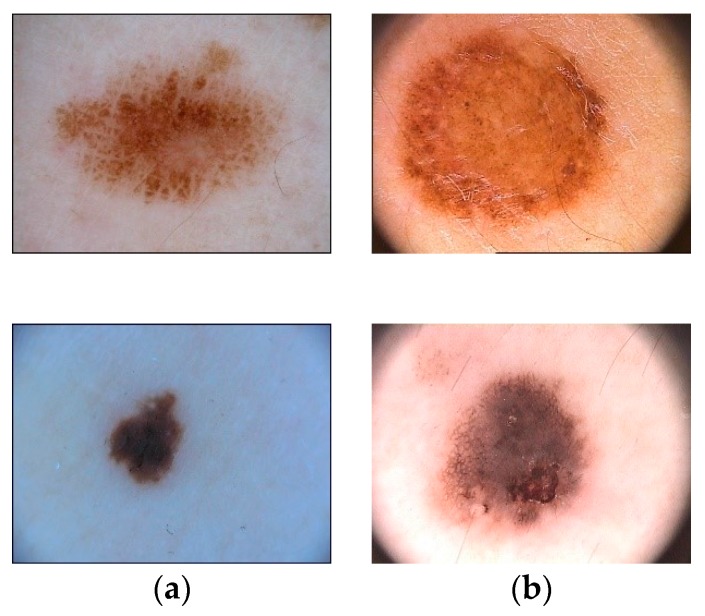
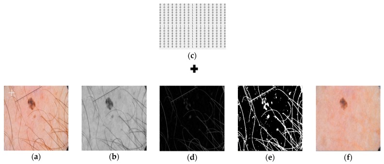
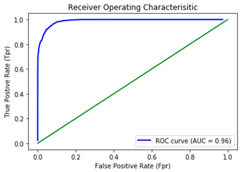
Clinical treatment of skin lesion is primarily dependent on timely detection and delimitation of lesion boundaries for accurate cancerous region localization. Prevalence of skin cancer is on the higher side, especially that of melanoma, which is aggressive in nature due to its high metastasis rate. Therefore, timely diagnosis is critical for its treatment before the onset of malignancy. To address this problem, medical imaging is used for the analysis and segmentation of lesion boundaries from dermoscopic images. Various methods have been used, ranging from visual inspection to the textural analysis of the images. However, accuracy of these methods is low for proper clinical treatment because of the sensitivity involved in surgical procedures or drug application. This presents an opportunity to develop an automated model with good accuracy so that it may be used in a clinical setting. This paper proposes an automated method for segmenting lesion boundaries that combines two architectures, the U-Net and the ResNet, collectively called Res-Unet. Moreover, we also used image inpainting for hair removal, which improved the segmentation results significantly. We trained our model on the ISIC 2017 dataset and validated it on the ISIC 2017 test set as well as the PH2 dataset. Our proposed model attained a Jaccard Index of 0.772 on the ISIC 2017 test set and 0.854 on the PH2 dataset, which are comparable results to the current available state-of-the-art techniques.
Keywords: Jaccard Index; ROC curve; ResNet; U-Net; convolutional neural networks; dermoscopic images; image inpainting; melanoma.
Conflict of interest statement
The authors declare no conflict of interest.
Figures


Similar articles
-
Efficient skin lesion segmentation using separable-Unet with stochastic weight averaging.Comput Methods Programs Biomed. 2019 Sep;178:289-301. doi: 10.1016/j.cmpb.2019.07.005. Epub 2019 Jul 8. Comput Methods Programs Biomed. 2019. PMID: 31416556
-
LAMA: Lesion-Aware Mixup Augmentation for Skin Lesion Segmentation.J Imaging Inform Med. 2024 Aug;37(4):1812-1823. doi: 10.1007/s10278-024-01000-5. Epub 2024 Feb 26. J Imaging Inform Med. 2024. PMID: 38409610 Free PMC article.
-
Skin lesion segmentation in dermoscopy images via deep full resolution convolutional networks.Comput Methods Programs Biomed. 2018 Aug;162:221-231. doi: 10.1016/j.cmpb.2018.05.027. Epub 2018 May 19. Comput Methods Programs Biomed. 2018. PMID: 29903489
-
Deep Learning Approaches Towards Skin Lesion Segmentation and Classification from Dermoscopic Images - A Review.Curr Med Imaging. 2020;16(5):513-533. doi: 10.2174/1573405615666190129120449. Curr Med Imaging. 2020. PMID: 32484086 Review.
-
Computational neural network in melanocytic lesions diagnosis: artificial intelligence to improve diagnosis in dermatology?Eur J Dermatol. 2019 Apr 1;29(S1):4-7. doi: 10.1684/ejd.2019.3538. Eur J Dermatol. 2019. PMID: 31017580 Review.
Cited by
-
Skin Cancer Image Segmentation Based on Midpoint Analysis Approach.J Imaging Inform Med. 2024 Oct;37(5):2581-2596. doi: 10.1007/s10278-024-01106-w. Epub 2024 Apr 16. J Imaging Inform Med. 2024. PMID: 38627267 Free PMC article.
-
New Trends in Melanoma Detection Using Neural Networks: A Systematic Review.Sensors (Basel). 2022 Jan 10;22(2):496. doi: 10.3390/s22020496. Sensors (Basel). 2022. PMID: 35062458 Free PMC article.
-
Advances in Medical Image Segmentation: A Comprehensive Review of Traditional, Deep Learning and Hybrid Approaches.Bioengineering (Basel). 2024 Oct 16;11(10):1034. doi: 10.3390/bioengineering11101034. Bioengineering (Basel). 2024. PMID: 39451409 Free PMC article. Review.
-
The Role of Machine Learning and Deep Learning Approaches for the Detection of Skin Cancer.Healthcare (Basel). 2023 Feb 1;11(3):415. doi: 10.3390/healthcare11030415. Healthcare (Basel). 2023. PMID: 36766989 Free PMC article.
-
Medical Image Segmentation with Learning Semantic and Global Contextual Representation.Diagnostics (Basel). 2022 Jun 25;12(7):1548. doi: 10.3390/diagnostics12071548. Diagnostics (Basel). 2022. PMID: 35885454 Free PMC article.
References
-
- Macià F., Pumarega J., Gallén M., Porta M. Time from (clinical or certainty) diagnosis to treatment onset in cancer patients: The choice of diagnostic date strongly influences differences in therapeutic delay by tumor site and stage. J. Clin. Epidemiol. 2013;66:928–939. doi: 10.1016/j.jclinepi.2012.12.018. - DOI - PubMed
-
- Matthews N.H., Li W.-Q., Qureshi A.A., Weinstock M.A., Cho E. Cutaneous Melanoma: Etiology and Therapy. Codon Publications; Brisbane, Australia: 2017. Epidemiology of melanoma; pp. 3–22. - PubMed
-
- Colditz G.A. Google Books. SAGE Publications, Inc.; Thousand Oaks, CA, USA: 2015. Encyclopedia of Cancer and Society.
MeSH terms
LinkOut - more resources
Full Text Sources
Other Literature Sources
