Review
. 2020 Mar 13;12(3):762.
doi: 10.3390/nu12030762.

A Guide to Human Zinc Absorption: General Overview and Recent Advances of In Vitro Intestinal Models

Affiliations
Review

A Guide to Human Zinc Absorption: General Overview and Recent Advances of In Vitro Intestinal Models

Maria Maares et al. Nutrients. .

Abstract

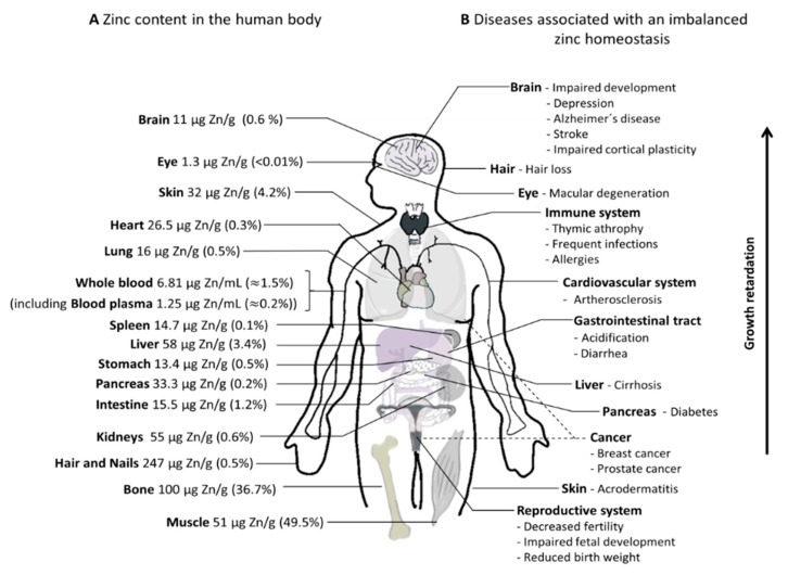
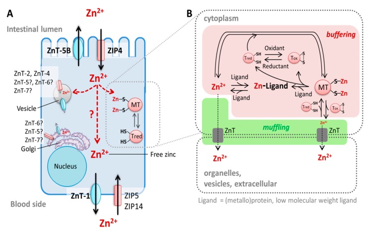
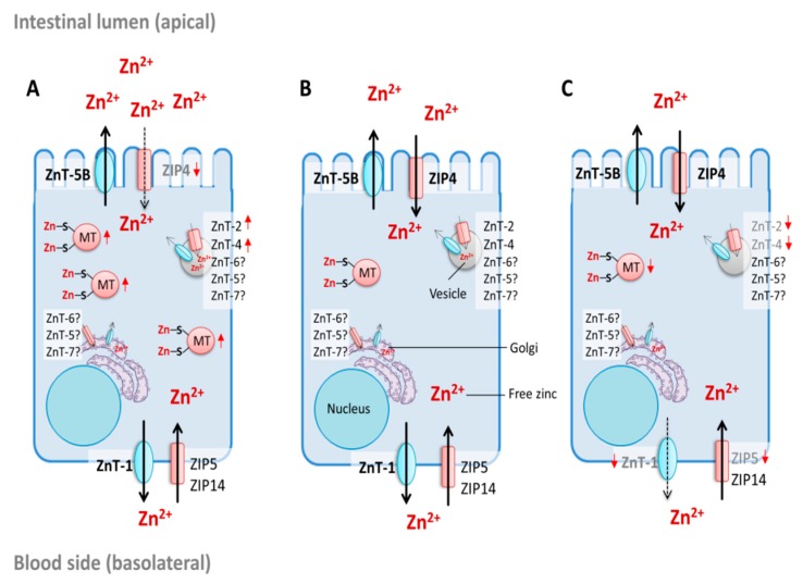
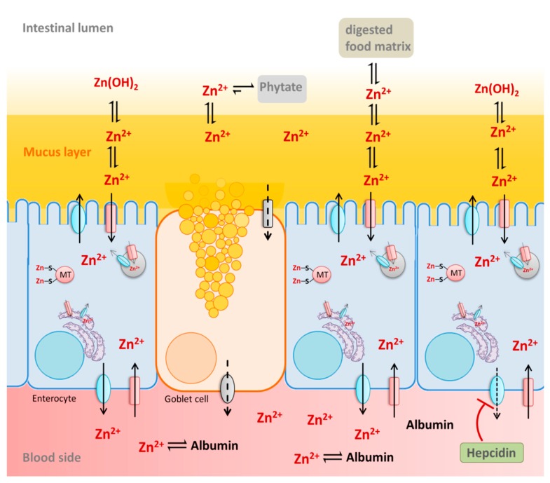
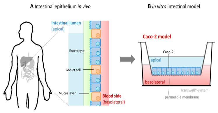
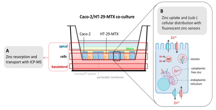
Zinc absorption in the small intestine is one of the main mechanisms regulating the systemic homeostasis of this essential trace element. This review summarizes the key aspects of human zinc homeostasis and distribution. In particular, current knowledge on human intestinal zinc absorption and the influence of diet-derived factors on bioaccessibility and bioavailability as well as intrinsic luminal and basolateral factors with an impact on zinc uptake are discussed. Their investigation is increasingly performed using in vitro cellular intestinal models, which are continually being refined and keep gaining importance for studying zinc uptake and transport via the human intestinal epithelium. The vast majority of these models is based on the human intestinal cell line Caco-2 in combination with other relevant components of the intestinal epithelium, such as mucin-secreting goblet cells and in vitro digestion models, and applying improved compositions of apical and basolateral media to mimic the in vivo situation as closely as possible. Particular emphasis is placed on summarizing previous applications as well as key results of these models, comparing their results to data obtained in humans, and discussing their advantages and limitations.

Keywords: Caco-2; in vitro intestinal model; intestinal; intestinal absorption; zinc; zinc bioavailability; zinc homeostasis; zinc uptake.


Conflict of interest statement

The authors declare no conflict of interest.

Figures

Figure 1
Figure 1
Overview of zinc distribution and disease association in the human body. (A) Approximate zinc content (µg per g wet weight) of the respective tissues and the resulting proportion of total body zinc. Detailed estimation of the tissues’ zinc content and references are depicted in Supplementary Table S1. (B) Diseases of the respective organ systems associated with imbalanced zinc homeostasis.
Figure 2
Figure 2
Enterocyte zinc homeostasis. (A) Zinc homeostasis in enterocytes during zinc absorption. Three main zinc pools in enterocytes have been described: (i) cytoplasmic-free zinc, which is only complexed by low molecular weight ligands, (ii) protein-bound zinc, depicted here as metallothionein (MT)-bound zinc, and (iii) free zinc stored in vesicles [104]. The vesicular [102,103] and cytoplasmic-free zinc pools [101] are recognized to be involved in zinc absorption by enterocytes [105]. Cellular zinc homeostasis is maintained by three main groups of proteins: the zinc transporter (ZnT)-and the Zrt-, Irt-like protein (ZIP)-family as well as the zinc-binding metallothioneins [99]. They regulate the cytoplasmic-free zinc concentration and provide its distribution into organelles and vesicles. Exporters of zinc from vesicular stores in enterocytes remain to be identified and transfer of the divalent cation through the enterocytes after its uptake by the cells (illustrated by red arrows) is not yet fully understood. (B) Zinc buffering and muffling role of metallothioneins (MTs). MTs and other ligands (such as proteins) bind free zinc and, thereby, buffer its cytoplasmic concentration. In addition to zinc transporters, MTs represent zinc muffling moieties, which decrease free zinc content in the cytoplasm by transferring the cation to transporters, sequestering it into organelles, vesicles, or outside the cell. Notably, free zinc itself can also be transported into organelles, whereby, in this process, the ZnT solely undertakes the muffling [100]. Moreover, MTs re-distribute intracellular zinc by transferring it to other ligands, such as metalloproteins [106]. This zinc transfer may be enforced by a redox-active mechanism in which the apo-protein Thionein (Tred) binds the cation, which results in its metal-loaded form, MT, which releases zinc upon its oxidation to Thionin (Tox) (reviewed in Reference [107]).
Figure 3
Figure 3
Regulation of intestinal zinc absorption. Potential regulatory mechanisms of zinc absorption into enterocytes during (A) zinc excess, (B) adequate supply, and (C) zinc deficiency, based on experimental data on the zinc-dependent expression pattern of the intestinal zinc transporters (ZnT) and the Zrt-, Irt-like protein (ZIP)-transporters as well as metallothioneins (MT). Enterocyte zinc homeostasis is controlled by these proteins, regulating the amount of intestinally absorbed and basolaterally exported zinc [150]. The subcellular localization of ZnT-5, ZnT-6, and ZnT-7 in enterocytes is not yet fully investigated. Zinc-dependent up-regulation or downregulation of the respective protein and/or messenger ribonucleic acid (mRNA) are indicated by red arrows.
Figure 4
Figure 4
Luminal and serosal factors recognized to influence zinc absorption. Food-derived macromolecules and low molecular weight ligands positively or negatively influence the speciation of the ion as well as its luminal free and available concentration, consequently affecting its absorption by the intestinal epithelium [3]. Phytate forms stable complexes with zinc at intestinal pH, which diminishes its availability for enterocytes [153]. Conversely, the protein content of the consumed food has a positive effect on zinc absorption due to the release of amino acids and peptides upon degradation. Presumably, these increase luminal solubility of the metal, and, consequently, enhance its availability to enterocytes [154,155]. Serum albumin is an important serosal factor, acting as a basolateral zinc-acceptor and enhancing enterocytic zinc release into the blood circulation [102]. Additionally, systemic humoral factors, such as hepcidin, seem to influence ZnT-1-mediated export of zinc by intestinal cells [156], which indicates that the liver might play an important role in secreting humoral factors regulating intestinal zinc absorption.
Figure 5
Figure 5
Schematic representation of the three-dimensional in vitro cellular intestinal model Caco-2. (A) The intestinal epithelium in vivo is mainly composed of enterocytes and goblet cells [255], which represents about 90% of intestinal cells of the brush border membrane [256,257]. These are covered by a viscoelastic gel: the mucus layer. This physical barrier is synthesized and secreted by goblet cells and serves as a protective layer for the underlying epithelium. (B) Three-dimensional Caco-2 monoculture in the “Transwell® system”. The intestinal cell line Caco-2 is cultured in inserts on a permeable membrane, and, in most cases, composed of polycarbonate. This results in three compartments: an apical compartment representing the intestinal lumen, a basolateral side corresponding to the serosal surface of enterocytes, and the intestinal barrier formed by differentiated Caco-2 cells.
Figure 6
Figure 6
Application of in vitro cellular intestinal models to study intestinal zinc transport. Schematic representation of the three-dimensional intestinal Caco-2/HT-29-MTX co-culture model. (A) Zinc is quantified in all three compartments (apical, cellular, basolateral) with conventional analytical approaches, such as inductively coupled mass spectrometry (ICP-MS) or flame atomic absorption spectrometry (FAAS). (B) The application of chemical-based or protein-based fluorescent zinc sensors in enterocytes provides additional information about the subcellular distribution of the micronutrient upon its uptake into the cell. These sensors bind intracellular free zinc and track small changes of this zinc moiety. Depending on the subcellular localization of the sensor, the cytoplasmic free zinc pool or free zinc in organelles, such as vesicles and the endoplasmic reticulum (circled in red) can be investigated.

References

    1. Maret W. Zinc in cellular regulation: The nature and significance of “zinc signals”. Int. J. Mol. Sci. 2017;18:2285. doi: 10.3390/ijms18112285. - DOI - PMC - PubMed
    1. Rink L., Gabriel P. Zinc and the immune system. Proc. Nutr. Soc. 2000;59:541. doi: 10.1017/S0029665100000781. - DOI - PubMed
    1. Krebs N.F. Overview of zinc absorption and excretion in the human gastrointestinal tract. J. Nutr. 2000;130:1374S–1377S. doi: 10.1093/jn/130.5.1374S. - DOI - PubMed
    1. Ford D. Intestinal and placental zinc transport pathways. Proc. Nutr. Soc. 2004;63:21–29. doi: 10.1079/PNS2003320. - DOI - PubMed
    1. King J.C. Zinc: An essential but elusive nutrient. Am. J. Clin. Nutr. 2011;94:6795–6845. doi: 10.3945/ajcn.110.005744. - DOI - PMC - PubMed

LinkOut - more resources