Review
. 2020 Mar 14;21(6):1985.
doi: 10.3390/ijms21061985.

Potential Role of Venular Amyloid in Alzheimer's Disease Pathogenesis

Affiliations
Review

Potential Role of Venular Amyloid in Alzheimer's Disease Pathogenesis

Christopher D Morrone et al. Int J Mol Sci. .

Abstract

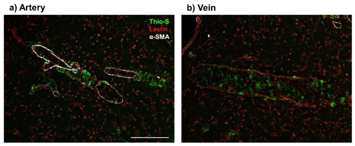
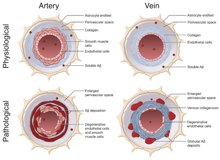
Insurmountable evidence has demonstrated a strong association between Alzheimer's disease (AD) and cerebral amyloid angiopathy (CAA), along with various other cerebrovascular diseases. One form of CAA, which is the accumulation of amyloid-beta peptides (Aβ) along cerebral vessel walls, impairs perivascular drainage pathways and contributes to cerebrovascular dysfunction in AD. To date, CAA research has been primarily focused on arterial Aβ, while the accumulation of Aβ in veins and venules were to a lesser extent. In this review, we describe preclinical models and clinical studies supporting the presence of venular amyloid and potential downstream pathological mechanisms that affect the cerebrovasculature in AD. Venous collagenosis, impaired cerebrovascular pulsatility, and enlarged perivascular spaces are exacerbated by venular amyloid and increase Aβ deposition, potentially through impaired perivascular clearance. Gaining a comprehensive understanding of the mechanisms involved in venular Aβ deposition and associated pathologies will give insight to how CAA contributes to AD and its association with AD-related cerebrovascular disease. Lastly, we suggest that special consideration should be made to develop Aβ-targeted therapeutics that remove vascular amyloid and address cerebrovascular dysfunction in AD.

Keywords: Alzheimer’s disease; Aβ; TgF344-AD rat model; cerebral amyloid angiopathy; perivascular clearance; vein/venule; venous collagenosis; venular amyloid.


Conflict of interest statement

The authors declare no conflict of interest.

Figures

Figure 1
Figure 1
Presence of venular amyloid in 16-month old TgF344-AD rats. Immunofluorescence imaging of vascular amyloid deposition, stained by Thioflavin S (Thio-S, green), in cortical penetrating vessels (Lectin, red). Amyloid beta peptide (Aβ) deposits present a ‘double-barreling’ morphology, forming cyclical rings surrounding the arteries (a). Venular Aβ is present as smaller, globular deposits along the veins (b). Arteries were distinguished from veins by presence of alpha smooth muscle actin (α-SMA, white). Scale bar = 100 μm.
Figure 2
Figure 2
The pathological role of vascular Aβ deposition on arteries and veins. This schematic summarizes the effects of Aβ (red) accumulation on cerebral vessels, relative to physiological conditions. In physiological conditions, soluble Aβ can be cleared from the brain into the vessel lumen and along the perivascular spaces of arteries and veins. In pathological arteries, Aβ is deposited along the smooth muscle cells and basement membranes, forming a ‘double-barreling’ morphology surrounding the vessel, resulting in arterial smooth muscle and endothelial cell loss. In pathological veins, the Aβ deposition is distinct, forming globular deposits in perivascular spaces. Venular Aβ contributes to extensive venous collagenosis (dark blue) in the form of concentric rings along venular walls, significant enlargement of the perivascular space, and endothelial cell loss. These pathological features in arteries and veins exacerbate Aβ accumulation, ultimately causing vascular and cognitive dysfunction in cases of cerebral amyloid angiopathy (CAA) and Alzheimer’s disease (AD). Image not to scale. Image by Sherry Lai.

References

    1. Iadecola C., Gorelick P.B. Mechanisms in Vascular and Neurodegenerative Dementia. Stroke. 2003;34:335–337. doi: 10.1161/01.STR.0000054050.51530.76. - DOI - PubMed
    1. Saito S., Ihara M. New Therapeutic Approaches for Alzhiemer’s disease and Cerebral Amyloid Angiopathy. Front. Aging Neurosci. 2014;6:290. doi: 10.3389/fnagi.2014.00290. - DOI - PMC - PubMed
    1. Querfurth H.W., LaFerla F.M. Alzheimer′s Disease. N. Engl. J. Med. 2010;362:329–344. doi: 10.1056/NEJMra0909142. - DOI - PubMed
    1. Weller R.O., Subash M., Preston S.D., Mazanti I., Carare R.O. SYMPOSIUM: Clearance of Aβ from the Brain in Alzheimer′s Disease: Perivascular Drainage of Amyloid-β Peptides from the Brain and its Failure in Cerebral Amyloid Angiopathy and Alzheimer′s Disease. Brain Pathol. 2008;18:253–266. doi: 10.1111/j.1750-3639.2008.00133.x. - DOI - PMC - PubMed
    1. Cai Y., An S.S., Kim S. Mutations in presenilin 2 and its implications in Alzheimer’s disease and other dementia-Associated disorders. Clin. Interv. Aging. 2015;10:1163–1172. doi: 10.2147/CIA.S85808. - DOI - PMC - PubMed

MeSH terms

Substances