Review
. 2020 Mar 14;8(3):59.
doi: 10.3390/biomedicines8030059.

Aptamers Increase Biocompatibility and Reduce the Toxicity of Magnetic Nanoparticles Used in Biomedicine

Affiliations
Review

Aptamers Increase Biocompatibility and Reduce the Toxicity of Magnetic Nanoparticles Used in Biomedicine

Galina S Zamay et al. Biomedicines. .

Abstract

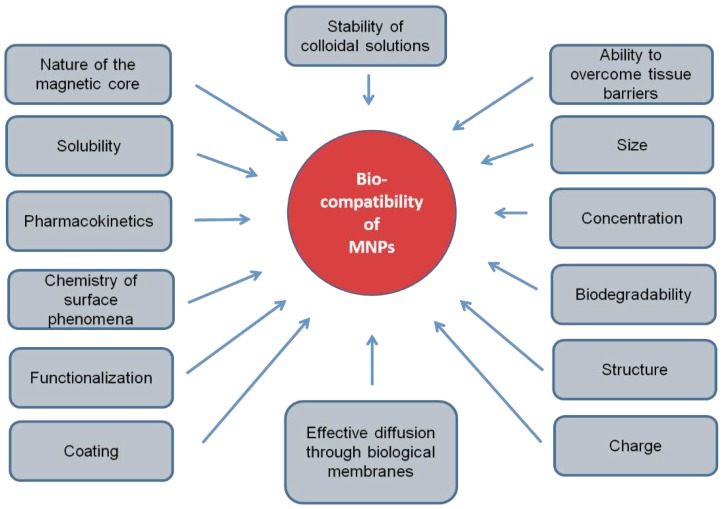
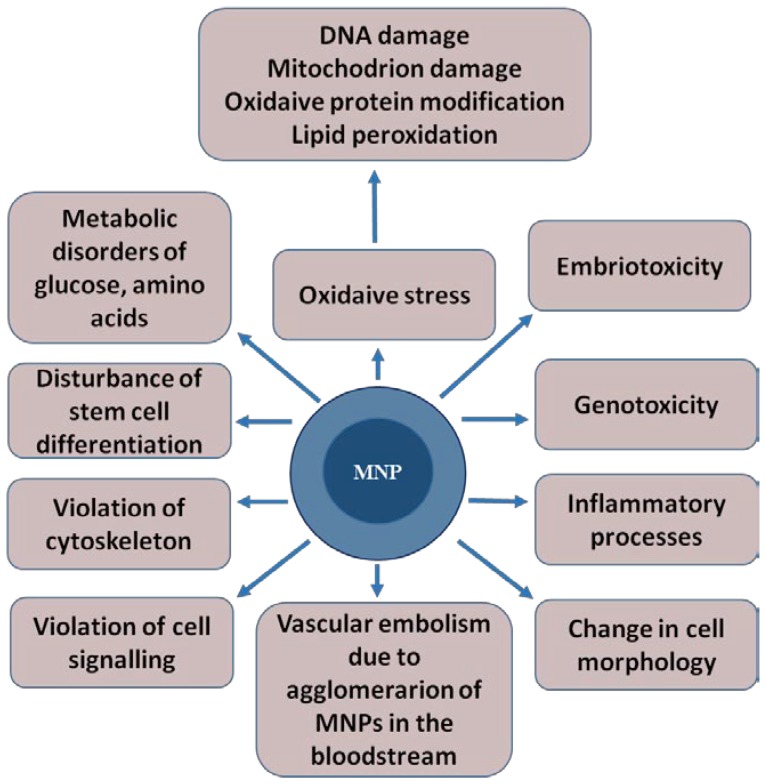
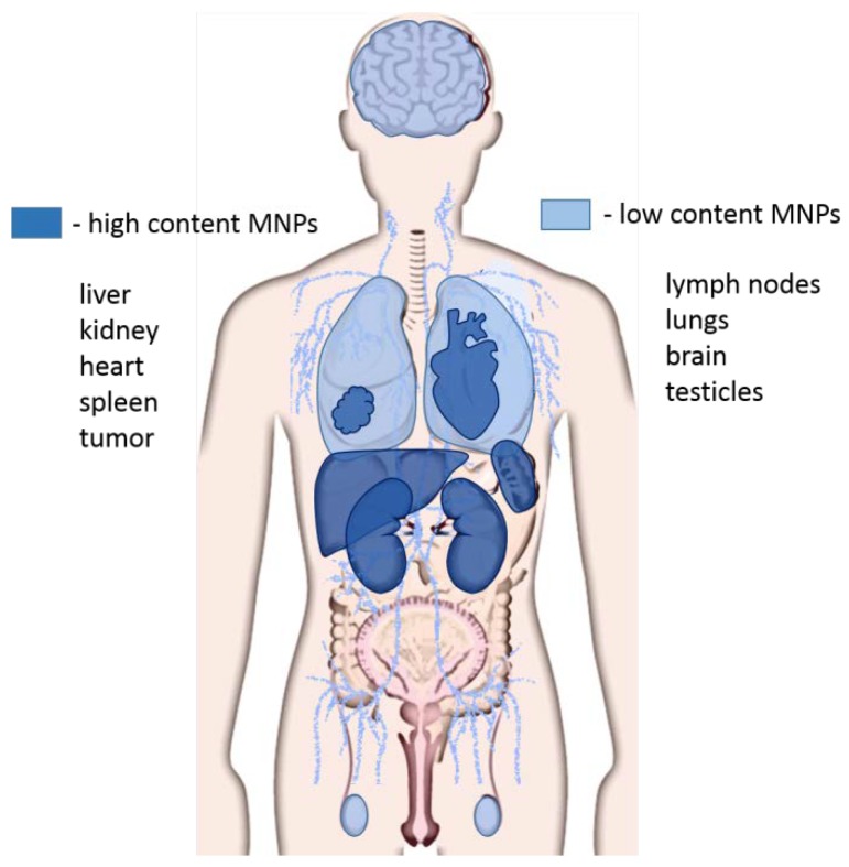
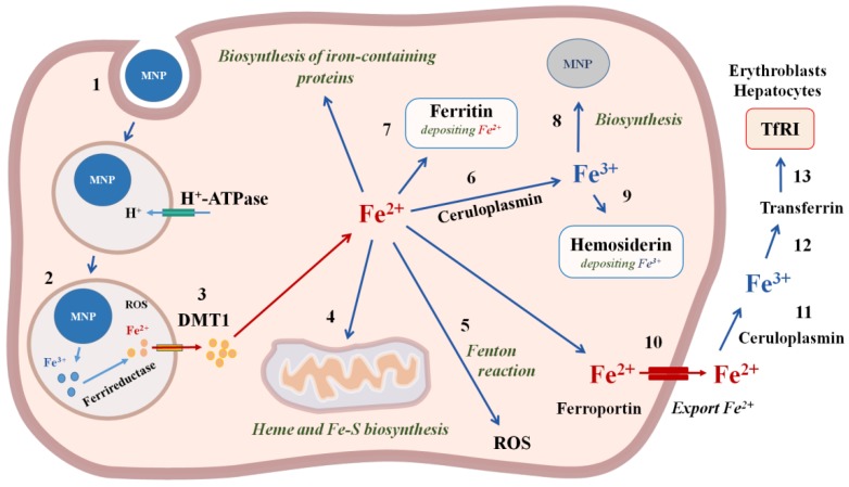
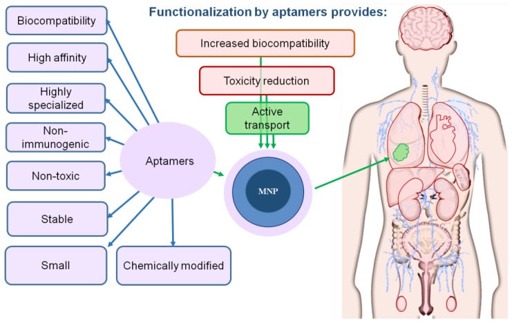
Aptamer-based approaches are very promising tools in nanomedicine. These small single-stranded DNA or RNA molecules are often used for the effective delivery and increasing biocompatibility of various therapeutic agents. Recently, magnetic nanoparticles (MNPs) have begun to be successfully applied in various fields of biomedicine. The use of MNPs is limited by their potential toxicity, which depends on their biocompatibility. The functionalization of MNPs by ligands increases biocompatibility by changing the charge and shape of MNPs, preventing opsonization, increasing the circulation time of MNPs in the blood, thus shielding iron ions and leading to the accumulation of MNPs only in the necessary organs. Among various ligands, aptamers, which are synthetic analogs of antibodies, turned out to be the most promising for the functionalization of MNPs. This review describes the factors that determine MNPs' biocompatibility and affect their circulation time in the bloodstream, biodistribution in organs and tissues, and biodegradation. The work also covers the role of the aptamers in increasing MNPs' biocompatibility and reducing toxicity.

Keywords: aptamers; biocompatibility; magnetic nanoparticles; toxicity.


Conflict of interest statement

The authors declare no conflict of interest.

Figures

Figure 1
Figure 1
Physical and chemical factors of magnetic nanoparticles, determining their biocompatibility.
Figure 2
Figure 2
The toxic effects that magnetic nanoparticles can cause in a living organism. The magnetic core of the nanoparticle is shown in dark blue, and shell is represented in light blue.
Figure 3
Figure 3
Distribution of magnetic nanoparticles in organs and tissues. The high concentration of magnetic nanoparticles (MNPs) accumulates in the liver, kidneys, spleen, heart, and tumors. Lower concentrations of MNPs are observed in the brain, lungs, lymph nodes, and testes.
Figure 4
Figure 4
The mechanisms of iron magnetic nanoparticles incorporation and biodegradation in the cell. 1—MNP incorporation using endocytosis; 2—degradation of MNP and reduction in iron with ferrireductase; 3—Fe2+ transfer through the membrane with divalent metal transporter DMT1; 4—transport of Fe2+ into mitochondria for heme and Fe-S biosynthesis; 5—ROS formation in the Fenton reaction; 6—oxidation of Fe2+ with ceruloplasmin to Fe3+; 7—deposition of Fe2+ with ferritin; 8—biosinthesis of cellular MNPs; 9—deposition of Fe3+ with hemosiderin; 10—export of Fe2+ from the cell with ferroportin; 11—oxidation of Fe2+ with ceruloplasmin to Fe3+; 12—binding to transferrin; 13—Transferrin entry into the cells, which contain Transferrin receptors TfR1on the cell membranes, in particular, erythroblasts and hepatocytes.
Figure 5
Figure 5
Aptamers increase the biocompatibility of magnetic nanoparticles. The magnetic core of the nanoparticle is shown in dark blue; the shell is represented in light blue; aptamer coating is light violet. The tumor is depicted in green.

References

    1. Schwenk M.H. Ferumoxytol: A new intravenous iron preparation for the treatment of iron deficiency anemia in patients with chronic kidney disease. Pharmacotherapy. 2010;30:70–79. doi: 10.1592/phco.30.1.70. - DOI - PubMed
    1. Du V., Luciani N., Richard S., Mary G., Gay C., Mazuel F., Reffay M., Menasche P., Agbulut O., Wilhelm C. A 3D magnetic tissue stretcher for remote mechanical control of embryonic stem cell differentiation. Nat. Commun. 2017;8:400. doi: 10.1038/s41467-017-00543-2. - DOI - PMC - PubMed
    1. Souza G.R., Molina J.R., Raphael R.M., Ozawa M.G., Stark D.J., Levin C.S., Bronk L.F., Ananta J.S., Mandelin J., Georgescu M.M., et al. Three-Dimensional tissue culture based on magnetic cell levitation. Nat. Nanotechnol. 2010;5:291–296. doi: 10.1038/nnano.2010.23. - DOI - PMC - PubMed
    1. Yamamoto Y., Ito A., Fujita H., Nagamori E., Kawabe Y., Kamihira M. Functional evaluation of artificial skeletal muscle tissue constructs fabricated by a magnetic force-based tissue engineering technique. Tissue Eng. Part A. 2011;17:107–114. doi: 10.1089/ten.tea.2010.0312. - DOI - PubMed
    1. Zhang H., Xia J., Pang X., Zhao M., Wang B., Yang L., Wan H., Wu J., Fu S. Magnetic nanoparticle-loaded electrospun polymeric nanofibers for tissue engineering. Mater. Sci. Eng. C Mater. Biol. Appl. 2017;73:537–543. doi: 10.1016/j.msec.2016.12.116. - DOI - PubMed

LinkOut - more resources