. 2020 Mar 14;21(6):1994.
doi: 10.3390/ijms21061994.

Oxidative Stress and Mitochondrial Abnormalities Contribute to Decreased Endothelial Nitric Oxide Synthase Expression and Renal Disease Progression in Early Experimental Polycystic Kidney Disease

Affiliations

Oxidative Stress and Mitochondrial Abnormalities Contribute to Decreased Endothelial Nitric Oxide Synthase Expression and Renal Disease Progression in Early Experimental Polycystic Kidney Disease

Alp S Kahveci et al. Int J Mol Sci. .

Abstract

Vascular abnormalities are the most important non-cystic complications in Polycystic Kidney Disease (PKD) and contribute to renal disease progression. Endothelial dysfunction and oxidative stress are evident in patients with ADPKD, preserved renal function, and controlled hypertension. The underlying biological mechanisms remain unknown. We hypothesized that in early ADPKD, the reactive oxygen species (ROS)-producing nicotinamide adenine dinucleotide phosphate hydrogen (NAD(P)H)-oxidase complex-4 (NOX4), a major source of ROS in renal tubular epithelial cells (TECs) and endothelial cells (ECs), induces EC mitochondrial abnormalities, contributing to endothelial dysfunction, vascular abnormalities, and renal disease progression. Renal oxidative stress, mitochondrial morphology (electron microscopy), and NOX4 expression were assessed in 4- and 12-week-old PCK and Sprague-Dawley (wild-type, WT) control rats (n = 8 males and 8 females each). Endothelial function was assessed by renal expression of endothelial nitric oxide synthase (eNOS). Peritubular capillaries were counted in hematoxylin-eosin (H&E)-stained slides and correlated with the cystic index. The enlarged cystic kidneys of PCK rats exhibited significant accumulation of 8-hydroxyguanosine (8-OHdG) as early as 4 weeks of age, which became more pronounced at 12 weeks. Mitochondria of TECs lining cysts and ECs exhibited loss of cristae but remained preserved in non-cystic TECs. Renal expression of NOX4 was upregulated in TECs and ECs of PCK rats at 4 weeks of age and further increased at 12 weeks. Contrarily, eNOS immunoreactivity was lower in PCK vs. WT rats at 4 weeks and further decreased at 12 weeks. The peritubular capillary index was lower in PCK vs. WT rats at 12 weeks and correlated inversely with the cystic index. Early PKD is associated with NOX4-induced oxidative stress and mitochondrial abnormalities predominantly in ECs and TECs lining cysts. Endothelial dysfunction precedes capillary loss, and the latter correlates with worsening of renal disease. These observations position NOX4 and EC mitochondria as potential therapeutic targets in PKD.

Keywords: NOX4; endothelial dysfunction; mitochondria; oxidative stress; polycystic kidney disease.


Conflict of interest statement

The authors declare no conflict of interest.

Figures

Figure 1
Figure 1
Renal oxidative stress worsens PKD progression. Representative immunofluorescence (IF) staining for 8-hydroxyguanosine (8-OHdG, green) in renal tissue sections of WT and PCK rats (A) and its quantification (B), showing increased 8-OHdG immunoreactivity in PCK vs. WT rats at 4 weeks, which further increased at 12 weeks; 8-OHdG was quantified as % stained area and adjusted to DAPI-stained % area. ** p < 0.01, **** p < 0.001. (n = 16/group).
Figure 2
Figure 2
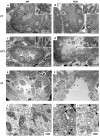
PKD is associated with mitochondrial structural abnormalities in cyst-lining tubular cells. Transmission electron micrograph (TEM) displaying representative tubular structures from WT (left column) and PCK (right column) kidneys at 4 weeks. In PCK kidneys, the cells of proximal tubules (B) had normal appearance and were characterized by a tall brush border and extensive invaginations of the basolateral plasma membrane containing abundant elongated mitochondria (×2500 and ×80,000) compared to WT kidneys (A). Similarly, the cells from distal tubules in PCK animals (D) presented numerous long mitochondria arranged between the foldings of the basal lamina that resembled normal appearing mitochondria as in WT kidneys (C). On the other hand, mitochondria from CD principal cells (P), characterized by a light appearance, extensive infoldings of the basal plasma membrane, and intercalated cells (I), characterized by a denser cytoplasm, numerous apical projections, and more abundant mitochondria, lining micro cysts on PCK animals (F), showed cristae remodeling and loss (arrow heads) (×2500 and × 80,000) compared to WT CD (E). PT, proximal tubule, DCT, distal convoluted tubule; CD, collecting duct. The panels below E and F are high-magnification images of the P and I cells in the dotted squares.
Figure 3
Figure 3
PKD is associated with mitochondrial structural abnormalities in endothelial cells. TEM displaying representative peritubular capillary endothelial cells (EC) from WT (left column) and PCK (right column) kidneys at 4 and 12 weeks ((A,B) and (C,D) respectively). In PCK, peritubular capillary EC exhibited morphological abnormalities (cristae remodeling and loss) not only in the capillaries surrounding cystic structures but also in the capillaries surrounding normal appearing tubules (×2500 and ×80,000).
Figure 4
Figure 4
Renal NOX4 is upregulated in tubular epithelial cells in PKD. Representative immunofluorescence staining for NOX4 (red) in renal tissue sections of WT and PCK rats (A) and its quantification (B), showing increased NOX4 immunoreactivity in PCK vs. WT rats at 4 weeks, which further increased at 12 weeks. **** p < 0.0001. NOX4 was quantified as % stained area and adjusted to DAPI-stained % area. (n = 16/group).
Figure 5
Figure 5
Renal NOX4 is upregulated in peritubular capillary endothelial cells in PKD. Representative IF staining for NOX4 (red) and endothelial cell marker CD31 (green) in renal tissue sections (40×) (A) and its quantification (B), showing increased number of double-positive cells (NOX4/CD31+) in PCK vs. WT rats at 4 weeks, which further increased at 12 weeks. *** p < 0.001; **** p < 0.0001. The number of double-positive NOX4/CD31cells was adjusted to the number of positive CD31 cells. (n = 16/group).
Figure 6
Figure 6
Renal eNOS immunoreactivity is downregulated in PKD. Representative IF staining for eNOS (red) in WT and PCK rats at 4 and 12 weeks (A) and its quantification (B) showing decreased eNOS immunoreactivity in PCK animals at 4 weeks, which further decreased at 12 weeks. **** p < 0.000; eNOS was quantified as % stained area and adjusted to DAPI-stained % area; (n = 16/group).
Figure 7
Figure 7
Peritubular capillary loss in PKD. Representative hematoxylin–eosin-stained kidney sections from WT and PCK rats showing capillary loss at 12 weeks. Capillaries were identified by the presence of lumen, red blood cells, and/or an endothelial cell lining (arrowheads), and the ratio of capillary number to non-cystic parenchyma was calculated; (n = 16/group).

References

    1. Helal I., Reed B., Mettler P., Mc Fann K., Tkachenko O., Yan X.-D., Schrier R.W. Prevalence of cardiovascular events in patients with autosomal dominant polycystic kidney disease. Am. J. Nephrol. 2012;36:362–370. doi: 10.1159/000343281. - DOI - PMC - PubMed
    1. Helal I., Al-Rowaie F., Abderrahim E., Kheder A. Update on pathogenesis, management, and treatment of hypertension in autosomal dominant polycystic kidney disease. Saudi J. kidney Dis. Transplant. 2017;28:253–260. doi: 10.4103/1319-2442.202774. - DOI - PubMed
    1. Schrier R.W. Blood pressure in early autosomal dominant polycystic kidney disease. N. Engl. J. Med. 2015;372:976–977. doi: 10.1056/NEJMoa1402685. - DOI - PubMed
    1. Spithoven E.M., Kramer A., Meijer E., Orskov B., Wanner C., Caskey F., Collart F., Finne P., Fogarty D., Groothoff J.W., et al. Analysis of data from the ERA-EDTA Registry indicates that conventional treatments for chronic kidney disease do not reduce the need for renal replacement therapy in autosomal dominant polycystic kidney disease. Kidney Int. 2014;86:1244–1252. doi: 10.1038/ki.2014.120. - DOI - PubMed
    1. Orskov B., Sorensen V.R., Feldt-Rasmussen B., Strandgaard S. Changes in causes of death and risk of cancer in Danish patients with autosomal dominant polycystic kidney disease and end-stage renal disease. Nephrol. Dial. Transplant. 2011;27:1607–1613. doi: 10.1093/ndt/gfr467. - DOI - PubMed

LinkOut - more resources