. 2020 Mar 17;21(1):14.
doi: 10.1186/s12860-020-00259-0.

ILK supports RhoA/ROCK-mediated contractility of human intestinal epithelial crypt cells by inducing the fibrillogenesis of endogenous soluble fibronectin during the spreading process

Affiliations

ILK supports RhoA/ROCK-mediated contractility of human intestinal epithelial crypt cells by inducing the fibrillogenesis of endogenous soluble fibronectin during the spreading process

David Gagné et al. BMC Mol Cell Biol. .

Abstract

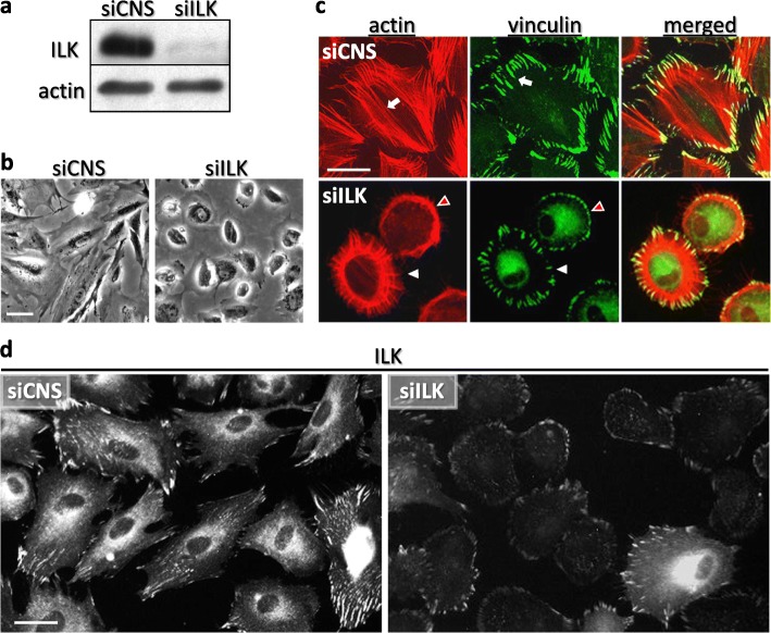
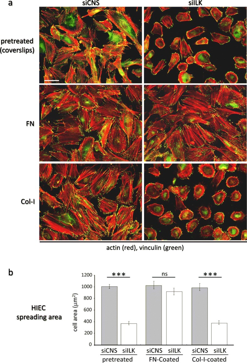
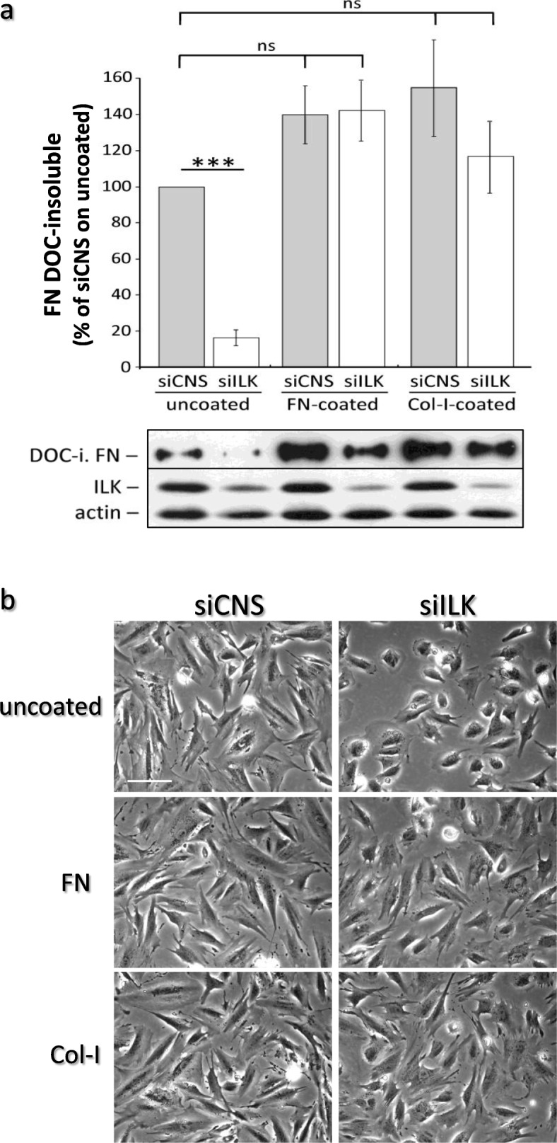
Background: Fibronectin (FN) assembly into an insoluble fibrillar matrix is a crucial step in many cell responses to extracellular matrix (ECM) properties, especially with regards to the integrin-related mechanosensitive signaling pathway. We have previously reported that the silencing of expression of integrin-linked kinase (ILK) in human intestinal epithelial crypt (HIEC) cells causes significant reductions in proliferation and spreading through concomitantly acquired impairment of soluble FN deposition. These defects in ILK-depleted cells are rescued by growth on exogenous FN. In the present study we investigated the contribution of ILK in the fibrillogenesis of FN and its relation to integrin-actin axis signaling and organization.

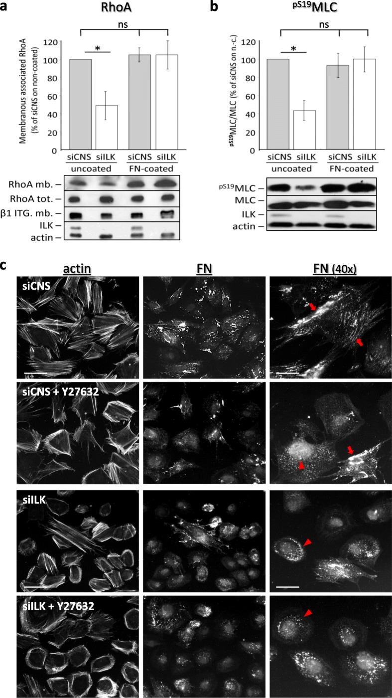
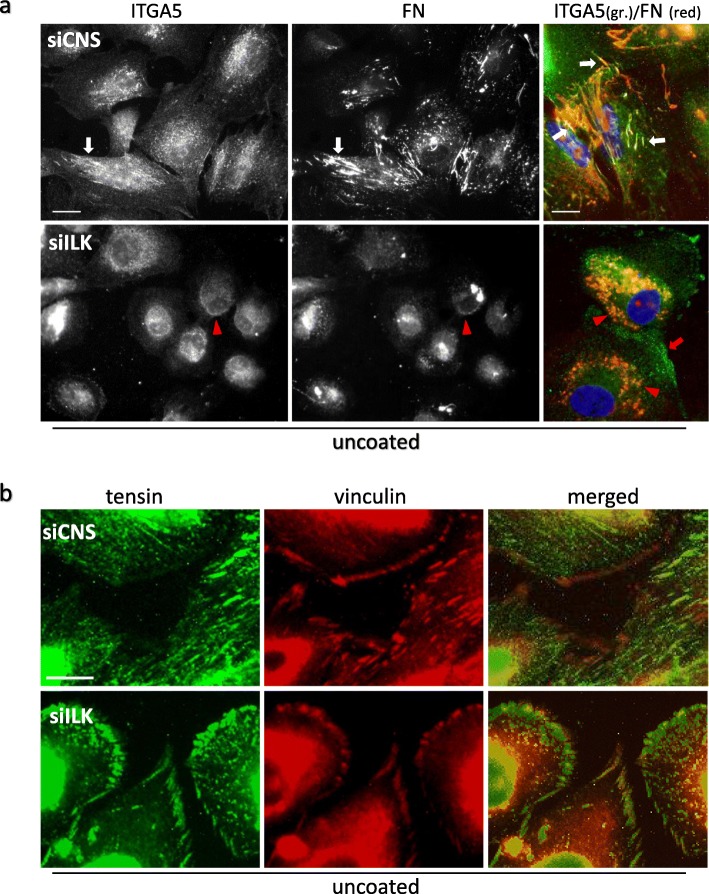
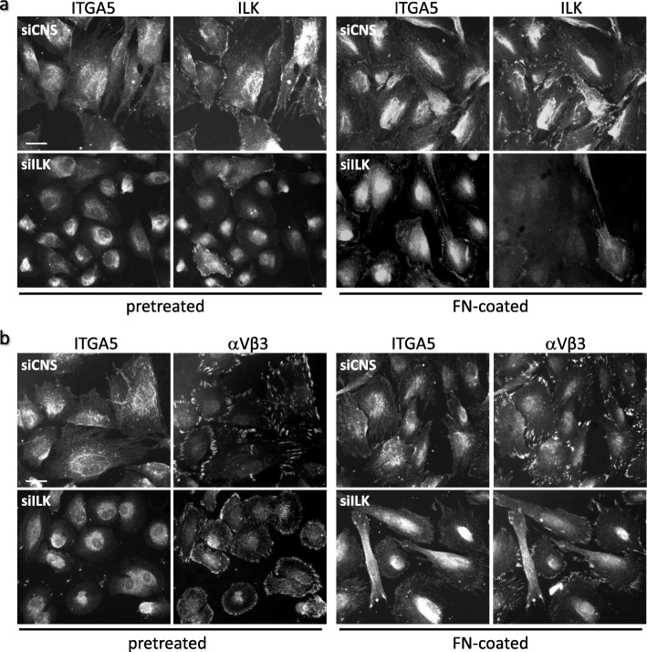
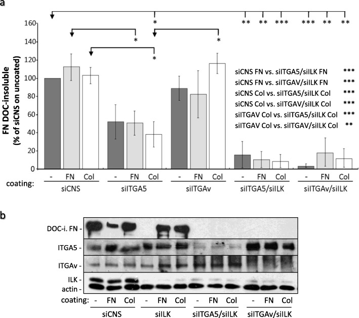
Results: We show that de novo fibrillogenesis of endogenous soluble FN is ILK-dependent. This function seemingly induces the assembly of an ECM that supports increased cytoskeletal tension and the development of a fully spread contractile cell phenotype. We observed that HIEC cell adhesion to exogenous FN or collagen-I (Col-I) is sufficient to restore fibrillogenesis of endogenous FN in ILK-depleted cells. We also found that optimal engagement of the Ras homolog gene family member A (RhoA) GTPase/Rho-associated kinase (ROCK-1, ROCK-2)/myosin light chain (MLC) pathway, actin ventral stress fiber formation, and integrin adhesion complex (IAC) maturation rely primarily upon the cell's capacity to execute FN fibrillogenesis, independent of any significant ILK input. Lastly, we confirm the integrin α5β1 as the main integrin responsible for FN assembly, although in ILK-depleted cells αV-class integrins expression is needed to allow the rescue of FN fibrillogenesis on exogenous substrate.

Conclusion: Our study demonstrates that ILK specifically induces the initiation of FN fibrillogenesis during cell spreading, which promotes RhoA/ROCK-dependent cell contractility and maturation of the integrin-actin axis structures. However, the fibrillogenesis process and its downstream effect on RhoA signaling, cell contractility and spreading are ILK-independent in human intestinal epithelial crypt cells.

Keywords: Actin stress fibers; Cell contractility; Epithelial cells; Fibrillogenesis; Fibronectin; ILK; IPP complex; Integrin; RhoA; α5β1.


Conflict of interest statement

The authors declare no potential conflict of interest.

Figures

Fig. 1
Fig. 1
Effect of ILK silencing on structural and morphological characteristics of HIEC cells. (a to d) HIEC were transfected with 20 nM of control, non-interfering siRNA (siCNS) or an siRNA directed against ILK expression (siILK). a Western blot (WB) analysis of ILK protein levels in whole-cell lysates of siCNS and siILK cells 48 h after the transfection. β-actin protein levels were used as the loading control. b Inverted contrast microscopy images of siCNS and siILK cells grown on plastic dishes. c Immunofluorescence (IF) microscopy images of the actin cytoskeleton (TRITC-phalloidin; red) and vinculin (anti-vinculin antibody; green) staining in siCNS and siILK cells grown for 24 h on serum-pretreated glass coverslips (4% serum-containing medium). Arrows in siCNS point to ventral stress fibers (actin) and elongated mature FA (vinculin). White arrowheads in siILK point to a cell with an orthogonal type of actin fiber network (actin) associated with FA (vinculin), while red arrowheads point to a cell with a cortical actin ring (actin) and associated FX (vinculin). d IF microscopy images of ILK staining detected in siCNS and siILK cells grown for 24 h on serum-pretreated glass coverslips. Scale bar in (b, c, and d): 15 μm
Fig. 2
Fig. 2
Adhesion to exogenous FN coating supports the restoration of the contractile phenotype in ILK-depleted HIEC. a IF microscopy images representative of the actin cytoskeleton (detected with TRITC-phalloidin; in red) and vinculin-positive ECM adhesion structures (anti-vinculin antibody; green) stained in siCNS and siILK cells grown for 24 h on serum-pretreated, FN-coated (FN; 3 μg/cm2) or Col-I-coated (Col-I;10 μg/cm2) glass coverslips. b Histograms of the average surface spreading area (μm2) measured numerically (Metamorph Imaging System software) on cells cultured under the same conditions as in (a). Results are expressed as the mean ± SEM (n = 3; > 50 cells have been analyzed for each condition). *** p < 0.001 in a one-way ANOVA using Tukey’s correction for multiple group comparisons. Scale bar in (a) 25 μm
Fig. 3
Fig. 3
ILK silencing hinders normal FN-dependant RhoA/ROCK/MLC pathway stimulation and FN fibrillar assembly in HIEC. (a and b) siCNS and siILK cells were grown for 48 h on uncoated or FN-coated (FN-coated; 3 μg/cm2) plastic dishes. a Histograms represent the relative levels (membranous/total) of membrane-associated active RhoA (mb) compared to RhoA and β-actin detected in whole-cell lysates (tot). Values obtained by densitometry quantification analysis of WB bands were normalized to β1 integrin (from mb fractions) and β-actin (from cell lysate). The lower panels show representative corresponding protein immunoblots. b Histograms of the relative phosphorylation status of serine19 of the myosin light chain (pS19MLC/MLC) in cells cultured under the same conditions as described in (a). The values were obtained from densitometry quantification of pS19MLC and MLC bands detected by WB in whole cell lysates. β-actin protein levels were used as the loading control. (a and b) Results are expressed as the percentage (%) of the values obtained for the siCNS cells on uncoated dishes ± SEM (n ≥ 3). * p < 0.05, in a one-way ANOVA using Tukey’s correction for multiple group comparisons. ns: not significant. The lower panel in (a) and (b) shows representative corresponding protein immunoblots. c IF microscopy images representative of the actin cytoskeleton (TRITC-phalloidin) and human FN (detected with the HFN 7.1 antibody) staining in siCNS and siILK cells grown for 24 h on serum-pretreated coverslips with or without 20 uM of the specific inhibitor Y-27632 to inhibit ROCK-mediated cell contractility. The right panels show magnification (40x objective) of FN staining, red arrows point to examples of cell-assembled FN fibrils, while the red arrowheads point to examples of non-fibrillar FN aggregates. Scale bar in (c) left panels: 20 μm; right panels: 10 μm
Fig. 4
Fig. 4
Adhesion to exogenous FN/ECM-coated substrates restores DOC-insoluble FN assembly capacity in ILK-depleted HIEC. siCNS and siILK cells were grown for 36 to 48 h on uncoated, FN-coated (3 μg/cm2) or Col-I-coated (10 μg/cm2) plastic dishes and then harvested for DOC-insoluble proteins. The histograms present the levels of isolated human DOC-insoluble FN quantified from WB densitometry analysis for each condition. The β-actin levels detected in the corresponding soluble fractions were used to normalize the DOC-insoluble levels. Results are expressed as the percentage (%) of the siCNS cells on uncoated dishes ± SEM (n ≥ 9). *** p < 0.001 in a one-way ANOVA test using Tukey’s correction for multiple group comparisons. ns: not significant. The lower panel shows representative protein immunoblots of human DOC-insoluble (DOC-i.) FN, as well as corresponding ILK and β-actin, detected from the DOC-soluble fractions. b Inverted contrast microscopy images representative of the same cells grown for 24 h on the same uncoated and coated plastic dish culture conditions. Scale bar in (b) 30 μm
Fig. 5
Fig. 5
Adhesion to exogenous FN/ECM-coated substrates prevents peripheral membrane detachment and retraction following serum-induced cell contractility. siCNS and siILK cells were grown for 24 h on uncoated (a and b), FN-coated (b) or Col-I-coated (b) plastic dishes. Cells were then starved for 24 h without serum before RhoA-mediated cell contractility was stimulated by adding serum (FBS, 10% final concentration) to the culture medium. a Time-lapse inverted contrast microscopy images of the siCNS and siILK cells on uncoated dishes taken at a time interval of 15 s showing cells immediately before (− 15 s) and after (90 s, 5 min, and 55 min) adding the serum to the starved cells. b Histograms of the percentage of cells (collapsed vs total cells) that show detachment and retraction of their peripheral plasma membrane before (serum: -) and 20 to 30 min after adding the serum (+). Assays on uncoated dishes were also performed with cells pretreated with 20 μM of the ROCK inhibitor Y-27632. Results are expressed as mean ± SEM. *** p < 0.001 in paired t-test on the average values (siCNS vs siILK; only the statistically significant difference is indicated; n ≥ 3, 363 ± 151 cells counted for every specific condition in each experiment). s: second; min: minute. Scale bar in (a): 20 μm
Fig. 6
Fig. 6
Inhibition of FN synthesis with cycloheximide prevents FN fibrillogenesis in ILK-depleted HIEC on exogenous FN. (a, b and c) siCNS and siILK cells were grown for 24 h on uncoated or FN-coated plastic dishes, with or without cycloheximide (10 μg/ml) and were harvested for DOC-insoluble proteins. a Representative WB of human FN (HFN 7.1 antibody) detected in DOC-soluble (DOC-s.) and DOC-insoluble (DOC-i.) fractions, as well as the corresponding ILK and β-actin protein immunoblots detected from the DOC-s. fractions. The order of WB bands in (a) follows the order of the histograms presented in (b and c). (b and c) Histograms of the levels of human FN quantified from WB densitometry analysis of siCNS and siILK cells b DOC-soluble and c DOC-insoluble fractions. β-actin detected in the soluble fractions was used to normalize the results for each experiment. Results are expressed as the percentage (%) of the siCNS cells on uncoated dishes ± SEM. (n = 4). *** p < 0.001, ** p < 0.01 and * p < 0.05 in a one-way ANOVA test using Newman-Keuls correction for multiple group comparisons. ns: not significant
Fig. 7
Fig. 7
Effect of ILK silencing on α5β1 integrin, FN, and tensin distribution in HIEC. (a and b) siCNS and siILK cells were grown for 24 h on serum-pretreated glass coverslips and then fixed and prepared for IF staining. a Co-IF microscopy images representative of α5β1 integrin (ITGA5; α5 subunit detected with the AB1928 antibody) and cell-associated human FN (HFN 7.1 antibody) staining distribution in siCNS and siILK cells. The right panels show merged and magnified co-detection (ITGA5 in green, FN red, DAPI staining of the nucleus in blue). White arrows in siCNS points to ITGA5 and FN colocalization in fibrillar structures. Red arrows and red arrowheads in siILK point respectively to subcortical staining of α5 integrin and its colocalization with small punctuated aggregates of FN. b Co-IF microscopy images representative of tensin (green) and vinculin (red) staining distribution in siCNS and siILK cells. Scale bar in (a): 10 μm; (b): 5 μm
Fig. 8
Fig. 8
Adhesion to exogenous FN restores normal α5β1 and αVβ3 integrin distribution in ILK-depleted HIEC. siCNS and siILK cells were grown for 24 h on serum-pretreated or FN-coated glass coverslips and then fixed and prepared for IF staining. (a and b) Co-IF microscopy images representative of α5β1 integrin (ITGA5; α5 subunit/AB1928 antibody) staining with a ILK staining, and b αVβ3 staining distribution in siCNS and siILK cells. Scale bar in (a and b): 15 μm
Fig. 9
Fig. 9
Effect of α5 or αV integrins and concomitant ILK silencing on FN assembly in HIEC. (a and b) HIEC cells were transfected with 20 nM of siCNS, siILK or siRNA directed against the expression of α5 (siITGA5) and αV (siITGAV) integrin subunits alone or in combination with 20 nM of siILK (e.g. siITGA5/siILK). The cells were subsequently plated and grown for 48 h on uncoated, FN-coated and Col-I coated plastic dishes before being harvested for DOC-insoluble protein extraction. a The histograms present the levels of human DOC-insoluble FN quantified from densitometry analysis of WB bands. β-actin detected in the soluble fractions was used to normalize the results for each experiment. Results are expressed as the percentage (%) of the siCNS on uncoated dishes ± SEM (n ≥ 3). * p < 0.05, ** p < 0.01 and *** p < 0.001 in a one-way ANOVA test using Tukey’s correction for multiple group comparisons (only the statistically significant differences are indicated). b Representative protein immunoblots of DOC-insoluble (DOC-i.) human FN and the corresponding ILK, β-actin, α5 (ITGA5) and αV (ITGAV) integrin subunits detected from the DOC-soluble fractions of siCNS, siILK, siITGA5/siILK and siITGAV/siILK cells

Similar articles

Cited by

References

    1. Humphries JD, Chastney MR, Askari JA, Humphries MJ. Signal transduction via integrin adhesion complexes. Curr Opin Cell Biol. 2019;56:14–21. doi: 10.1016/j.ceb.2018.08.004. - DOI - PubMed
    1. Bachmann M, Kukkurainen S, Hytonen VP, Wehrle-Haller B. Cell adhesion by Integrins. Physiol Rev. 2019;99(4):1655–1699. doi: 10.1152/physrev.00036.2018. - DOI - PubMed
    1. Kechagia JZ, Ivaska J, Roca-Cusachs P. Integrins as biomechanical sensors of the microenvironment. Nat Rev Mol Cell Biol. 2019;20(8):457–473. doi: 10.1038/s41580-019-0134-2. - DOI - PubMed
    1. Wolfenson H, Yang B, Sheetz MP. Steps in Mechanotransduction pathways that control cell morphology. Annu Rev Physiol. 2019;81:585–605. doi: 10.1146/annurev-physiol-021317-121245. - DOI - PMC - PubMed
    1. Geiger B, Yamada KM. Molecular architecture and function of matrix adhesions. Cold Spring Harb Perspect Biol. 2011;3(5):a005033. - PMC - PubMed

LinkOut - more resources