

A unique temporary collateral pathway between carotid-vertebrobasilar arteries in a carotid dissection patient
- PMID: 32183730
- PMCID: PMC7076917
- DOI: 10.1186/s12883-020-01651-1
A unique temporary collateral pathway between carotid-vertebrobasilar arteries in a carotid dissection patient
Abstract
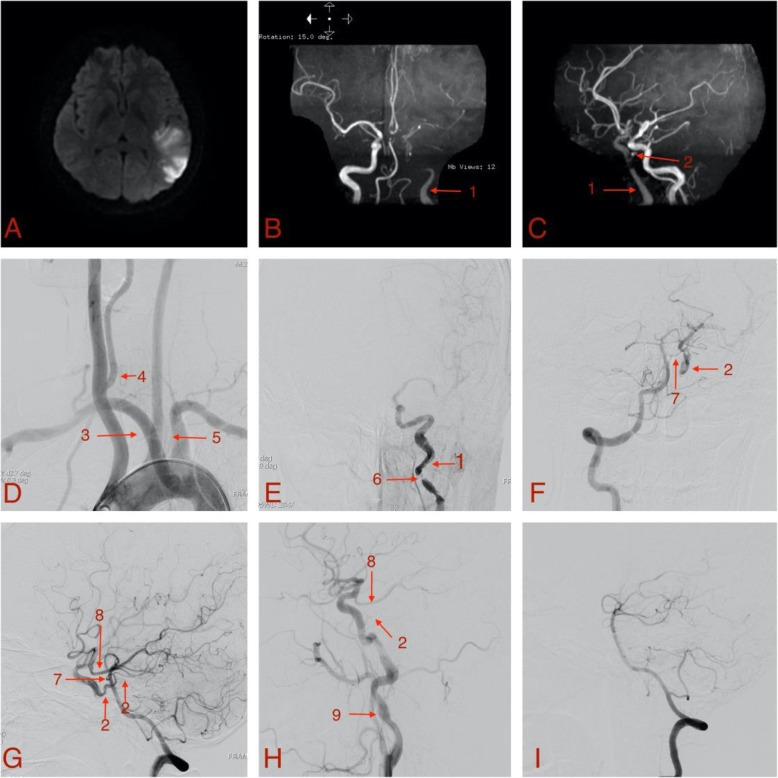
Background: In adults, the anastomosis between carotid and vertebrobasilar arteries is usually the posterior communicating artery, sometimes the primitive trigeminal artery. In this case, the basilar artery fed the internal carotid artery through the pontine-to-tentorial artery anastomosis after severe stenosis from traumatic carotid dissection.
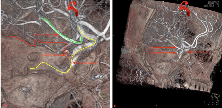
Case presentation: A 32-year-old female was diagnosed with ischemic stroke caused by traumatic carotid artery dissection. Aspirin (100 mg/day) and clopidogrel (75 mg/day) were prescribed. Digital subtraction angiography performed 6 days after stroke onset showed a dissection in the cervical segment of left internal carotid artery with severe local stenosis, and a collateral pathway from BA to the cavernous segment of internal carotid artery through the lateral pontine and tentorial artery. Without interventional therapy, clinical symptoms improved significantly within 10 days after onset. At 3-month follow-up, left common carotid artery angiography showed the stenosis had been significantly improved with a residual aneurysm. There was no collateral pathway between carotid-vertebrobasilar arteries, and a residual small artery originated from the posterior vertical segment of cavernous internal carotid artery. The small artery was clearly visualized by 3-dimensional rotational angiography and identified the tentorial artery.
Conclusion: To the author's knowledge, this is the first report of a collateral pathway between carotid vertebrobasilar arteries through the pontine-to-tentorial artery anastomosis. Meanwhile, tentorial artery origination directly from the cavernous segment of internal carotid artery is rare and easily mistaken for persistent primitive trigeminal artery. 3-dimensional rotational angiography can provide sensitive and accurate diagnostic assessment of the small artery, and may be a useful tool for screening of abnormal small arteries.
Keywords: 3-dimensional rotational angiography; Collateral circulations; Internal carotid artery dissection; Ischemic stroke.
Conflict of interest statement
The authors declare that they have no competing interests.
Figures
Similar articles
-
Persistent Trigeminal Artery as Collateral Circulation in Ischemic Stroke.World Neurosurg. 2021 Apr;148:67-69. doi: 10.1016/j.wneu.2021.01.034. Epub 2021 Jan 18. World Neurosurg. 2021. PMID: 33476776
-
Internal Carotid Artery Dissecting Aneurysm Associated with Persistent Trigeminal Artery: A Case Report.Curr Med Imaging. 2024;20:1-6. doi: 10.2174/0115734056263907231127170341. Curr Med Imaging. 2024. PMID: 38389372
-
Complete ophthalmoplegia, complete ptosis and dilated pupil due to internal carotid artery dissection: as the first manifestation of Takayasu arteritis.BMC Cardiovasc Disord. 2017 Jul 25;17(1):201. doi: 10.1186/s12872-017-0638-7. BMC Cardiovasc Disord. 2017. PMID: 28743241 Free PMC article.
-
Persistent primitive trigeminal artery associated with a cavernous carotid aneurysm. Case report and literature review.J Radiol Case Rep. 2018 Nov 30;12(11):1-11. doi: 10.3941/jrcr.v12i11.3500. eCollection 2018 Nov. J Radiol Case Rep. 2018. PMID: 30647831 Free PMC article. Review.
-
[A case of carotid superior cerebellar artery anastomosis associated with bilateral hypoplasia of the internal carotid artery represented as the rupture of posterior cerebral artery-posterior communicating artery aneurysm].No Shinkei Geka. 1988 Sep;16(10):1211-7. No Shinkei Geka. 1988. PMID: 3060752 Review. Japanese.
References
Publication types
MeSH terms
LinkOut - more resources
Full Text Sources
