. 2020 Mar 17;18(1):46.
doi: 10.1186/s12964-020-00535-8.

Angiopoietin-2 induces angiogenesis via exosomes in human hepatocellular carcinoma

Affiliations

Angiopoietin-2 induces angiogenesis via exosomes in human hepatocellular carcinoma

Ji-Yan Xie et al. Cell Commun Signal. .

Abstract

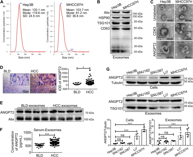
Background: Hepatocellular carcinoma (HCC) is the most common primary liver cancer and is a highly vascularized solid tumor. Angiopoietin-2 (ANGPT2) has been described as an attractive target for antiangiogenic therapy. Exosomes are small extracellular vesicles secreted by most cell types and contribute to cell-to-cell communication by delivering functional cargo to recipient cells. The expression of ANGPT2 in tumor-derived exosomes remains unknown.

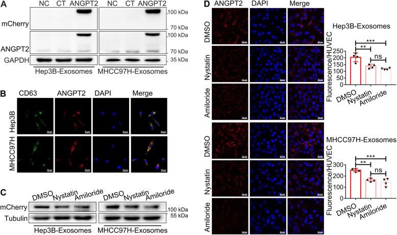
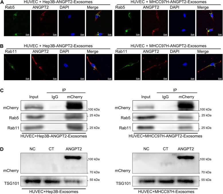
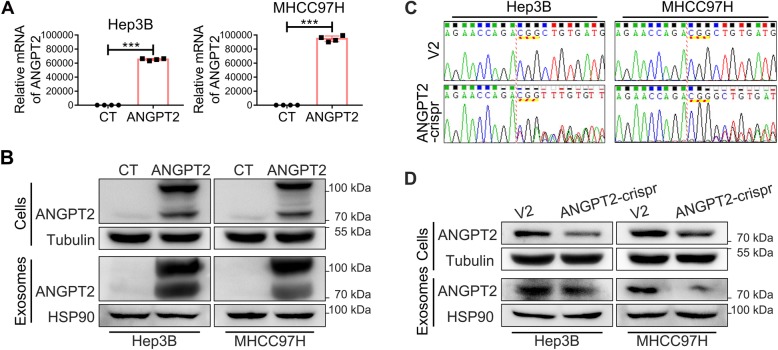
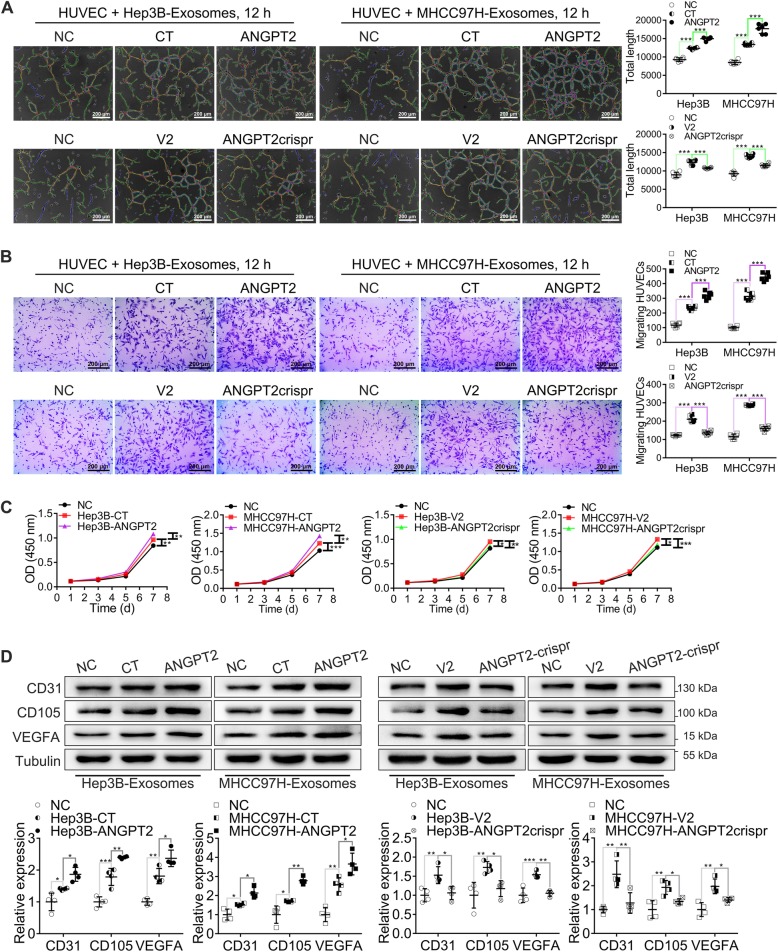
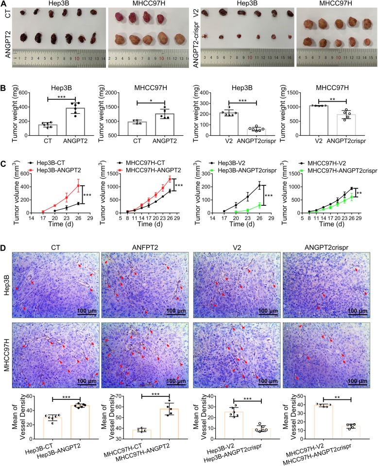
Methods: We detected the ANGPT2 expression in HCC-derived exosomes by immunoblotting, enzyme-linked immunosorbent assay and immunogold labeling, then observed exosomal ANGPT2 internalization and recycling by confocal laser scanning microscopy, co-immunoprecipitation and immunoblotting. We used two HCC cell lines (Hep3B and MHCC97H) to overexpress ANGPT2 by lentivirus infection or knockdown ANGPT2 by the CRISPR/Cas system, then isolated exosomes to coculture with human umbilical vein endothelial cells (HUVECs) and observed the angiogenesis by Matrigel microtubule formation assay, transwell migration assay, wound healing assay, cell counting kit-8 assay, immunoblotting and in vivo tumorigenesis assay.

Results: We found that HCC-derived exosomes carried ANGPT2 and delivered it into HUVECs by exosome endocytosis, this delivery led to a notable increase in angiogenesis by a Tie2-independent pathway. Concomitantly, we observed that HCC cell-secreted exosomal ANGPT2 was recycled by recipient HUVECs and might be reused. In addition, the CRISPR-Cas systems to knock down ANGPT2 significantly inhibited the angiogenesis induced by HCC cell-secreted exosomal ANGPT2, and obviously suppressed the epithelial-mesenchymal transition activation in HCC.

Conclusions: Taken together, these results reveal a novel pathway of tumor angiogenesis induced by HCC cell-secreted exosomal ANGPT2 that is different from the classic ANGPT2/Tie2 pathway. This way may be a potential therapeutic target for antiangiogenic therapy. Video Abstract.

Keywords: Angiogenesis; Angiopoietin-2; CRISPR-Cas systems; Endocytosis; Epithelial-mesenchymal transition; Exosomes; Hepatocellular carcinoma; Recycling.


Conflict of interest statement

The authors declare that they have no competing interests.

Figures

Fig. 1
Fig. 1
ANGPT2 exists on HCC-derived exosomes. a NTA displayed that the majority of isolated exosomes were within 30–150 nm, which is the typical size of exosomes. b Immunoblotting showed the typical exosomal markers (Alix, HSP90, TSG101 and CD63) in isolated exosomes. c Transmission electron microscopic view of isolated exosomes. The isolated exosomes had cup-shaped morphology, were labeled with exosomal marker CD63 (immunogold = 5 nm), and ANGPT2 was also labeled on isolated exosomes by immunogold (immunogold = 10 nm). Scale bar = 100 nm. d IHC demonstrated that the expression of ANGPT2 in HCC tissues (IOD = 270.6 ± 29.36, n = 96) was higher than that in BLD tissues (IOD = 157.3 ± 34.9, n = 11). Scale bar = 100 μm. *P < 0.05, Welch’s t-tests. e Immunoblotting showed that ANGPT2 was positive in exosomes isolated from the sera of both HCC and BLD patients. f ELISA showed that the level of exosomal ANGPT2 isolated from the sera of HCC patients (756.5 ± 20.3 pg/mL, n = 67) was significantly higher than that from the sera of BLD patients (541.3 ± 18.82 pg/mL, n = 26). ***P < 0.001, Welch’s t-tests. g Immunoblotting showed the levels of ANGPT2 in different HCC cell lines and their exosomes (Hep3B, SNU182, SNU387, Li7 and MHCC97H), and the levels of HCC cell-secreted exosomal ANGPT2 were consistent with their corresponding cells. n = 4 for cell groups, n = 5 for exosome groups, **P < 0.01, ***P < 0.001, one-way ANOVA with Tukey’s multiple comparison tests
Fig. 2
Fig. 2
HCC cell-secreted exosomal ANGPT2 is delivered into HUVECs via exosome endocytosis. a HUVECs were cultured with or without exosomes derived from ANGPT2-mCherry-expressing HCC cells and their matched control cells for 6 h. Immunoblotting showed that the ANGPT2-mCherry fusion protein was detected in HUVECs. b ANGPT2-mCherry-expressing HCC cells were transfected with the pLV-EGFP-CD63 plasmid for 48–72 h to obtain cells that coexpressed the ANGPT2-mCherry and CD63-EGFP fusion proteins. Then, exosomes were isolated from the above HCC cells to be cocultured with HUVECs for 6 h. The confocal laser scanning microscopy observed that ANGPT2-mCherry and CD63-EGFP coexisted and mostly colocalized in HUVECs. Scale bar = 30 μm. c, d HUVECs were treated with the endocytosis inhibitors nystatin (25 μmol/L), amiloride (100 μmol/L) or control DMSO for 30 min and then cultured with exosomes derived from ANGPT2-mCherry-expressing HCC cells for 6 h. Immunoblotting (c) and confocal laser scanning (d) showed that the ANGPT2-mCherry level in HUVECs treated with nystatin or amiloride was significantly lower than that in the DMSO control group. Scale bar = 30 nm. n = 4 for each group, **P < 0.01, ***P < 0.001, one-way ANOVA with Tukey’s multiple comparison tests
Fig. 3
Fig. 3
HCC cell-secreted exosomal ANGPT2 is recycled by recipient HUVECs. a HUVECs were transfected with the pLV-EGFP-hRab5 plasmid for 48–72 h to express the Rab5-EGFP fusion protein and then cultured with ANGPT2-mCherry-expressing exosomes derived from HCC cells for 11 h. The confocal laser scanning revealed that ANGPT2-mCherry and Rab5-EGFP colocalized near the nucleus in HUVECs. Scale bar = 15 μm. b HUVECs were cultured with ANGPT2-mCherry-expressing exosomes derived from HCC cells for 12 h. The confocal laser scanning microscopy observed that ANGPT2-mCherry and Rab11-EGFP also colocalized in HUVECs by immunofluorescent staining using Rab11 antibody. Scale bar = 15 μm. c HUVECs were cultured with ANGPT2-mCherry-expressing exosomes derived from HCC cells for 12 h. The co-IP assay showed that exosomal ANGPT2-mCherry had interaction with Rab5 and Rab11 in HUVECs. d HUVECs were cultured with or without HCC cell-secreted exosomes for 6 h, then washed with PBS for 3 times and cultured with fresh medium supplemented with 10% exosome-depleted FBS for 12 h. Immunoblotting showed that ANGPT2-mCherry was positive in exosomes derived from HUVECs which had been cultured with ANGPT2-mCherry-expressing exosomes
Fig. 4
Fig. 4
The overexpression or knockdown of ANGPT2 in HCC cells and their exosomes in vitro. a, b Recombinant lentiviruses that carried the pLV-hANGPT2-mCherry vector or control lentiviruses (only carried pLV-mCherry) were used to infect Hep3B and MHCC97H cells to obtain stable cell lines that overexpressed ANGPT2 fused with mCherry (named Hep3B-ANGPT2 and MHCC97H-ANGPT2, respectively) and matched control stable cell lines (named Hep3B-CT and MHCC97H-CT, respectively). Real-time qPCR detected the relative mRNA levels of ANGPT2 in the stable cell lines (a). Immunoblotting showed the expression levels of ANGPT2 in the stable cell lines and their exosomes, which showed that the levels of exosomal ANGPT2 were increased correspondingly (b). n = 4 for each group, ***P < 0.001, Student’s t-tests. c, d Recombinant lentiviruses that carried lentiCRISPRv2-ANGPT2gRNA or control lentiviruses (only carried lentiCRISPRv2) were used to infect Hep3B and MHCC97H cells to obtain stable cell lines that knocked down ANGPT2 (named Hep3B-ANGPT2crispr and MHCC97H-ANGPT2crispr, respectively) and matched control stable cell lines (named Hep3B-V2 and MHCC97H-V2, respectively). Genomic DNA sequencing detected mutations in the ANGPT2 gene in stable cell lines (c). Immunoblotting showed the expression levels of ANGPT2 in the stable cell lines and their exosomes, which showed that the levels of exosomal ANGPT2 were decreased correspondingly (d)
Fig. 5
Fig. 5
HCC cell-secreted exosomal ANGPT2 promotes the angiogenesis capability of HUVECs in vitro. a HUVECs were cultured with or without HCC cell-secreted exosomes for 12 h. The Matrigel microtubule formation assay showed that ANGPT2-overexpressing exosomes significantly promoted tubule formation of HUVECs, and compared with control exosomes, ANGPT2-deficient exosomes abrogated exosome-induced tubule formation. b HUVECs were cultured with or without HCC cell-secreted exosomes for 12 h. The transwell migration assay showed that ANGPT2-overexpressing exosomes significantly promoted migration of HUVECs, and compared with control exosomes, ANGPT2-deficient exosomes abrogated exosome-induced migration. c HUVECs were cultured with or without HCC cell-secreted exosomes for 7 d and were counted by measuring the OD at 450 nm at 1, 3, 5, and 7 d. CCK-8 showed that HUVEC proliferation was significantly increased after coculture with ANGPT2-overexpressing exosomes, and compared with the coculture with control exosomes, ANGPT2-deficient exosomes abrogated exosome-induced proliferation. d HUVECs were cultured with or without HCC cell-secreted exosomes for 72 h. Immunoblotting showed that ANGPT2-overexpressing exosomes notably increased the levels of tumor angiogenesis-related proteins (CD31, CD105 and VEGFA) in HUVECs, and the promotional effect of ANGPT2-deficient exosomes on these angiogenesis-related proteins was notably reduced compared to the promotional effect of control exosomes. Scale bar = 200 μm. n = 6 for each group (a, b), n = 4 for each group (c, d), *P < 0.05, **P < 0.01, ***P < 0.001, one-way ANOVA with Tukey’s multiple comparison tests
Fig. 6
Fig. 6
ANGPT2 promotes growth and angiogenesis of HCC in vivo. a 1 × 107 ANGPT2-overexpressing or ANGPT2-deficient HCC cells (Hep3B-ANGPT2 and MHCC97H-ANGPT2 or Hep3B-ANGPT2crispr and MHCC97H-ANGPT2crispr, respectively) and their matched control cells (Hep3B-CT, MHCC97H-CT; Hep3B-V2, MHCC97H-V2) were resuspended in 100 μL PBS and were injected subcutaneously into the right flank of 5-week-old male BALB/c nude mice; tumorigenesis was then observed for 26 d. b Tumor weight at 26 d after HCC cell implantation. The weight of the ANGPT2-overexpressing group was significantly increased compared with that of the control group, and compared with the control group, the ANGPT2-deficient group had a significant decrease in weight. c Tumor volume was measured once every 3 d. The overexpression of ANGPT2 led to a notable increase in tumor volume, and the knockdown of ANGPT2 resulted in an obvious decrease in tumor volume. d IHC analysis of the vascular density of tumors was performed by labeling CD31. The overexpression of ANGPT2 led to a notable increase in vascular density, and knockdown of ANGPT2 resulted in a significant decrease in vascular density. n = 6 for all Hep3B groups, n = 4 for MHCC97H-CT group, n = 5 for other MHCC97H groups, *P < 0.05, **P < 0.01, ***P < 0.001, Student’s t-tests

Similar articles

Cited by

References

    1. Forner A, Reig M, Bruix J. Hepatocellular carcinoma. Lancet. 2018;391:1301–1314. doi: 10.1016/S0140-6736(18)30010-2. - DOI - PubMed
    1. Bray F, Ferlay J, Soerjomataram I, Siegel RL, Torre LA, Jemal A. Global cancer statistics 2018: GLOBOCAN estimates of incidence and mortality worldwide for 36 cancers in 185 countries. CA Cancer J Clin. 2018;68:394–424. doi: 10.3322/caac.21492. - DOI - PubMed
    1. Kulik L, El-Serag HB. Epidemiology and Management of Hepatocellular Carcinoma. Gastroenterology. 2019;156:477–491. doi: 10.1053/j.gastro.2018.08.065. - DOI - PMC - PubMed
    1. Yang ZF, Poon RTP. Vascular changes in hepatocellular carcinoma. Anat Rec (Hoboken) 2008;291:721–734. doi: 10.1002/ar.20668. - DOI - PubMed
    1. Mossenta M, Busato D, Baboci L, Di Cintio F, Toffoli G, Dal Bo M. New insight into therapies targeting angiogenesis in hepatocellular carcinoma. Cancers. 2019;11:1086–1106. doi: 10.3390/cancers11081086. - DOI - PMC - PubMed

Publication types

MeSH terms