

Characterization of VDR and CYP27B1 expression in the endometrium during the menstrual cycle before embryo transfer: implications for endometrial receptivity
- PMID: 32183826
- PMCID: PMC7079352
- DOI: 10.1186/s12958-020-00579-y
Characterization of VDR and CYP27B1 expression in the endometrium during the menstrual cycle before embryo transfer: implications for endometrial receptivity
Abstract
Background: Molecular analyses of vitamin D in a typical cycling endometrium has received minimal research attention in the reproductive field. This study was designed to assess how expression of the endometrial vitamin D receptor (VDR) and CYP27B1, a vitamin D metabolizing enzyme, change during the menstrual cycle in women of reproductive age. In addition, this study explores the association between expression of vitamin D-VDR system and endometrial receptivity during the implantation window.
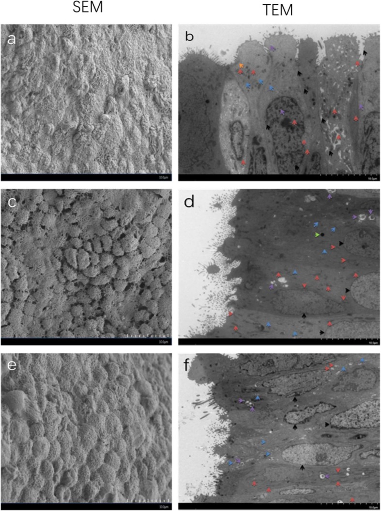
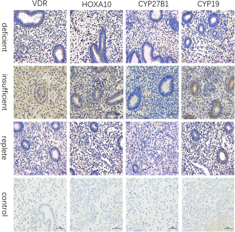
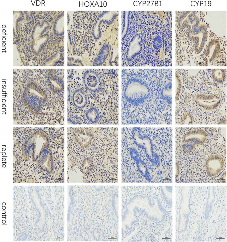
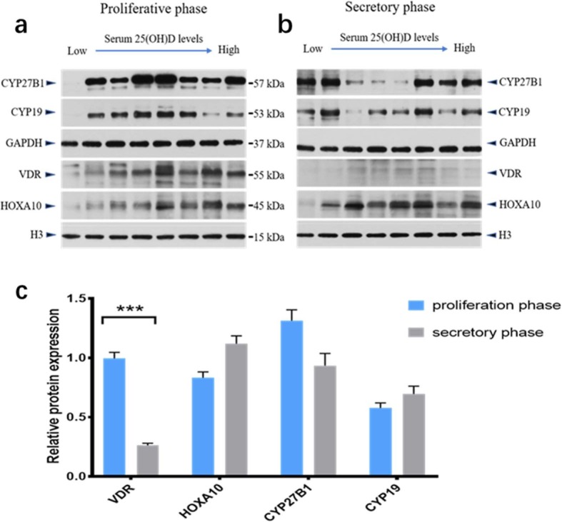
Methods: Sixteen patients underwent standardized in vitro fertilization (IVF) treatment and freeze-all techniques. Before embryo transfer, total serum 25(OH) D levels were determined through blood samples and VDR, CYP27B1, HOXA10, and CYP19 expression were determined through endometrial samples. Endometrial receptivity was also assessed using an electron microscope.
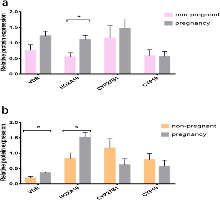
Results: We found that VDR protein expression was significantly lower throughout the endometrial secretory phase compared to the proliferative phase, while CYP27B1 expression remained constant during the menstrual cycle. During the implantation window, ultrastructural evaluation showed that higher serum vitamin D levels were associated with more mature pinopodes; VDR and HOXA10 protein expression were substantially elevated in pregnant women compared to non-pregnant women; and VDR protein levels were positively correlated with HOXA10 levels. In addition, serum vitamin D levels were positively correlated with VDR and HOXA10 protein levels in the endometrium.
Conclusions: Women with increased VDR expression in the endometrium, especially during the implantation window of the menstrual cycle, were significantly more likely to be pregnant than women with decreased expression. Our results support the hypothesis that the Vitamin D-VDR system performs a role during the development of endometrial receptivity.
Keywords: CYP27B1; Endometrial receptivity; In-vitro fertilization; Vitamin D; Vitamin D receptor.
Conflict of interest statement
The authors declare that they have no conflict of interest.
Figures
Similar articles
-
Anti-phospholipid antibody may reduce endometrial receptivity during the window of embryo implantation.J Gynecol Obstet Hum Reprod. 2021 Jun;50(6):101912. doi: 10.1016/j.jogoh.2020.101912. Epub 2020 Sep 17. J Gynecol Obstet Hum Reprod. 2021. PMID: 32950746
-
Regulation of endometrial receptivity by the highly expressed HOXA9, HOXA11 and HOXD10 HOX-class homeobox genes.Hum Reprod. 2014 Apr;29(4):781-90. doi: 10.1093/humrep/deu004. Epub 2014 Feb 18. Hum Reprod. 2014. PMID: 24549215
-
Decreased PECAM1-mediated TGF-β1 expression in the mid-secretory endometrium in women with recurrent implantation failure.Hum Reprod. 2018 May 1;33(5):832-843. doi: 10.1093/humrep/dey022. Hum Reprod. 2018. PMID: 29617817
-
Endometrial receptivity: changes in cell-surface morphology.Semin Reprod Med. 2000;18(3):229-35. doi: 10.1055/s-2000-12561. Semin Reprod Med. 2000. PMID: 11299962 Review.
-
[Endometrial biopsy in the evaluation of endometrial receptivity].J Gynecol Obstet Biol Reprod (Paris). 2004 Feb;33(1 Pt 2):S13-7. doi: 10.1016/s0368-2315(04)96397-1. J Gynecol Obstet Biol Reprod (Paris). 2004. PMID: 14968038 Review. French.
Cited by
-
Supplementation with vitamin D improves the embryo quality in in vitro fertilization (IVF) programs, independently of the patients' basal vitamin D status.Arch Gynecol Obstet. 2024 Jun;309(6):2881-2890. doi: 10.1007/s00404-024-07473-7. Epub 2024 Apr 5. Arch Gynecol Obstet. 2024. PMID: 38580857 Free PMC article.
-
Association of Bioavailable 25(OH)D Levels With Endometriosis in Infertile Bangladeshi Women: A Cross-Sectional Study.Health Sci Rep. 2025 Jul 18;8(7):e71070. doi: 10.1002/hsr2.71070. eCollection 2025 Jul. Health Sci Rep. 2025. PMID: 40687539 Free PMC article.
-
Glyphosate and a glyphosate-based herbicide dysregulate the epigenetic landscape of Homeobox A10 (Hoxa10) gene during the endometrial receptivity in Wistar rats.Front Toxicol. 2024 Sep 13;6:1438826. doi: 10.3389/ftox.2024.1438826. eCollection 2024. Front Toxicol. 2024. PMID: 39345349 Free PMC article.
-
Vitamin D Supplementation Improves Uterine Receptivity in a Rat Model of Vitamin D Deficiency: A Possible Role of HOXA-10/FKBP52 Axis.Front Physiol. 2021 Nov 25;12:744548. doi: 10.3389/fphys.2021.744548. eCollection 2021. Front Physiol. 2021. PMID: 34899377 Free PMC article.
-
Influence of Vitamin D supplementation on reproductive outcomes of infertile patients: a systematic review and meta-analysis.Reprod Biol Endocrinol. 2023 Feb 3;21(1):17. doi: 10.1186/s12958-023-01068-8. Reprod Biol Endocrinol. 2023. PMID: 36737817 Free PMC article.
References
-
- Pilz S, Tomaschitz A, Pieber TR, Lafer I, Drechsler C, Meinitzer A, Grammer TB, Marz W. Vitamin D: clinical implications beyond musculoskeletal diseases. LABORATORIUMSMEDIZIN. 2011;35:211–216. doi: 10.1515/jlm.2011.025. - DOI
-
- Voulgaris N, Papanastasiou L, Piaditis G, Angelousi A, Kaltsas G, Mastorakos G, Kassi E. Vitamin D and aspects of female fertility. Hormones (Athens) 2017;16:5–21. - PubMed
-
- Monastra G, De Grazia S, De Luca L, Vittorio S, Unfer V. Vitamin D: a steroid hormone with progesterone-like activity. Eur Rev Med Pharmacol Sci. 2018;22:2502–2512. - PubMed
MeSH terms
Substances
Grants and funding
LinkOut - more resources
Full Text Sources
