

Adipose stem cells isolated from diabetic mice improve cutaneous wound healing in streptozotocin-induced diabetic mice
- PMID: 32183899
- PMCID: PMC7079496
- DOI: 10.1186/s13287-020-01621-x
Adipose stem cells isolated from diabetic mice improve cutaneous wound healing in streptozotocin-induced diabetic mice
Abstract
Background: Adipose-derived mesenchymal stem cells (ASCs) therapy is emerging as a novel therapeutic option for the treatment of a variety of diseases including diabetes and diabetic wound healing. Multiple studies indicate that ASCs could promote wound healing and reverse diabetes. However, whether ASCs from diabetic donors retain their therapeutic functions and the mechanisms of how ASCs contribute to wound healing remain largely unknown. In this study, we explored the cutaneous wound healing ability of ASCs collected from C57BL/6 mice that had been rendered diabetic with streptozotocin (STZ).
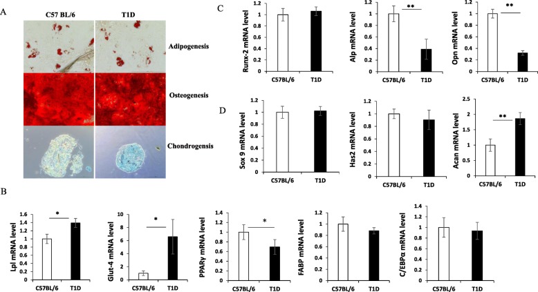
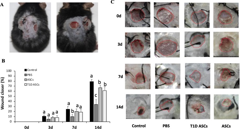
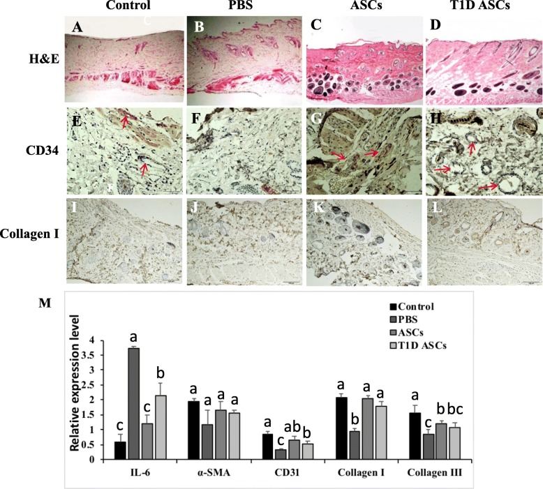
Methods: ASCs were harvested from adipose tissues of type 1 diabetic (T1D) or normal C57BL/6 mice. Cell phenotypes were evaluated by flow cytometry analysis, and cell differentiation into adipocytes, chondrocytes, and osteocytes was compared. Secretions of transforming growth factor β (TGF-β1), basic fibroblast growth factor (bFGF), and vascular endothelial growth factor (VEGF) by ASCs were assessed by ELISA. Migration and proliferation of fibroblasts co-cultured with T1D ASCs or control ASCs were also compared. The therapeutic effects of T1D and control ASCs in promoting wound closure were measured in vivo in a T1D wound mouse model. Granulation tissues were collected and stained with H&E at 14th day. CD34 and collagen I were detected by immunohistochemistry. Expressions of IL-6, α-SMA, CD31, collagen I, and collagen III were quantified by real-time PCR. GFP-expressing ASCs were used to trace in vivo cell differentiation.
Results: T1D ASCs and control ASCs showed similar expression of cell surface markers (CD29, CD34, CD105) and proliferation pattern. They can both differentiate into different cell types. T1D ASCs secreted similar amounts of VEGF and bFGF, but less TGF-β compared with control ASCs. Like control ASCs, T1D ASCs promoted the proliferation and migration of skin fibroblast cells. When injected in cutaneous wound of T1D mice, T1D ASCs increased wound closure and hair follicle regeneration at a comparable extent as ASCs. Mice receiving T1D ASCs or ASCs exhibited significantly higher expressions of collagen I, collagen III, and CD31 and reduced expression of IL-6 in wound tissues. Immunohistochemistry staining showed increased angiogenesis in mice receiving ASCs as was evident by increased CD34+ cells and collagen I staining. GFP+ ASCs injection showed that ASCs differentiated into fibroblasts and endothelial cells in vivo.
Conclusions: Our results suggest that T1D ASCs could accelerate cutaneous wound healing. Mechanisms may include increasing fibroblast growth and migration, skin angiogenesis, and differentiation into fibroblasts and endothelial cells. This study provides evidence that diabetic ASCs may be used as a therapeutic option in cutaneous wound healing in diabetic recipients.
Keywords: Adipose stem cells; Cutaneous wound healing; Diabetes.
Conflict of interest statement
The authors declare that they have no competing interests.
Figures


Similar articles
-
Adipose stem cells from type 2 diabetic mice exhibit therapeutic potential in wound healing.Stem Cell Res Ther. 2020 Jul 17;11(1):298. doi: 10.1186/s13287-020-01817-1. Stem Cell Res Ther. 2020. PMID: 32680569 Free PMC article.
-
[Preliminary evaluation and mechanism of adipose-derived stem cell transplantation from allogenic diabetic rats in the treatment of diabetic rat wounds].Zhonghua Shao Shang Za Zhi. 2019 Sep 20;35(9):645-654. doi: 10.3760/cma.j.issn.1009-2587.2019.09.002. Zhonghua Shao Shang Za Zhi. 2019. PMID: 31594182 Chinese.
-
Paracrine effects of adipose-derived stem cells in cutaneous wound healing in streptozotocin-induced diabetic rats.J Wound Care. 2022 Mar 1;31(Sup3):S29-S38. doi: 10.12968/jowc.2022.31.Sup3.S29. J Wound Care. 2022. PMID: 35199561
-
Adipose-Derived Stem Cells for the Treatment of Diabetic Wound: From Basic Study to Clinical Application.Front Endocrinol (Lausanne). 2022 Jul 11;13:882469. doi: 10.3389/fendo.2022.882469. eCollection 2022. Front Endocrinol (Lausanne). 2022. PMID: 35898452 Free PMC article. Review.
-
Angiogenic Effects and Crosstalk of Adipose-Derived Mesenchymal Stem/Stromal Cells and Their Extracellular Vesicles with Endothelial Cells.Int J Mol Sci. 2021 Oct 8;22(19):10890. doi: 10.3390/ijms221910890. Int J Mol Sci. 2021. PMID: 34639228 Free PMC article. Review.
Cited by
-
3D-bioprinted human lipoaspirate-derived cell-laden skin constructs for healing of full-thickness skin defects.Int J Bioprint. 2023 Mar 23;9(4):718. doi: 10.18063/ijb.718. eCollection 2023. Int J Bioprint. 2023. PMID: 37323499 Free PMC article.
-
Progress and application of adipose-derived stem cells in the treatment of diabetes and its complications.Stem Cell Res Ther. 2024 Jan 2;15(1):3. doi: 10.1186/s13287-023-03620-0. Stem Cell Res Ther. 2024. PMID: 38167106 Free PMC article. Review.
-
Exosomes from circ-Astn1-modified adipose-derived mesenchymal stem cells enhance wound healing through miR-138-5p/SIRT1/FOXO1 axis regulation.World J Stem Cells. 2023 May 26;15(5):476-489. doi: 10.4252/wjsc.v15.i5.476. World J Stem Cells. 2023. PMID: 37342222 Free PMC article.
-
Effects of Adra2α expression of adipose stem cells on the treatment of type 2 diabetic mice.Stem Cell Res Ther. 2025 Feb 14;16(1):72. doi: 10.1186/s13287-025-04192-x. Stem Cell Res Ther. 2025. PMID: 39948679 Free PMC article.
-
Endogenous electric field coupling Mxene sponge for diabetic wound management: haemostatic, antibacterial, and healing.J Nanobiotechnology. 2024 Sep 2;22(1):530. doi: 10.1186/s12951-024-02799-5. J Nanobiotechnology. 2024. PMID: 39218901 Free PMC article.
References
-
- Stadelmann WK, Digenis AG, Tobin GR. Physiology and healing dynamics of chronic cutaneous wounds. Am J Surg. 1998;176(2A Suppl):26S–38S. - PubMed
-
- Fahey TJ, 3rd, Sadaty A, Jones WG, 2nd, Barber A, Smoller B, Shires GT. Diabetes impairs the late inflammatory response to wound healing. J Surg Res. 1991;50(4):308–313. - PubMed
-
- Peplow PV, Baxter GD. Gene expression and release of growth factors during delayed wound healing: a review of studies in diabetic animals and possible combined laser phototherapy and growth factor treatment to enhance healing. Photomed Laser Surg. 2012;30(11):617–636. - PubMed
Publication types
MeSH terms
Substances
LinkOut - more resources
Full Text Sources
