. 2020 Mar 17;8(1):34.
doi: 10.1186/s40478-020-00909-6.

Loss of BICD2 in muscle drives motor neuron loss in a developmental form of spinal muscular atrophy

Affiliations

Loss of BICD2 in muscle drives motor neuron loss in a developmental form of spinal muscular atrophy

Alexander M Rossor et al. Acta Neuropathol Commun. .

Abstract

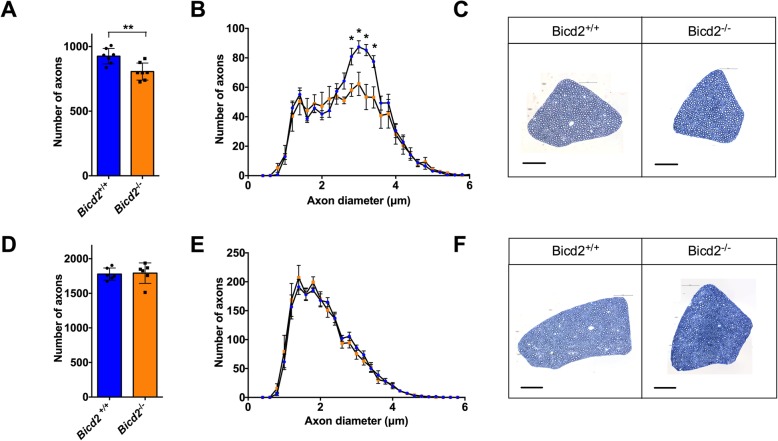
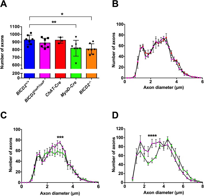
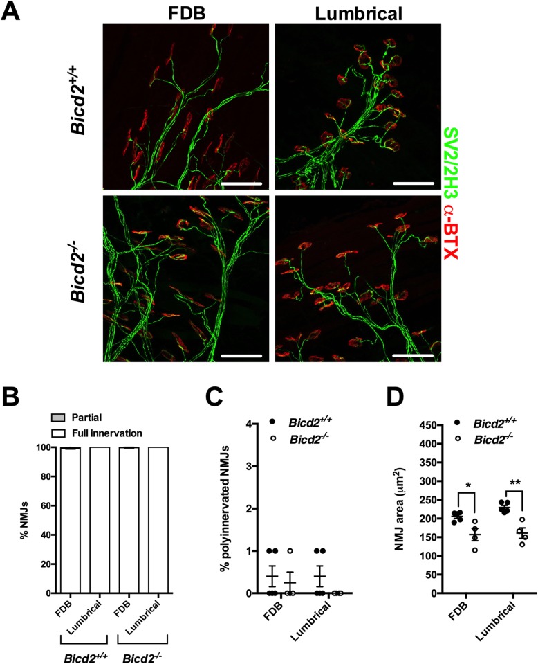
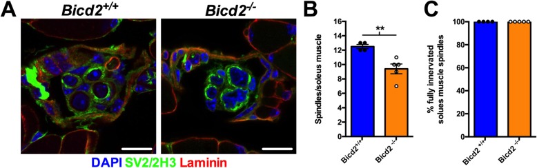
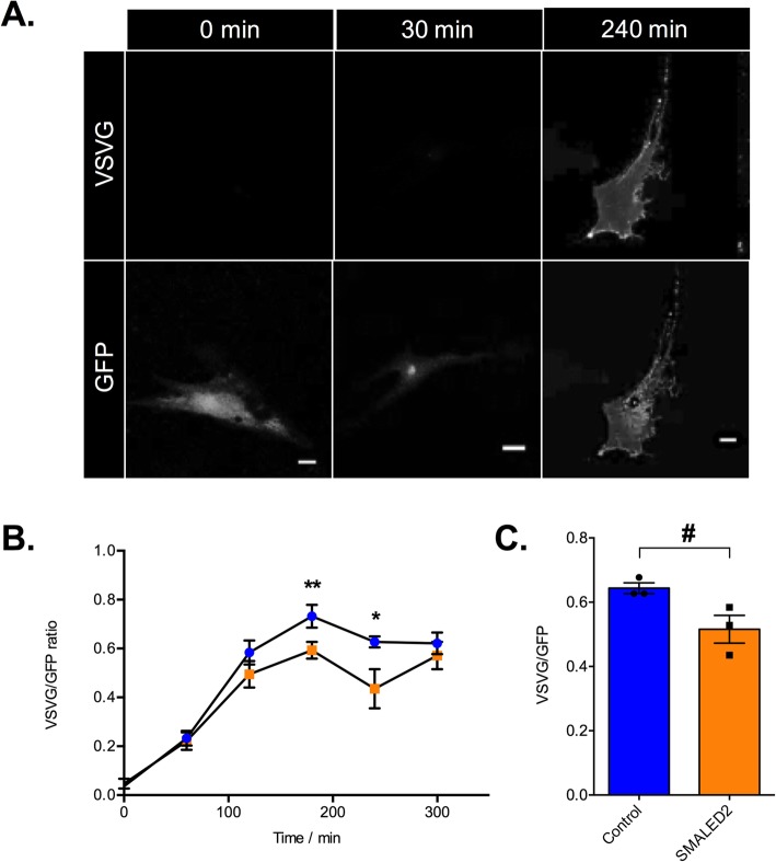
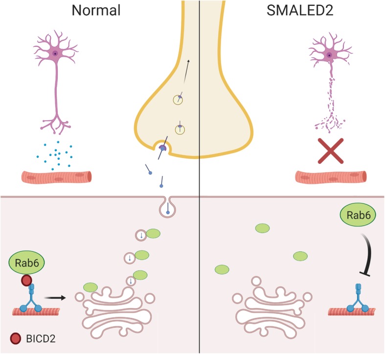
Autosomal dominant missense mutations in BICD2 cause Spinal Muscular Atrophy Lower Extremity Predominant 2 (SMALED2), a developmental disease of motor neurons. BICD2 is a key component of the cytoplasmic dynein/dynactin motor complex, which in axons drives the microtubule-dependent retrograde transport of intracellular cargo towards the cell soma. Patients with pathological mutations in BICD2 develop malformations of cortical and cerebellar development similar to Bicd2 knockout (-/-) mice. In this study we sought to re-examine the motor neuron phenotype of conditional Bicd2-/- mice. Bicd2-/- mice show a significant reduction in the number of large calibre motor neurons of the L4 ventral root compared to wild type mice. Muscle-specific knockout of Bicd2 results in a similar reduction in L4 ventral axons comparable to global Bicd2-/- mice. Rab6, a small GTPase required for the sorting of exocytic vesicles from the Trans Golgi Network to the plasma membrane is a major binding partner of BICD2. We therefore examined the secretory pathway in SMALED2 patient fibroblasts and demonstrated that BICD2 is required for physiological flow of constitutive secretory cargoes from the Trans Golgi Network to the plasma membrane using a VSV-G reporter assay. Together, these data indicate that BICD2 loss from muscles is a major driver of non-cell autonomous pathology in the motor nervous system, which has important implications for future therapeutic approaches in SMALED2.

Keywords: BICD2; DYNC1H1; Hereditary motor neuropathy; Muscle; SMALED2; Spinal muscular atrophy.


Conflict of interest statement

The authors declare that they have no competing interests.

Figures

Fig. 1
Fig. 1
Loss of motor axons in Bicd2−/− mice at 21 days of age. a shows the number of motor axons in the L4 ventral root of Bicd2+/+ (wild type) and Bicd2−/− (knockout) mice (**unpaired 2-sided t-test, p = 0.0041). b shows a histogram of the total number of L4 motor axons classified using 0.2 μm bins (blue = Bicd2+/+, orange = Bicd2−/−), *p < 0.001; multiple t-tests corrected for multiple comparisons using the Holm-Sidak method. c Representative images from 0.8 μm cross-sections of 1% toluene blue stained L4 ventral roots of Bicd2+/+ and Bicd2−/− mice. d shows the number of sensory axons in the L4 dorsal root of Bicd2+/+ and Bicd2−/− mice. e shows a histogram of the total number of L4 sensory axons classified as in b (blue = Bicd2+/+, orange = Bicd2−/−). f Representative images from 0.8 μm cross sections of 1% toluene blue stained L4 dorsal roots of Bicd2+/+ and Bicd2−/− mice. Error bars = standard error of the mean (SEM). Scale bars = 50 μm
Fig. 2
Fig. 2
Motor axon loss in muscle specific Bicd2 knockout mice. a shows the total number of axons at 21 days of age in Bicd2+/+ mice (blue), Bicd2loxP/loxP mice in which both Bicd2 alleles are flanked by loxP sites (pink), mice with motor neuron specific Bicd2 knockout (ChAT-Cre, red), mice with muscle-specific Bicd2 knockout (Myod-Cre, green), and Bicd2−/− mice (orange). A significant reduction in the total number of L4 ventral axons was found in Myod-Cre and Bicd2−/− mice compared to wild type (one-way ANOVA p = 0.0187, Dunnett’s 2-sided t-test with Bicd2+/+ as control, **p = 0.019, *p = 0.034). b shows a histogram of the total number of L4 ventral axons at 21 days of age classified using 0.2 μm bins (pink = Bicd2loxP/loxP, red = ChAT-Cre). c shows a histogram of the total number of L4 motor axons at 21 days of age classified using 0.2 μm bins (pink = Bicd2loxP/loxP, green = Myod-Cre); multiple t-tests corrected for multiple comparisons using the Holm-Sidak method, ***p = 0.0009. d shows a histogram of the total number of L4 motor axons at 42 days of age classified using 0.4 μm bins (pink = Bicd2loxP/loxP (n = 4), green = Myod-Cre (n = 6); multiple t-tests corrected for multiple comparisons using the Holm-Sidak method,****p = 0.0002). Error bars = SEM
Fig. 3
Fig. 3
Normal NMJ analysis of Bicd2−/− mice at 21 days of age. a shows representative images of the NMJs of the FDB (flexor digitorum brevis) and feet lumbrical muscles stained with anti-SV2/2H3 antibodies (green) to visualise motor neurons and fluorescent alpha-bungarotoxin (red) to identify post-synaptic acetylcholine receptors on the muscle fibre surface. Scale bars = 50 μm. b shows the percentage of fully and partially innervated NMJs in Bicd2+/+ (n = 4) and Bicd2−/− (n = 4) mice. c shows the percentage of poly-innervated (measure of immaturity) NMJs between Bicd2+/+ (n = 4) and Bicd2−/− (n = 4) mice. d shows the area of the NMJ (area occupied by each single AchR cluster) in the FDB and lumbrical muscles in Bicd2+/+ (mean 205 and 230 μm2, respectively; n = 4) and Bicd2−/− (mean 157 and 161 μm2, respectively; n = 4) mice, (multiple t-tests corrected for multiple comparisons using the Holm-Sidak method, *p = 0.05, **p = 0.004). Error bars = SEM. The normal NMJ analysis suggests that there is no active denervation in Bicd2−/− mice at 21 days of age
Fig. 4
Fig. 4
Loss of muscle spindles in Bicd2−/− mice at 21 days of age. (a) shows example images of a cross section through a muscle spindle in Bicd2+/+ and Bicd2−/− mice stained for nuclei (DAPI, blue), the neuronal marker SV2/2H3 (green) and laminin (muscle membrane, red). Scale bars = 10 μm. The concentric SV2/SH3 staining around laminin positive muscle fibres indicates a muscle spindle (b) shows the total number of muscle spindles in the soleus muscle of Bicd2+/+ and Bicd2−/− mice, **p = 0.0065 (unpaired t-test, n = 4–5). (c) shows full innervation patterns in the muscle spindles of Bicd2+/+ and Bicd2−/− mice. Error bars = SEM. The loss of muscle spindles correlates with the loss of presumed gamma motor neurons in Bicd2−/− compared to Bicd2+/+ mice
Fig. 5
Fig. 5
SMALED2 patient fibroblasts show delayed VSV-G secretion compared to controls. a is an example of a human fibroblast transfected with a plasmid encoding for the thermo-sensitive-GFP vesicular stomatitis virus glycoprotein (ts0–45 VSV-G) at 32 °C. The time prior to fixation is indicated on the top; staining with an anti-VSV-G [8G5F11] against a surface epitope of VSV-G in non-permeabilised cells (top row of panels); GFP staining is shown in the bottom row. Scale bars = 20 μm. At 0 mins, all ts0–45 VSVG is retained within the ER with no surface staining. At 30mins all ts0–45 VSV-G has been trafficked to the Golgi. By 240 mins all ts045-VSV-g has been trafficked to the plasma membrane and is evident in both the GFP (bottom) and anti-VSV-G surface epitope antibody (top)_ panels. b Kinetics of VSV-G secretion in fibroblasts isolated from a patient with SMALED2 (I189F mutation, orange) and an age-matched control (blue) are quantified as the ratio of total surface VSV-G staining to total GFP. The x axis shows the time in minutes at 32 °C prior to fixation (n = 10 cells per condition; **p = 0.008, *p = 0.009; multiple unpaired t-tests corrected for multiple comparisons using the Holm-Sidak method). c shows the average ratio of surface VSV-G to total GFP at 240 min for three independent healthy control and three unrelated SMALED2 (S107L, I189F and R501P) fibroblast cell lines (# p = 0.052, unpaired t-test). Error bars = SEM. The impaired secretion in SMALED2 patient fibroblasts suggests that a similar impairment of secretion may be evident in the muscle of Bicd2−/− mice and SMALED2 patients
Fig. 6
Fig. 6
A summary of the proposed non-cell autonomous mechanism in SMALED2. In normal muscle, BICD2 ensures the correct localisation of Rab6 at the trans Golgi surface, required for the efficient trafficking of secretory vesicles (containing neurotrophins) to the plasma membrane. In SMALED2 muscle, loss of BICD2 function impairs the localisation of Rab6 to the trans Golgi surface resulting in impaired trafficking of secretory vesicles to the plasma membrane and a reduction in neurotrophin release required for motor neuron survival

Similar articles

Cited by

References

    1. Oates EC, Rossor AM, Hafezparast M, Gonzalez M, Speziani F, Macarthur DG, et al. Mutations in BICD2 cause dominant congenital spinal muscular atrophy and hereditary spastic paraplegia. Am J Hum Genet. 2013;92:965–973. doi: 10.1016/j.ajhg.2013.04.018. - DOI - PMC - PubMed
    1. Neveling K, Martinez-Carrera LA, Hölker I, Heister A, Verrips A, Hosseini-Barkooie SM, et al. Mutations in BICD2, which encodes a golgin and important motor adaptor, cause congenital autosomal-dominant spinal muscular atrophy. Am J Hum Genet. 2013;92:946–954. doi: 10.1016/j.ajhg.2013.04.011. - DOI - PMC - PubMed
    1. Scoto M, Rossor AM, Harms MB, Cirak S, Calissano M, Robb S, et al. Novel mutations expand the clinical spectrum of DYNC1H1-associated spinal muscular atrophy. Neurology. 2015;84:668–679. doi: 10.1212/WNL.0000000000001269. - DOI - PMC - PubMed
    1. Harms MB, Ori-McKenney KM, Scoto M, Tuck EP, Bell S, Ma D, et al. Mutations in the tail domain of DYNC1H1 cause dominant spinal muscular atrophy. Neurology. 2012;78:1714–1720. doi: 10.1212/WNL.0b013e3182556c05. - DOI - PMC - PubMed
    1. Peeters K, Litvinenko I, Asselbergh B, Almeida-Souza L, Chamova T, Geuens T, et al. Molecular defects in the motor adaptor BICD2 cause proximal spinal muscular atrophy with autosomal-dominant inheritance. Am J Hum Genet. 2013;92:955–964. doi: 10.1016/j.ajhg.2013.04.013. - DOI - PMC - PubMed

Publication types

MeSH terms

Substances