. 2020 Mar 17;11(2):e03250-19.
doi: 10.1128/mBio.03250-19.

Snake Deltavirus Utilizes Envelope Proteins of Different Viruses To Generate Infectious Particles

Affiliations

Snake Deltavirus Utilizes Envelope Proteins of Different Viruses To Generate Infectious Particles

Leonora Szirovicza et al. mBio. .

Abstract

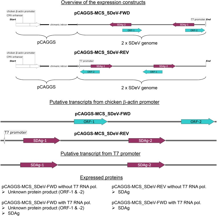
Satellite viruses, most commonly found in plants, rely on helper viruses to complete their replication cycle. The only known example of a human satellite virus is the hepatitis D virus (HDV), and it is generally thought to require hepatitis B virus (HBV) to form infectious particles. Until 2018, HDV was the sole representative of the genus Deltavirus and was thought to have evolved in humans, the only known HDV host. The subsequent identification of HDV-like agents in birds, snakes, fish, amphibians, and invertebrates indicated that the evolutionary history of deltaviruses is likely much longer than previously hypothesized. Interestingly, none of the HDV-like agents were found in coinfection with an HBV-like agent, suggesting that these viruses use different helper virus(es). Here we show, using snake deltavirus (SDeV), that HBV and hepadnaviruses represent only one example of helper viruses for deltaviruses. We cloned the SDeV genome into a mammalian expression plasmid, and by transfection could initiate SDeV replication in cultured snake and mammalian cell lines. By superinfecting persistently SDeV-infected cells with reptarenaviruses and hartmaniviruses, or by transfecting their surface proteins, we could induce production of infectious SDeV particles. Our findings indicate that deltaviruses can likely use a multitude of helper viruses or even viral glycoproteins to form infectious particles. This suggests that persistent infections, such as those caused by arenaviruses and orthohantaviruses used in this study, and recurrent infections would be beneficial for the spread of deltaviruses. It seems plausible that further human or animal disease associations with deltavirus infections will be identified in the future.IMPORTANCE Deltaviruses need a coinfecting enveloped virus to produce infectious particles necessary for transmission to a new host. Hepatitis D virus (HDV), the only known deltavirus until 2018, has been found only in humans, and its coinfection with hepatitis B virus (HBV) is linked with fulminant hepatitis. The recent discovery of deltaviruses without a coinfecting HBV-like agent in several different taxa suggested that deltaviruses could employ coinfection by other enveloped viruses to complete their life cycle. In this report, we show that snake deltavirus (SDeV) efficiently utilizes coinfecting reptarena- and hartmaniviruses to form infectious particles. Furthermore, we demonstrate that cells expressing the envelope proteins of arenaviruses and orthohantaviruses produce infectious SDeV particles. As the envelope proteins are responsible for binding and infecting new host cells, our findings indicate that deltaviruses are likely not restricted in their tissue tropism, implying that they could be linked to animal or human diseases other than hepatitis.

Keywords: coinfection; deltavirus; hepatitis; virology; zoonotic infections.


Figures

FIG 1
FIG 1
Isolation of SDeV from the brain of an infected snake using cultured boid kidney cells (I/1Ki). Mock-infected I/1Ki (top panels) and brain homogenate-inoculated I/1Ki cells (bottom panels) were stained for SDAg (anti-SDAg-AF488 [α-SDAg-AF488], left panels, green), reptarenavirus or hartmanivirus nucleoprotein [anti-NP-AF594 (α-NP-AF594) or α-HISV NP (AF594), middle panels, red], and Hoechst 33342 was used to visualize the nuclei. The panels on the right show an overlay of the three images. The images were taken at ×400 magnification using a Zeiss Axioplan 2 microscope. inf., infected; inoc., inoculated; dpi, days postinfection; mo, months.
FIG 2
FIG 2
Inserts in the SDeV expression plasmids with putative transcripts and proteins produced. The inserts in pCAGGS-MCS_SDeV-FWD and pCAGGS-MCS_SDeV-REV with the cytomegalovirus (CMV) enhancer, chicken β-actin promoter, and chimeric intron from pCAGGS are schematically depicted at the top of the figure. Below are the putative transcripts produced under chicken β-actin promoter following transfection to cells, and transcripts produced in the presence of T7 RNA polymerase (or transcription from T7 promoter). The expressed proteins are listed at the bottom.
FIG 3
FIG 3
Transfection of I/1Ki and Vero E6 cells with pCAGGS-SDeV-FWD and pCAGGS-SDeV-REV constructs results in SDeV replication. (A) I/1Ki (top) and Vero E6 (bottom) cells transfected with Δ-fwd (pCAGGS-SDeV-FWD) and Δ-rev (pCAGGS-SDeV-REV) were stained for SDAg (anti-SDAg antiserum [1:7,500] and Alexa Fluor 594-labeled donkey anti-rabbit immunoglobulin [1:1,000]) at 1, 2, 3, and 4 days posttransfection (from left to right). The images were taken at ×400 magnification using a Zeiss Axioplan 2 microscope. (B) Western blot of I/1Ki (left panel) and Vero E6 (right panel) cell pellets at 1, 2, 3, and 4 days posttransfection with Δ-fwd and Δ-rev constructs. Precision Plus Protein Dual Color Standards (Bio-Rad) served as the marker, and the results were recorded using the Odyssey infrared imaging system (Li-Cor).
FIG 4
FIG 4
SDeV replicates in human and reptilian cell lines. (A) A549 (human lung carcinoma), HEK293FT (human embryonic kidney), HeLa (human cervical cancer), and HepG2 (human hepatocellular carcinoma) cells transfected with Δ-fwd (pCAGGS-SDeV-FWD) were stained at 5 days posttransfection for SDAg (anti-SDAg-AF594 [α-SDAg-AF594], left panels, red). Hoechst 33342 was used to visualize the nuclei. The panels on the right show an overlay of the three images. (B) Boid cell lines I/1Ki (kidney), V/1Ki (kidney), V/2Hz (heart), V/1Liv (liver), and V/5Lu (lung) transfected with Δ-fwd (pCAGGS-SDeV-FWD) were stained at 5 days posttransfection for SDAg (α-SDAg-AF594, left panels, red), and Hoechst 33342 was used to visualize the nuclei. The panels on the right show an overlay of the three images. (C) The transfected boid cells from panel B were allowed to grow, passaged three times, and stained for SDAg (α-SDAg-AF488, left panels, green), and Hoechst 33342 was used to visualize the nuclei. The panels on the right show an overlay of the two images. All images were taken at ×400 magnification using a Zeiss Axioplan 2 microscope, and a 30-μm bar is shown in the bottom right corner of each panel.
FIG 5
FIG 5
SDeV-infected cells can be superinfected with reptarenaviruses (UHV-2 and UGV-1) and hartmanivirus (HISV-1). (A) Mock-infected I/1Ki cells (boa kidney) and mock-, UGV-1-, and UHV-2-infected I/1Ki-Δ cells were stained for SDAg (anti-SDAg-AF488 [α-SDAg-AF488], left panels, green) and reptarenavirus NP (α-NP-AF594, second column, red). Hoechst 33342 was used to visualize the nuclei. The panels on the right show an overlay of the three images. (B) Mock-, UGV-1-, and HISV-1-infected V/1Ki-Δ cells (boa kidney) were stained for SDAg (α-SDAg-AF488, left panels, green), reptarenavirus NP (α-NP-AF594, second column, except bottom, red), or hartmanivirus NP (anti-HISV NP [1:3,000] and Alexa Fluor 594-labeled donkey anti-rabbit immunoglobulin [1:1,000], second column bottom panel, red). Hoechst 33342 was used to visualize the nuclei. The panels on the right show an overlay of the three images. (C) Mock-, UGV-1-, and HISV-1-infected V/1Liv-Δ cells (boa liver) were stained for SDAg (α-SDAg-AF488, left panels, green), reptarenavirus NP (α-NP-AF594, second column, except bottom, red), or hartmanivirus NP (anti-HISV NP [1:3,000] and Alexa Fluor 594-labeled donkey anti-rabbit immunoglobulin [1:1,000], second column bottom panel, red). Hoechst 33342 was used to visualize the nuclei. The panels on the right show an overlay of the three images. (D) Mock-, UGV-1-, and HISV-1-infected V/2Hz-Δ cells (boa heart) were stained for SDAg (α-SDAg-AF488, left panels, green), reptarenavirus NP (α-NP-AF594, second column, except bottom, red), or hartmanivirus NP (anti-HISV NP [1:3,000] and Alexa Fluor 594-labeled donkey anti-rabbit immunoglobulin [1:1,000], second column bottom panel, red). Hoechst 33342 was used to visualize the nuclei. The panels on the right show an overlay of the three images. All images were taken at ×400 magnification using a Zeiss Axioplan 2 microscope, and a 30-μm bar is shown in the bottom right corner of each panel.
FIG 6
FIG 6
Superinfection of permanently SDeV-infected boid cells (I/1Ki-Δ and V/2Hz-Δ) induced production of infectious SDeV particles. (A) Supernatant collected from mock-, UHV-2 (top), UGV-1 (middle), and HISV-1 (bottom) superinfected I/1Ki-Δ cells at 2, 4, 6, and 8 days postinfection (dpi) were titrated on clean I/1Ki cells. The y axis shows the number of fluorescent focus-forming units (FFFUs) per milliliter of culture medium. (B) Supernatants collected from mock-, UHV-2, UGV-1, and HISV-1 superinfected I/1Ki-Δ cells and UGV-1 and HISV-1 superinfected V/2Hz-Δ cells were pelleted by ultracentrifugation and analyzed by Western blotting. The left panel shows anti-SDAg staining, and the right panel shows anti-SDAg, anti-reptarenavirus NP, and anti-hartmanivirus NP staining. (C) Pelleted supernatants collected from mock-, UGV-1, and HISV-1 superinfected I/1Ki-Δ cells and UGV-1 and HISV-1 superinfected V/2Hz-Δ cells were subjected to density gradient ultracentrifugation, and the fractions collected from the bottom of the tubes were analyzed by Western blotting using anti-SDAg and anti-reptarenavirus NP staining (for mock and UGV-1) or anti-SDAg and anti-hartmanivirus NP staining (for HISV-1). The arrows indicate the locations of SDAg and reptarenavirus or hartmanivirus NP. Precision Plus Protein Dual Color Standards (Bio-Rad) served as the markers for both panels B and C, and the results were recorded using the Odyssey infrared imaging system (Li-Cor).
FIG 7
FIG 7
Transmission electron microscopy (TEM) of pelleted supernatants. Persistently SDeV-infected I/1Ki-Δ cells were inoculated with medium collected from clean I/1Ki cells (mock) or superinfected with UGV-1 or HISV-1. V/2Hz-Δ cells were superinfected either with UGV-1 or HISV-1. The cell culture medium was collected at 2- to 3-day intervals until 14 days postinfection, after which the supernatants were pooled and filtered, followed by ultracentrifugation to pellet the virus particles. After resuspending the pelleted material, an aliquot of the pelleted material was prepared for TEM with negative staining. The top panels show the material pelleted from mock-infected I/1Ki-Δ cells, the second row of panels show the material pelleted from UGV-1-infected I/1Ki-Δ cells, the third row of panels show the material pelleted from HISV-1-infected I/1Ki-Δ cells, and the bottom panels show the material pelleted from UGV-1-infected V/2Hz-Δ cells. The black arrows in the figure panels point to UGV-1 particles, and the white arrows show putative SDeV particles as judged by size. The images were taken by using a JEOL JEM-1400 transmission electron microscope at ×200,000 magnification.
FIG 8
FIG 8
Infectious SDeV particles are formed when I/1Ki-Δ cells are transfected with viral glycoproteins. (A) I/1Ki-Δ cells transfected with HISV-1 GPC (top row), Puumala virus glycoproteins (PUUV Gn&Gc, second row), UGV-1 GPC (third row), UGV-1 ZP and GPC (fourth row), HBV S-Ag (fifth row), and empty pCAGGS-MCS plasmid (bottom row) were stained for HA tag (anti-HA [1:4,000] and Alexa Fluor 594-labeled donkey anti-mouse immunoglobulin [1:1,000], left panels, red) and SDAg (α-SDAg-AF488, middle panels, green). Hoechst 33342 was used to visualize the nuclei. The panels on the right show an overlay of the two images. A 30-μm bar is shown in the bottom right corner. All images were taken at ×400 magnification using a Zeiss Axioplan 2 microscope. (B) Supernatants collected from I/1Ki-Δ cells transfected with empty pCAGGS-MCS plasmid, UGV-1 ZP, UGV-1 GPC and ZP, UGV-1 GPC, HISV-1 GPC, LCMV GPC, JUNV GPC, PUUV glycoproteins, and HBV S-Ag were pelleted by ultracentrifugation and analyzed by Western blotting. The left panel shows anti-SDAg staining, and the right panel shows anti-SDAg and anti-HA staining. (C) Supernatants collected from I/1Ki-Δ cells transfected with empty pCAGGS-MCS plasmid, UGV-1 ZP, UGV-1 GPC and ZP, UGV-1 GPC, HISV-1 GPC, LCMV GPC, JUNV GPC, PUUV glycoproteins, and HBV S-Ag were titrated on clean I/1Ki cells. The plasmid used for transfection is shown in the left column, and the corresponding SDeV titer (in fluorescent focus-forming units [fffus] per milliliter) is shown in the right column.

References

    1. Flores R, Owens RA, Taylor J. 2016. Pathogenesis by subviral agents: viroids and hepatitis delta virus. Curr Opin Virol 17:87−94. doi:10.1016/j.coviro.2016.01.022. - DOI - PubMed
    1. Diener TO. 1971. Potato spindle tuber “virus”. IV. A replicating, low molecular weight RNA. Virology 45:411−438. doi:10.1016/0042-6822(71)90342-4. - DOI - PubMed
    1. Rizzetto M, Canese MG, Aricò S, Crivelli O, Trepo C, Bonino F, Verme G. 1977. Immunofluorescence detection of new antigen-antibody system (delta/anti-delta) associated to hepatitis B virus in liver and in serum of HBsAg carriers. Gut 18:997−1003. doi:10.1136/gut.18.12.997. - DOI - PMC - PubMed
    1. Flores R, Ruiz-Ruiz S, Serra P. 2012. Viroids and hepatitis delta virus. Semin Liver Dis 32:201−210. doi:10.1055/s-0032-1323624. - DOI - PubMed
    1. Wille M, Netter HJ, Littlejohn M, Yuen L, Shi M, Eden JS, Klaassen M, Holmes EC, Hurt AC. 2018. A divergent hepatitis D-like agent in birds. Viruses 10:720. doi:10.3390/v10120720. - DOI - PMC - PubMed

Publication types