

Serum biomarkers in myelin oligodendrocyte glycoprotein antibody-associated disease
- PMID: 32184342
- PMCID: PMC7136043
- DOI: 10.1212/NXI.0000000000000708
Serum biomarkers in myelin oligodendrocyte glycoprotein antibody-associated disease
Abstract
Objective: To test the hypothesis that the pattern of serum biomarkers of disease activity and disability in myelin oligodendrocyte glycoprotein antibody-associated disease (MOGAD) will be different from those in neuromyelitis optica spectrum disorder (NMOSD) with anti-aquaporin-4 antibodies (AQP4-Abs).
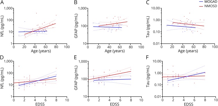
Methods: Using ultrasensitive single-molecule array assays, we measured neurofilament light chain (NfL), glial fibrillary acidic protein (GFAP), and tau in the sera of consecutive patients with MOGAD (n = 16) and NMOSD with AQP4-Ab (n = 33). Serum biomarker levels were compared between patients in relapse and remission states, and correlations between the levels of these biomarkers and Expanded Disability Status Scale (EDSS) scores were analyzed within each group.
Results: In the MOGAD group, the serum tau level was higher in a relapse state than in a remission state (relapse vs remission: 0.5 [0.4-0.5] vs 0.2 [0.1-0.3] pg/mL, p = 0.027). Both serum levels of NfL and tau correlated with the EDSS score (NfL: r = 0.684, p = 0.003; tau: r = 0.524, p = 0.045). Meanwhile, in the NMOSD group, serum NfL and GFAP levels were higher in a relapse state than in a remission state (relapse vs remission: NfL, 34.8 [12.2-62.3] vs 13.0 [11.3-20.0] pg/mL, p = 0.010; GFAP, 253.8 [150.6-303.0] vs 104.4 [93.9-127.9] pg/mL, p = 0.016). Only the serum GFAP level correlated with the EDSS score (r = 0.485, p = 0.012).
Conclusion: The pattern of serum biomarkers of disease activity and disability in MOGAD showed a distinct feature from those in NMOSD with AQP4-Ab, which implicates different pathogeneses between the 2 diseases.
Copyright © 2020 The Author(s). Published by Wolters Kluwer Health, Inc. on behalf of the American Academy of Neurology.
Figures

References
-
- Reindl M, Waters P. Myelin oligodendrocyte glycoprotein antibodies in neurological disease. Nat Rev Neurol 2019;15:89–102. - PubMed
-
- Juryńczyk M, Jacob A, Fujihara K, Palace J. Myelin oligodendrocyte glycoprotein (MOG) antibody-associated disease: practical considerations. Pract Neurol 2019;19:187–195. - PubMed
Publication types
MeSH terms
Substances
LinkOut - more resources
Full Text Sources
Miscellaneous