

Hair cortisol concentrations are associated with hippocampal subregional volumes in children
- PMID: 32184428
- PMCID: PMC7078215
- DOI: 10.1038/s41598-020-61131-x
Hair cortisol concentrations are associated with hippocampal subregional volumes in children
Abstract
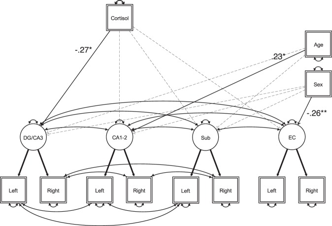
The human hippocampus, a brain structure crucial for memory across the lifespan, is highly sensitive to adverse life events. Stress exposures during childhood have been linked to altered hippocampal structure and memory performance in adulthood. Animal studies suggest that these differences are in part driven by aberrant glucocorticoid secretion during development, with strongest effects on the CA3 region and the dentate gyrus (CA3-DG) of the hippocampus, alongside associated memory impairments. However, only few pediatric studies have examined glucocorticoid associations with hippocampal subfield volumes and their functional relevance. In 84 children (age range: 6-7 years), we assessed whether volumes of hippocampal subregions were related to cumulative glucocorticoid levels (hair cortisol), parenting stress, and performance on memory tasks known to engage the hippocampus. We found that higher hair cortisol levels were specifically related to lower CA3-DG volume. Parenting stress did not significantly correlate with hair cortisol, and there was no evidence to suggest that individual differences in hippocampal subregional volumes manifest in memory performance. Our results suggest that the CA3-DG may be the hippocampal region most closely associated with hair cortisol levels in childhood. Establishing causal pathways underlying this association and its relation to environmental stress and memory development necessitates longitudinal studies.
Conflict of interest statement
The authors declare no competing interests.
Figures

Similar articles
-
Socioeconomic Disparities in Chronic Physiologic Stress Are Associated With Brain Structure in Children.Biol Psychiatry. 2019 Dec 15;86(12):921-929. doi: 10.1016/j.biopsych.2019.05.024. Epub 2019 Jun 12. Biol Psychiatry. 2019. PMID: 31409452 Free PMC article.
-
Effects of Variations in Daily Cortisol Pattern and Long-Term Cortisol Output on Hippocampal Subfield Volumes in the Adult Human Brain.Biol Psychiatry Glob Open Sci. 2025 Feb 6;5(3):100458. doi: 10.1016/j.bpsgos.2025.100458. eCollection 2025 May. Biol Psychiatry Glob Open Sci. 2025. PMID: 40201775 Free PMC article.
-
Stress-system genes and life stress predict cortisol levels and amygdala and hippocampal volumes in children.Neuropsychopharmacology. 2014 Apr;39(5):1245-53. doi: 10.1038/npp.2013.327. Epub 2013 Nov 25. Neuropsychopharmacology. 2014. PMID: 24304824 Free PMC article.
-
Hair cortisol concentration (HCC) as a measure for prenatal psychological distress - A systematic review.Psychoneuroendocrinology. 2018 Jun;92:21-28. doi: 10.1016/j.psyneuen.2018.03.019. Epub 2018 Mar 27. Psychoneuroendocrinology. 2018. PMID: 29609112
-
The Val66Met brain-derived neurotrophic factor gene variant interacts with early pain exposure to predict cortisol dysregulation in 7-year-old children born very preterm: Implications for cognition.Neuroscience. 2017 Feb 7;342:188-199. doi: 10.1016/j.neuroscience.2015.08.044. Epub 2015 Aug 28. Neuroscience. 2017. PMID: 26318333 Free PMC article. Review.
Cited by
-
Tentative Causes of Brain and Neuropsychological Alterations in Women Victims of Intimate Partner Violence.Brain Sci. 2024 Sep 30;14(10):996. doi: 10.3390/brainsci14100996. Brain Sci. 2024. PMID: 39452010 Free PMC article. Review.
-
Household socioeconomic status relates to specific hippocampal subfield volumes across development.Hippocampus. 2023 Sep;33(9):1067-1072. doi: 10.1002/hipo.23542. Epub 2023 May 3. Hippocampus. 2023. PMID: 37132590 Free PMC article.
-
Relationship between hair cortisol concentrations and cognitive functioning in adolescents with ADHD.Eur J Psychotraumatol. 2023;14(2):2281752. doi: 10.1080/20008066.2023.2281752. Epub 2023 Nov 21. Eur J Psychotraumatol. 2023. PMID: 38154075 Free PMC article.
-
Psychosocial factors and hippocampal subfields: The Medea-7T study.Hum Brain Mapp. 2023 Apr 1;44(5):1964-1984. doi: 10.1002/hbm.26185. Epub 2022 Dec 30. Hum Brain Mapp. 2023. PMID: 36583397 Free PMC article.
-
Relations of Lifetime Perceived Stress and Basal Cortisol With Hippocampal Volume Among Healthy Adolescents and Those at Clinical High Risk for Psychosis: A Structural Equation Modeling Approach.Biol Psychiatry. 2024 Sep 1;96(5):401-411. doi: 10.1016/j.biopsych.2023.11.027. Epub 2023 Dec 12. Biol Psychiatry. 2024. PMID: 38092185 Free PMC article.
References
-
- Tulving, E. Elements of episodic memory. (Clarendon, 1983).
Publication types
MeSH terms
Substances
LinkOut - more resources
Full Text Sources
Medical
Miscellaneous
