. 2020 Mar 17;3(1):128.
doi: 10.1038/s42003-020-0845-0.

Interaction of SHP-2 SH2 domains with PD-1 ITSM induces PD-1 dimerization and SHP-2 activation

Affiliations

Interaction of SHP-2 SH2 domains with PD-1 ITSM induces PD-1 dimerization and SHP-2 activation

Nikolaos Patsoukis et al. Commun Biol. .

Abstract

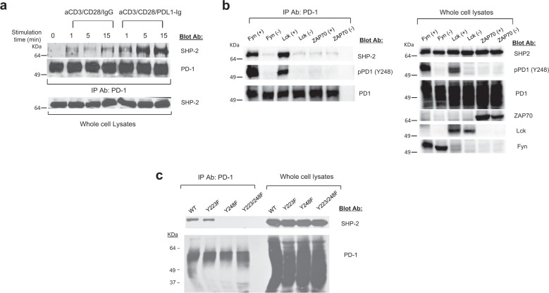
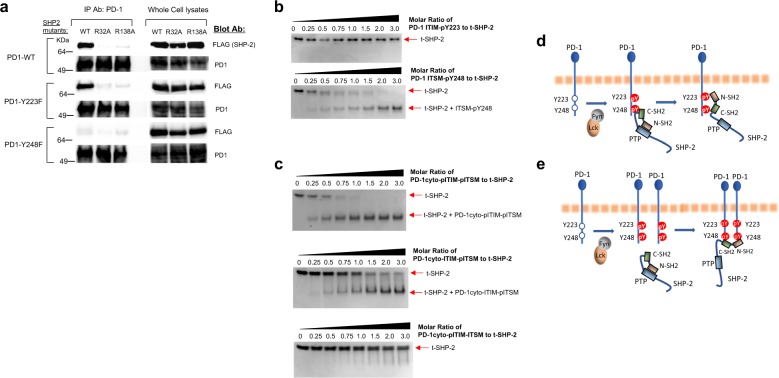
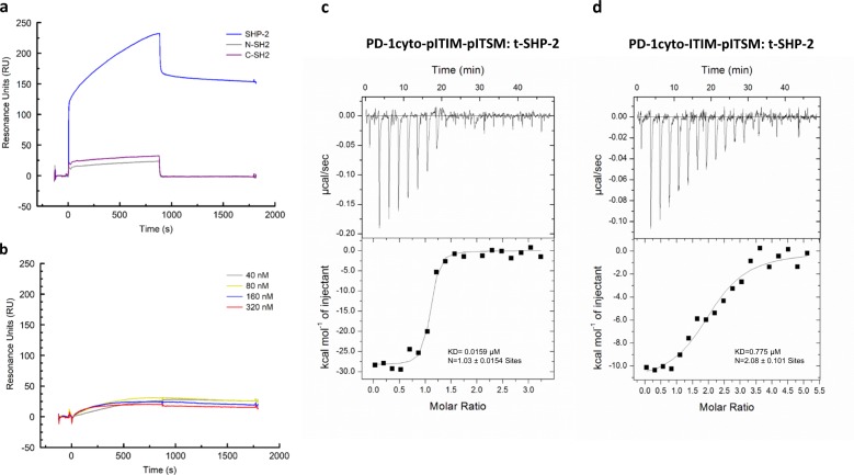
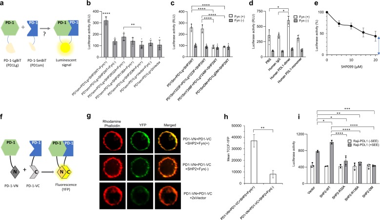
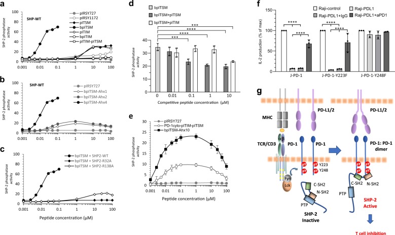
Programmed cell death-1 (PD-1) inhibits T cell responses. This function relies on interaction with SHP-2. PD-1 has one immunoreceptor tyrosine-based inhibitory motif (ITIM) at Y223 and one immunoreceptor tyrosine-based switch motif (ITSM) at Y248. Only ITSM-Y248 is indispensable for PD-1-mediated inhibitory function but how SHP-2 enzymatic activation is mechanistically regulated by one PD-1 phosphotyrosine remains a puzzle. We found that after PD-1 phosphorylation, SHP-2 can bridge phosphorylated ITSM-Y248 residues on two PD-1 molecules via its amino terminal (N)-SH2 and carboxyterminal (C)-SH2 domains forming a PD-1: PD-1 dimer in live cells. The biophysical ability of SHP-2 to interact with two ITSM-pY248 residues was documented by isothermal titration calorimetry. SHP-2 interaction with two ITSM-pY248 phosphopeptides induced robust enzymatic activation. Our results unravel a mechanism of PD-1: SHP-2 interaction that depends only on ITSM-Y248 and explain how a single docking site within the PD-1 cytoplasmic tail can activate SHP-2 and PD-1-mediated inhibitory function.


Conflict of interest statement

The authors declare no competing non-financial interests, but the following competing financial interests: V.A.B. and G.J.F. have patents on the PD-1 pathway licensed by Bristol-Myers Squibb, Roche, Merck, EMD-Serono, Boehringer Ingelheim, AstraZeneca, Novartis and Dako.

Figures

Fig. 1
Fig. 1. Phosphorylation of PD-1 ITSM Y248 by TCR proximal Src family kinases Fyn and Lck, but not ZAP-70 is required for interaction with SHP-2.
a Jurkat-PD1 cells were left unstimulated or stimulated with aCD3/CD28/IgG or aCD3/CD28/PDL1-Ig beads for the indicated times, lysates were prepared followed by immunoprecipitation with anti-PD-1 antibody, SDS-PAGE, and western blot with antibodies for SHP-2 and PD-1. SHP-2 expression in whole-cell lysates was examined. b COS cells were co-transfected with PD-1, SHP-2, and either the kinase active (+) or inactive (–) form of either Fyn, Lck, or ZAP-70. After PD-1 immunoprecipitation of cell lysates and SDS-PAGE, immunoblot was performed with antibodies for SHP-2, PD-1, or an antibody specific for phosphorylated tyrosine of PD-1 ITSM Y248. Expression of the indicated proteins in the same whole-cell lysates was assessed. c COS cells were co-transfected with SHP-2, Fyn kinase active and either PD-1 wild type, PD-1 Y223F, PD-1 Y248F, or PD-1 Y223F/Y248F. PD-1 immunoprecipitation was performed in cell lysates followed by SDS-PAGE and immunoblot with the indicated antibodies. Expression of the same proteins in the same whole-cell lysates was assessed. Results are representative of five independent experiments.
Fig. 2
Fig. 2. Interaction of both SH2 domains of SHP-2 is required for SHP-2 binding to PD-1.
a COS cells were transfected with kinase active Fyn, PD-1 WT and either SHP-2-WT, SHP-2-R32A, or SHP-2-R138A mutants FLAG-tagged; Fyn, PD-1-Y223F and either SHP-2-WT, SHP-2-R32A, or SHP-2-R138A mutants FLAG-tagged; or Fyn, PD-1-Y248F and either SHP-2-WT, SHP-2-R32A, or SHP-2-R138A mutants FLAG-tagged. Immunoprecipitation of cell lysates was performed with anti-PD-1 antibody followed by SDS-PAGE and immunoblot with FLAG- or PD-1-specific antibodies. Expression of the indicated proteins in the same whole-cell lysates was assessed. Results are representative of four separate experiments. b ITSM-pY248 but not ITIM-pY223 phosphopeptide binds with SHP-2 SH2 domains. For assessment of ITIM-pY223 and ITSM-pY248 binding to SHP-2 SH2 domains, SHP-2 amino acids 1–225, which only contains the tandem N-SH2 and C-SH2 domains in their natural sequence (referred to as t-SHP-2), was used. 2 µM of t-SHP-2 were mixed with either ITIM-pY223 or ITSM-pY248 phosphopeptides at the indicated molar ratios, incubated for 1 h at room temperature and binding was examined by Native PAGE followed by Coomassie blue staining. c t-SHP-2 was mixed with a phosphopeptide corresponding to the native sequence of PD-1 cytoplasmic tail in which either both ITIM-Y223 and ITSM-Y248, only the ITIM-Y223, or only the ITSM-Y248 tyrosines were phosphorylated (PD-1cyto-pITIM-pITSM, PD-1cyto-pITIM-ITSM, and PD-1cyto-ITIM-pITSM, respectively).  2μM of t-SHP-2 were mixed with each of the phosphopeptides at the indicated molar ratios, incubated for 1 h hour at room temperature and binding was examined by Native PAGE followed by Coomassie blue staining. d, e Tentative models of PD-1: SHP-2 interaction after PD-1 phosphorylation.
Fig. 3
Fig. 3. Surface plasmon resonance (SPR) and isothermal titration calorimetry analysis of SHP-2 interaction with PD-1.
a PD-1 phosphotyrosyl ITSM-Y248 peptide was immobilized on a CM5 BIAcore chip and binding of SHP-2 full-length SHP-2-N-SH2, and SHP-2-C-SH2, all at 320 nM, was determined by SPR. Full binding curves are presented in Supplementary Fig.  8. Sensograms of 900 s (15 min) association and 900 s dissociation time were analyzed and KD values were calculated. b PD-1 phosphotyrosyl ITIM-Y223 peptide was immobilized on a CM5 BIAcore chip and binding of SHP-2-full-length at the indicated concentrations, was determined by SPR. Results are representative of three experiments. c, d Isothermal titration calorimetry (ITC) experiments were performed on a MicroCal ITC200 instrument. PD-1 phosphopeptides (PD-1cyto-pITIM-pITSM and PD-1cyto-ITIM-pITSM) and t-SHP-2 protein were prepared in buffer containing 50 mM HEPES pH 7.4, 100 mM NaCl, 5 mM TCEP and stoichiometry of bisphosphorylated PD-1 peptide: t-SHP-2 interaction was assessed as described in Methods.
Fig. 4
Fig. 4. SHP-2 bridges two PD-1 molecules via PD-1 ITSM pY248.
a The NanoBiT proximity assay: PD-1-LgBiT and PD-1-SmBiT induce luciferase activity only if two PD-1 molecules stably interact resulting in the formation of an active luciferase enzyme. b HEK-293 cells were co-transfected as indicated and luciferase activity was assessed. Comparisons were performed by two-way ANOVA and Tukey’s multiple comparisons test (****P < 0.0001, PD1sm + PD1Lg + SHP2WT + Fyn(+) vs. all other samples, **P < 0.005, PD1sm + PD1Lg + SHP2R32A + Fyn(+) vs. PD1sm + PD1Lg + SHP2DM + Fyn(+), n = 6 experiments). c HEK-293 cells were co-transfected with the indicated constructs together with either kinase active (+) or inactive (-) Fyn and luciferase activity was assessed (****P < 0.0001, n = 3 experiments). d HEK-293 cells co-transfected with PD1SmBiT, PD1LgBiT and SHP-2 WT, together with either kinase active (+) or inactive (−) Fyn, were cultured in the presence of hPD-L1-Ig (dimer), hPD-L1 monomer or control IgG and luciferase activity was assessed (*P < 0.05, n = 3 experiments). e HEK-293 cells co-transfected with PD1SmBiT, PD1LgBiT, SHP-2 WT, and kinase active Fyn were cultured in the presence of increasing amounts of the allosteric SHP-2 inhibitor SHP099 or vehicle control (0) and luciferase activity was assessed (arrow indicates baseline luciferase activity). f The BiFC assay is based on structural complementation of two non-fluorescent N- and C-terminal fragments of Venus YFP-fluorescent protein fused to a pair of interacting proteins. g HEK-293 cells transfected with indicated constructs were analyzed by confocal microscopy. Representative fluorescent images of each indicated transfection condition. Polymerized actin was visualized with rhodamine-conjugated phalloidin. YFP signal from PD1-VN + PD1-VC complementation was visualized in the green channel. Intensity of yellow color in the merged images is indicative of co-localization of PD-1 dimer with the actin cytoskeleton. h Quantitative analysis of images from g and comparison of total corrected cellular YFP fluorescence (TCCF-YFP) in the indicated conditions after subtraction of TCCF-YFP from control transfection PD1-VN + PD1-VC + 2xVector, **P < 0.005, n = 50 from one of two independent experiments. i Primary human T cells co-transfected with PD1SmBiT, PD1LgBiT and the indicated SHP-2 constructs were cultured with Raji-PD-L1 with or without prior loading with SEE and luciferase activity was assessed 6 h later (****P < 0.0001, ***P < 0.0005, **P < 0.005, *P < 0.05, n = 3 experiments).
Fig. 5
Fig. 5. Interaction of SHP-2 SH2 domains with two PD-1 ITSM pY248 residues is required for activation of SHP-2 phosphatase activity.
a SHP-2-WT was incubated with 20 µM DiFMUP as substrate in the presence of the indicated phosphopeptides and phosphatase activity was assessed as described in Methods. b Phosphatase activity of SHP-2-WT incubated with bpITSM phosphotyrosyl peptides containing a 4-, 2-, or 1-Ahx spacer; pIRSY727 was used as negative control. c Phosphatase activity of SHP-2-WT, SHP-2-R32A, or SHP-2-R138A incubated with the indicated concentrations of bpITSM phosphotyrosyl peptide containing a 4-Ahx spacer. d Phosphatase activity of SHP-2-WT in the presence of phosphotyrosyl bpITSM peptide (0.01 µM) either alone or with increasing amounts of monophosphorylated pITIM or monophosphorylated pITSM peptide. Results are representative of three independent experiments. e Phosphatase activity of SHP-2-WT in the presence of a phosphopeptide corresponding to the native sequence of PD-1 cytoplasmic tail in which both ITIM-Y223 and ITSM-Y248 tyrosines were phosphorylated (PD-1cyto-pITIM-pITSM) or with a bpITSM phosphotyrosyl peptide containing a 10-Ahx spacer. pIRSY727 was used as negative control. In panels ae results from one representative of 3–10 independent experiments are shown. f Jurkat T cells stably expressing PD-1-WT (J-PD-1), PD-1-Y223F (J-PD-1-Y223F), or PD-1-Y248F (J-PD-1-Y248F) were co-cultured with Raji-control or Raji-PD-L1 cells loaded with SEE. Where indicated, anti-PD-1 blocking antibody or isotype control was added in the cultures. Culture supernatants were collected at 24 h and IL-2 production was measured. Results are expressed as % of maximum IL-2 production induced in each cell line by Raji-control cells loaded with SEE (****P < 0.0001, n = 3 experiments). g Model for PD-1: PD-1 bridging and activation of SHP-2 phosphatase activity: An “inside-out” regulation of PD-1: PD-1 dimer complex formation is initiated by TCR-mediated activation of TCR proximal Src family kinases, which induce phosphorylation of tyrosines in PD-1 cytoplasmic tail. The stabilization provided by PD-1 ligands allows the appropriate proximity of two phosphorylated PD-1 molecules to induce binding of both SH2 domains of SHP-2 to ITSM-pY248 and activation of SHP-2, leading to inhibition of T-cell responses.

Similar articles

Cited by

References

    1. Francisco LM, et al. PD-L1 regulates the development, maintenance, and function of induced regulatory T cells. J. Exp. Med. 2009;206:3015–3029. doi: 10.1084/jem.20090847. - DOI - PMC - PubMed
    1. Iwai Y, et al. Involvement of PD-L1 on tumor cells in the escape from host immune system and tumor immunotherapy by PD-L1 blockade. Proc. Natl Acad. Sci. USA. 2002;99:12293–12297. doi: 10.1073/pnas.192461099. - DOI - PMC - PubMed
    1. Barber DL, et al. Restoring function in exhausted CD8 T cells during chronic viral infection. Nature. 2006;439:682–687. doi: 10.1038/nature04444. - DOI - PubMed
    1. Petrovas C, et al. PD-1 is a regulator of virus-specific CD8+ T cell survival in HIV infection. J. Exp. Med. 2006;203:2281–2292. doi: 10.1084/jem.20061496. - DOI - PMC - PubMed
    1. Topalian SL, et al. Safety, activity, and immune correlates of anti-PD-1 antibody in cancer. N. Engl. J. Med. 2012;366:2443–2454. doi: 10.1056/NEJMoa1200690. - DOI - PMC - PubMed

Publication types

MeSH terms

Substances