. 2020 Mar 17;10(1):4890.
doi: 10.1038/s41598-020-61848-9.

Time course of bilateral microglial activation in a mouse model of laser-induced glaucoma

Affiliations

Time course of bilateral microglial activation in a mouse model of laser-induced glaucoma

Ana I Ramírez et al. Sci Rep. .

Abstract

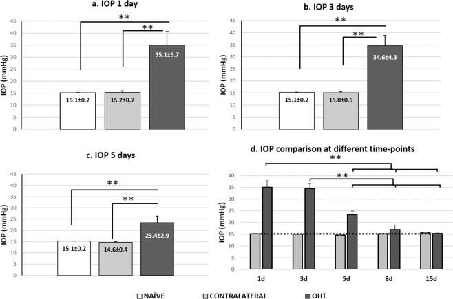
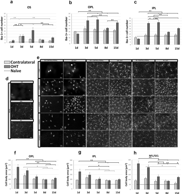
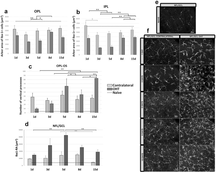
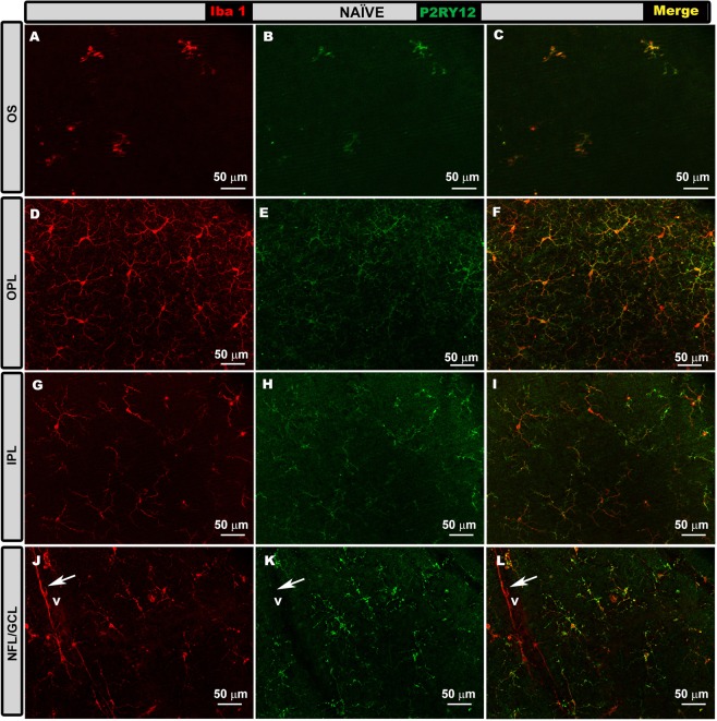
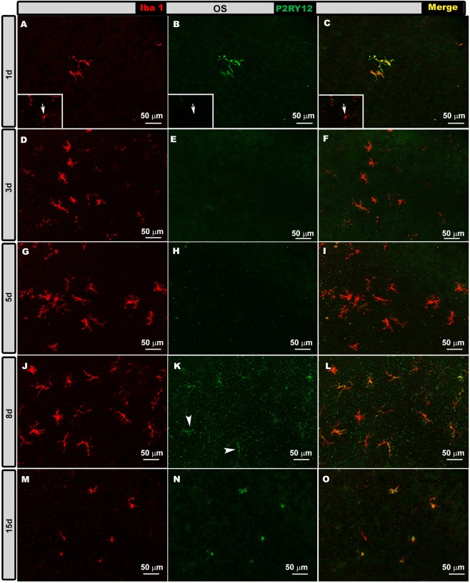
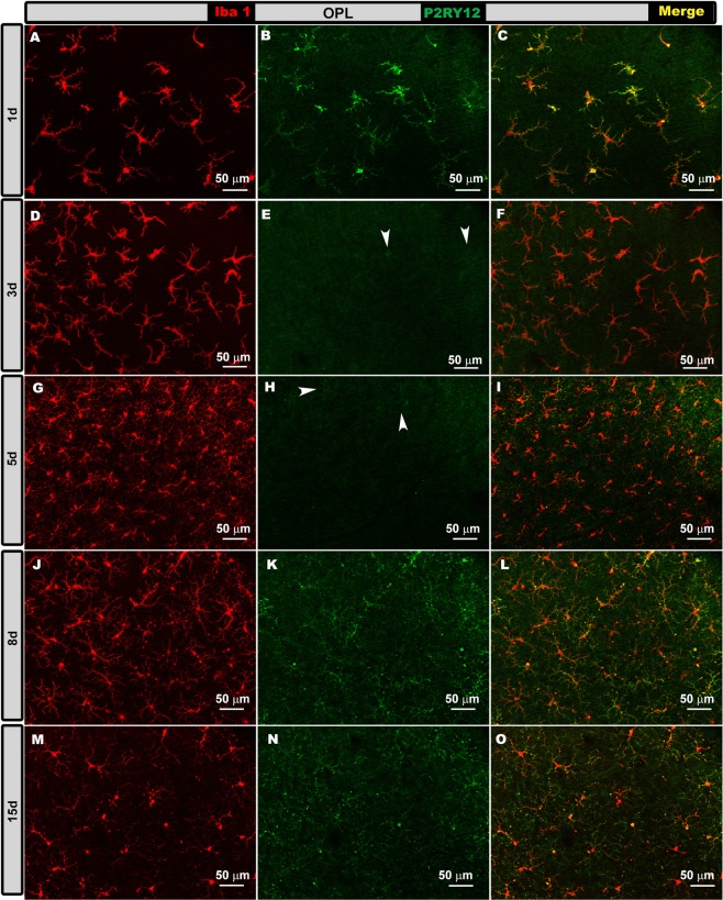
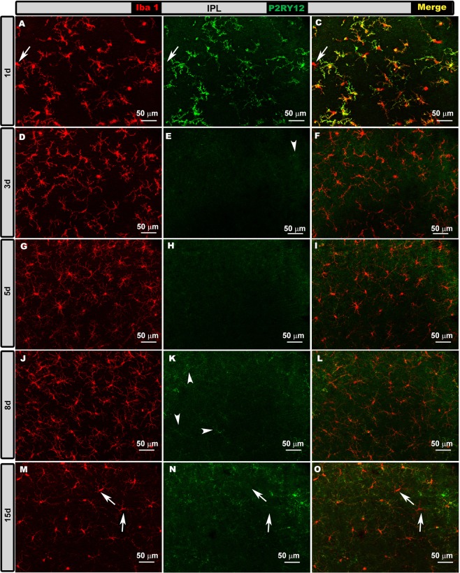
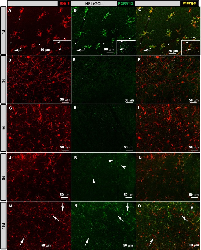
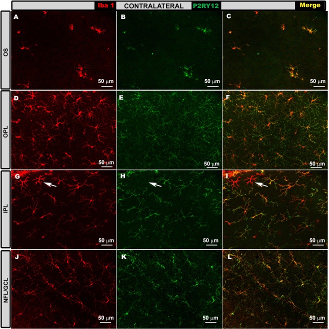
Microglial activation is associated with glaucoma. In the model of unilateral laser-induced ocular hypertension (OHT), the time point at which the inflammatory process peaks remains unknown. Different time points (1, 3, 5, 8, and 15 d) were compared to analyze signs of microglial activation both in OHT and contralateral eyes. In both eyes, microglial activation was detected in all retinal layers at all time points analyzed, including: i) increase in the cell number in the outer segment photoreceptor layer and plexiform layers (only in OHT eyes) from 3 d onward; ii) increase in soma size from 1 d onward; iii) retraction of the processes from 1 d in OHT eyes and 3 d in contralateral eyes; iv) increase in the area of the retina occupied by Iba-1+ cells in the nerve fiber layer/ganglion cell layer from 1 d onward; v) increase in the number of vertical processes from 1 d in contralateral eyes and 3 d in OHT eyes. In OHT eyes at 24 h and 15 d, most Iba-1+ cells were P2RY12+ and were down-regulated at 3 and 5 d. In both eyes, microglial activation was stronger at 3 and 5 d (inflammation peaked in this model). These time points could be useful to identify factors implicated in the inflammatory process.


Conflict of interest statement

The authors declare no competing interests.

Figures

Figure 1
Figure 1
Measurement of intraocular pressure. (a–d) Histograms show mean (±SD) intraocular pressure (IOP) for naïve, contralateral and ocular hypertensive (OHT) eyes at 1 d (a), 3 d (b), 5 d (c). d: Variation in IOP over time in OHT and contralateral eyes. The dotted line indicates mean IOP in naïve eyes. In OHT eyes, pressure rose and peaked at 1 and 3 d, remained stable at 5 d and then gradually returned to basal preoperative levels by 8 d. Contralateral eyes showed similar pressures as naïve eyes at all time points. *p < 0.05, **p < 0.01.
Figure 2
Figure 2
Iba-1+ cell number in the photoreceptor outer segment layer (OS), outer plexiform layer (OPL) and inner plexiform layer (IPL) and cell body area of Iba-1+ cells in the OPL, IPL and the nerve fiber-ganglion cell layer (NFL-GCL) at different times after laser-induced ocular hypertension (OHT). (a–c) Variation over time in the number of Iba-1+ cells. Histograms compare the mean (±SD) number of Iba-1+ cells per area of 0.1502 mm2 at 1, 3, 5, 8 and 15 d between eyes with ocular hypertension (OHT) and contralateral eyes in OS (a), OPL (b) and IPL (c). (d,e) Immunohistochemical images of anti-Iba-1-stained retinal whole-mounts (d: naïve; e: OHT and contralateral). OS (a,e) In OHT eyes, the number of Iba-1+ cells increased from 1 to 5 d, then decreased at 8 and 15 d. In contralateral eyes, the number of Iba-1+ cells increased from 1 to 3 d, then decreased slightly until 15 d. OPL and IPL (b,c,e) In OHT eyes, the largest increase in the number of microglial cells from 1 d occurred at 3 and 5 d. The number decreased from 5 d until 8 d and 15 d. (f–h) Variation over time in the cell body area of Iba-1+ cells after laser-induced OHT. Histograms show mean (±SD) cell body area of Iba-1+ cells in OHT eyes and contralateral eyes at 1, 3, 5, 8 and 15 d in OPL (f), IPL (g) and NFL-GCL (h). OPL-IPL (e,f,g) The cell body area in OHT eyes was significantly larger at 1 d, then significantly decrease at 5, 8 or 15 d. In contralateral eyes, the cell body area was greatest at 3 d, and then it decreased significantly at 8 and 15 d. NFL/GCL (h) In OHT eyes, the cell body area was largest at 1 d, and then it progressively decreased at 5, 8 and 15 d. In contralateral eyes, the cell body area was greater at 3 d, and then it decreased such that it was significantly smaller at 15 d than at 3 d. The dotted line indicates the mean values in naïve eyes. *p < 0.05, **p < 0.01.
Figure 3
Figure 3
Arbor area (AA) of Iba-1 + cells in the outer plexiform layer (OPL) and inner plexiform layer (IPL); Iba-1 + vertical processes (VP) between OPL and the photoreceptor outer segment layer (OS) and retinal area occupied by Iba-1+ cells (Iba1-RA) in the nerve fiber-ganglion cell layer (NFL-GCL) at different times after laser-induced ocular hypertension (OHT). (a,b) Variation over time in AA. Histograms show a mean (±SD) AA in OHT eyes and contralateral eyes at 1, 3, 5, 8 and 15 d in OPL (a) and IPL (b). In OHT eyes, AA in the OPL decreased marginally from 1 d to 3 d, and then it increased significantly at 8 d. AA in the IPL decreased significantly from 1 d to 5 d. Then the area increased from 5 d to 15 d. In contralateral eyes, AA in the OPL and IPL was smaller at 3 d than at 1 d, and it was significantly noticeable only in the IPL. From 3 d onward, the AA in OPL and IPL did not change significantly. (c) Variation over time in the number of microglial VP. The histogram shows a mean (±SD) number of microglial VP between OPL and OS in OHT eyes and contralateral eyes at 1, 3, 5, 8 and 15 d. In OHT eyes, the number of VP increased progressively from 1 to 15 d. In contrast, the number of vertical processes in contralateral eyes did not change significantly between 1 and 15 d. (d) Variation in the Iba1-RA over time after laser-induced OHT. The histogram shows a mean (±SD) Iba1-RA in OHT eyes and contralateral eyes at 1, 3, 5, 8 and 15 d. Iba1-RA was significantly greater at 5 d than at 1 d in OHT eyes (significantly) and in contralateral eyes (marginally), after which it decreased such that it was significantly lower at 15 d than at 5 d in both eyes. (e,f) Immunohistochemical images of anti-Iba-1 retinal whole-mounts (e) naïve; (f) OHT and contralateral). The dotted line indicates the mean values in naïve eyes. *p < 0.05, **p < 0.01.
Figure 4
Figure 4
P2RY12 expression in naïve eyes in the photoreceptor outer segment layer (OS), outer plexiform layer (OPL) inner plexiform layer (IPL) and in the nerve fiber-ganglion cell layer (NFL-GCL). Double immunostaining: Iba-1 (A,D,G,J), P2RY12 (B,E,H,K), merge (C,F,I,L). Retinal whole-mounts. In all retinal layers analyzed, Iba-1+ cells showed P2RY12 expression, except perivascular iba-1+ cells (j–l), which were P2RY12- (arrow). V: vessel.
Figure 5
Figure 5
P2RY12 expression in ocular hypertension (OHT) eyes in the photoreceptor outer segment layer (OS) at different times after laser-induced OHT (1, 3, 5, 8 and 15 d). Double immunostaining: Iba-1 (A,D,G,J,M), P2RY12 (B,E,H,K,N), merge (C,F,I,L,O). Retinal whole-mounts. At 1 and 15 d after laser induction, most Iba-1+ cells showed strong P2RY12 expression in OS. At 3 and 5 d, P2RY12 expression in Iba-1+ cells was substantially down-regulated in this layer; it increased slightly at 8 d (arrowheads), reaching values similar to those of naïve eyes at 15 d. Rounded cells were P2RY12- (arrow) (inset A–C).
Figure 6
Figure 6
P2RY12 expression in ocular hypertension (OHT) eyes in the outer plexiform layer (OPL) at different times after laser-induced OHT (1, 3, 5, 8 and 15 d). Double immunostaining: Iba-1 (A,D,G,J,M), P2RY12 (B,E,H,K,N), merge (C,F,I,L,O). Retinal whole-mounts. At 1 d after laser induction, most Iba-1+ cells intensely expressed P2RY12. This expression was strongly reduced at 3 and 5 d (arrowheads); it increased weakly at 8 d, reaching values similar to those of naïve eyes at 15 d.
Figure 7
Figure 7
P2RY12 expression in ocular hypertension (OHT) eyes in the inner plexiform layer (IPL) at different times after laser-induced OHT (1, 3, 5, 8 and 15 d). Double immunostaining: Iba-1 (A,D,G,J,M), P2RY12 (B,E,H,K,N), merge (C,F,I,L,O). Retinal whole-mounts. At 1 d after laser induction, most Iba-1+ cells strongly expressed P2RY12, but some amoeboid cells did not express this marker (arrow in A–C). At intermediate times (3 and 5 d), P2RY12 expression was much lower. At 8 d, this expression increased weakly (arrowheads) to reach values similar to those in naïve eyes at 15 d. At 15 d, a few ramified Iba-1+ cells were P2RY12- (arrows).
Figure 8
Figure 8
P2RY12 expression in ocular hypertension (OHT) eyes in the nerve fiber-ganglion cell layer (NFL-GCL) at different times after laser-induced OHT (1, 3, 5, 8 and 15 d). Double immunostaining: Iba-1 (A,D,G,J,M), P2RY12 (B,E,H,K,N), merge (C,F,I,L,O). Retinal whole-mounts. At 1 d, most Iba-1+ cells stained intensely as P2RY12+, while only a few rounded cells (inset A-C arrows) and perivascular cells (arrow) were P2RY12-. At 3 and 5 d, P2RY12 expression in Iba-1 + cells was dramatically reduced in this layer, then it increased slightly at 8 d (arrowheads) to reach values similar to those in naïve eyes at 15 d. At 15 d, a few ramified Iba-1+ cells did not express P2RY12 (arrows).
Figure 9
Figure 9
P2RY12 expression in contralateral eyes in the photoreceptor outer segment layer (OS), outer plexiform layer (OPL) inner plexiform layer (IPL) and in the nerve fiber-ganglion cell layer (NFL-GCL). Double immunostaining: Iba-1 (A,D,G,J), P2RY12 (B,E,H,K), merge (C,F,I,L). Retinal whole-mounts. In all retinal layers analyzed, most Iba-1+ cells were labelled with anti-P2RY12 antibody at all time points analyzed: 1 d (A–C), 3 d (G–H), 5 d, 8 d and 15 d (D–F,J–L). P2RY12 expression was similar in all instances. This was not the case for dendritic-like cells (arrow in G-I) or perivascular cells, which were P2RY12-.

Similar articles

Cited by

References

    1. Quigley HA, Broman AT. The number of people with glaucoma worldwide in 2010 and 2020. Br. J. Ophthalmol. 2006;90:262–7. doi: 10.1136/bjo.2005.081224. - DOI - PMC - PubMed
    1. Yang S, et al. Alpha 1-antitrypsin inhibits microglia activation and facilitates the survival of iPSC grafts in hypertension mouse model. Cell. Immunol. 2018;328:49–57. doi: 10.1016/j.cellimm.2018.03.006. - DOI - PubMed
    1. Agarwal R, Gupta SK, Agarwal P, Saxena R, Agrawal SS. Current concepts in the pathophysiology of glaucoma. Indian J. Ophthalmol. 2009;57:257–66. doi: 10.4103/0301-4738.53049. - DOI - PMC - PubMed
    1. Quigley HA. Ganglion cell death in glaucoma: pathology recapitulates ontogeny. Aust. N. Z. J. Ophthalmol. 1995;23:85–91. doi: 10.1111/j.1442-9071.1995.tb00135.x. - DOI - PubMed
    1. Soto I, Howell GR. The complex role of neuroinflammation in glaucoma. Cold Spring Harb. Perspect. Med. 2014;4:a017269–a017269. doi: 10.1101/cshperspect.a017269. - DOI - PMC - PubMed

Publication types

MeSH terms