. 2020 Feb 28:11:165.
doi: 10.3389/fgene.2020.00165. eCollection 2020.

An Upstream Open Reading Frame Represses Translation of Chicken PPARγ Transcript Variant 1

Affiliations

An Upstream Open Reading Frame Represses Translation of Chicken PPARγ Transcript Variant 1

Yankai Chu et al. Front Genet. .

Abstract

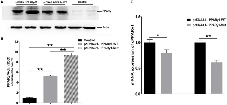
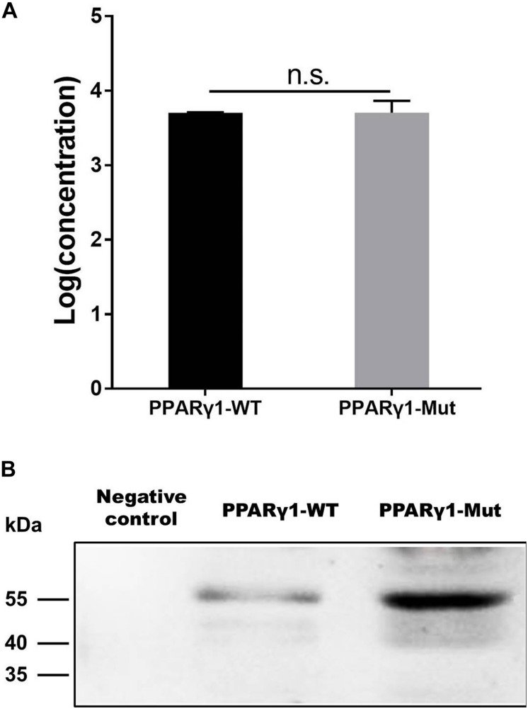
Peroxisome proliferator-activated receptor γ (PPARγ) is a master regulator of adipogenesis. The PPARγ gene produces various transcripts with different 5'-untranslated regions (5' UTRs) because of alternative promoter usage and splicing. The 5' UTR plays important roles in posttranscriptional gene regulation. However, to date, the regulatory role and underlying mechanism of 5' UTRs in the posttranscriptional regulation of PPARγ expression remain largely unclear. In this study, we investigated the effects of 5' UTRs on posttranscriptional regulation using reporter assays. Our results showed that the five PPARγ 5' UTRs exerted different effects on reporter gene activity. Bioinformatics analysis showed that chicken PPARγ transcript 1 (PPARγ1) possessed an upstream open reading frame (uORF) in its 5' UTR. Mutation analysis showed that a mutation in the uORF led to increased Renilla luciferase activity and PPARγ protein expression, but decreased Renilla luciferase and PPARγ1 mRNA expression. mRNA stability analysis using real-time RT-PCR showed that the uORF mutation did not interfere with mRNA stability, but promoter activity analysis of the cloned 5' UTR showed that the uORF mutation reduced promoter activity. Furthermore, in vitro transcription/translation assays demonstrated that the uORF mutation markedly increased the translation of PPARγ1 mRNA. Collectively, our results indicate that the uORF represses the translation of chicken PPARγ1 mRNA.

Keywords: 5′-untranslated region; PPARγ; gene expression; translational repression; upstream open reading frame.


Figures

FIGURE 1
FIGURE 1
Effects of PPARγ 5′ UTR isoforms on reporter gene activity. (A) The luciferase activity of each of the PPARγ 5′ UTR reporter constructs was measured in DF1 cells. (B) The luciferase activity of each of the PPARγ 5′ UTR reporter constructs was measured in ICP1 cells. Data are expressed as mean ± SEM (n ≥ 3 independent experiments). Lower case letters above bars indicate the order of expression levels, “a” represents the highest expression level, “e” represents the lowest expression level. Bars with different superscripts are mutually statistically different. ANOVA followed by Tukey’s multiple comparison test was used to determine significance.
FIGURE 2
FIGURE 2
Schematic representation of PPARγ1 5′ UTR and effects of the PPARγ1 uORF mutation on Rluc luciferase activity and mRNA expression. (A) A schematic diagram of the 117-nucleotide-long PPARγ1 5′ UTR, the uORF is from nucleotides –78 to –25 of the 5′ UTR, and indicated by a striped rectangle. All positions are numbered relative to the initiation codon AUG of PPARγ transcript 1, where A is +1. The uORF encodes a 17-amino acid peptide with the amino acid sequence shown in the bottom. (B) The effect of uORF mutation on the luciferase reporter gene activity. The wild-type (PPARγ1-5′UTR-WT) and uORF mutant (PPARγ1-5′UTR-Mut) PPARγ1 5′ UTR reporter constructs were transfected into ICP1 and DF1 cells, respectively, and reporter gene activity was measured. Compared with the wild-type PPARγ1 5′ UTR reporter, the luciferase activity of PPARγ1-5′UTR-Mut was significantly higher than that of PPARγ1-5′UTR-WT in both ICP1 and DF1 cells (n ≥ 3, **P < 0.01, Student’s t-test). (C) The Rluc mRNA quantification by real-time RT-PCR in the ICP1 and DF1 cells transfected with the indicated reporter constructs. The relative Rluc mRNA levels are normalized to the expression levels of the cells transfected with the reporter PPARγ1-5′UTR-WT. Data were expressed as the mean ± SEM, NONO was used as the internal mRNA control. n ≥ 3, *P < 0.05; **P < 0.01, Student’s t-test.
FIGURE 3
FIGURE 3
PPARγ1 translation is inhibited by its 5′ UTR uORF. (A) Detection of PPARγ1 protein levels. Equal amounts of the total cell lysates from the ICP1 cells transfected with either pcDNA3.1-PPARγ-WT or pcDNA3.1-PPARγ-Mut were separated and immunoblotted with an anti-PPARγ antibody. Actin was used as a loading control. (B) Quantification of PPARγ1 protein expression. Band intensities were measured by ImageJ software normalized to actin loading control. Data represent mean ± SEM. PPARγ1 protein expression was higher in the cells transfected with pcDNA3.1-PPARγ-Mut than in the cells transfected with the pcDNA3.1-PPARγ-WT (**P < 0.01, Student’s t-test). (C) Quantification of PPARγ1 mRNA by real-time RT-PCR in the ICP1 and DF1 cells transfected with the indicated constructs. PPARγ1 mRNA levels were normalized to the expression of the cells transfected with pcDNA3.1-PPARγ-WT. Data were expressed as the mean ± SEM, NONO was used as the internal mRNA control. n ≥ 3, *P < 0.05; **P < 0.01, Student’s t-test.
FIGURE 4
FIGURE 4
Effect of uORF mutation on Rluc mRNA stability. ICP1 cells were transiently transfected with PPARγ1-5′UTR-WT or PPARγ1-5′UTR-Mut, 48 h post-transfection, Rluc mRNA remaining after a 12 h time-course treatment with Actinomycin D was measured by real-time RT-PCR and calculated as a percentage of the level measured at time zero (0 h). Linear regression analysis was used to determine the half-life of the Rluc mRNA (t1/2), the time required for degrading 50% of the existing Rluc mRNA molecules at 0 h. No differences in relative mRNA decay rate were observed between the cells transfected with PPARγ1-5′UTR-WT and PPARγ1-5′UTR-Mut. Data were expressed as the mean ± SEM relative to NONO expression.
FIGURE 5
FIGURE 5
The promoter activity analysis of the DNA sequences corresponding wild-type and uORF-mutated 5′ UTRs of PPARγ1. The DNA sequences corresponding wild-type and uORF-mutated 5′ UTRs of PPARγ1 were cloned into luciferase reporter vector pGL3-basic to yield pGL3-PPARγ1-WT and pGL3-PPARγ1-Mut, respectively. The indicated reporters along with the pRL-TK Renilla luciferase vector were transiently transfected into DF1 (A) and ICP1 cells (B), and the luciferase activity was determined at 48 h after transfection. The pRL-TK vector was used for normalization of transfection efficiency. All data represent the mean ± SEM. *P < 0.05, **P < 0.01, Student’s t-test.
FIGURE 6
FIGURE 6
The uORF represses in vitro PPARγ1 translation. (A) In vitro transcribed PPARγ1 mRNAs from the wild-type and uORF-mutant PPARγ1 expression vectors (pcDNA3.1-PPARγ1-WT and pcDNA3.1-PPARγ1-Mut) were analyzed by quantitative real-time RT-PCR. No difference in PPARγ1 mRNA was observed. Data were expressed as the mean ± SEM, n.s., not significant, Student’s t-test. (B) Equal amounts of the in vitro transcribed mRNAs (2 μg) were used for in vitro translation. Note that the uORF strongly represses PPARγ1 translation. A in vitro translation reaction without RNA template was used as a negative control.
FIGURE 7
FIGURE 7
Translation can be initiated at the uAUG of the PPARγ1 uORF. (A) The pcDNA3.1-EGFP and pcDNA3.1-uORF-EGFP were respectively transiently transfected into ICP1 cells, 48 h post-transfection, the green fluorescence signal was visualized under a fluorescence microscope. (B) Lysates from the cells transfected with pcDNA3.1-EGFP and pcDNA3.1-uORF-EGFP, EGFP or uORF-EGFP fusion protein was immunoblotted with an anti-EGFP antibody. Actin was used as a loading control.
FIGURE 8
FIGURE 8
Potential models for uORF-mediated PPARγ1 translational inhibition. Translational inhibition of PPARγ1 may be due to uORF-mediated ribosome stalling (A) or inefficient reinitiation at the authentic start codon of PPARγ1 (B).

Similar articles

References

    1. Ahmadian M., Suh J. M., Hah N., Liddle C., Atkins A. R., Downes M., et al. (2013). PPARgamma signaling and metabolism: the good, the bad and the future. Nat. Med. 19 557–566. 10.1038/nm.3159 - DOI - PMC - PubMed
    1. Aprile M., Ambrosio M. R., D’Esposito V., Beguinot F., Formisano P., Costa V., et al. (2014). PPARG in human adipogenesis: differential contribution of canonical transcripts and dominant negative isoforms. PPAR Res. 2014:537865. 10.1155/2014/537865 - DOI - PMC - PubMed
    1. Araujo P. R., Yoon K., Ko D., Smith A. D., Qiao M., Suresh U., et al. (2012). Before it gets started: regulating translation at the 5′ UTR. Comp. Funct. Genomics 2012:475731. 10.1155/2012/475731 - DOI - PMC - PubMed
    1. Barbosa C., Peixeiro I., Romao L. (2013). Gene expression regulation by upstream open reading frames and human disease. PLoS Genet. 9:e1003529. 10.1371/journal.pgen.1003529 - DOI - PMC - PubMed
    1. Brandman O., Stewart-Ornstein J., Wong D., Larson A., Williams C. C., Li G. W., et al. (2012). A ribosome-bound quality control complex triggers degradation of nascent peptides and signals translation stress. Cell 151 1042–1054. 10.1016/j.cell.2012.10.044 - DOI - PMC - PubMed

LinkOut - more resources