Review
. 2019 Sep 30;13(9):8-27.
doi: 10.3941/jrcr.v13i9.3746. eCollection 2019 Sep.

Aortoenteric fistula secondary to an Inflammatory Abdominal Aortic Aneurysm

Affiliations
Review

Aortoenteric fistula secondary to an Inflammatory Abdominal Aortic Aneurysm

Shuhui Melissa Lee et al. J Radiol Case Rep. .

Abstract

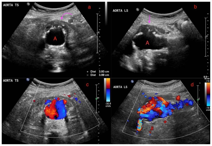
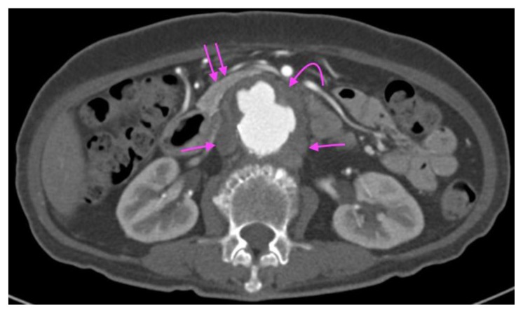
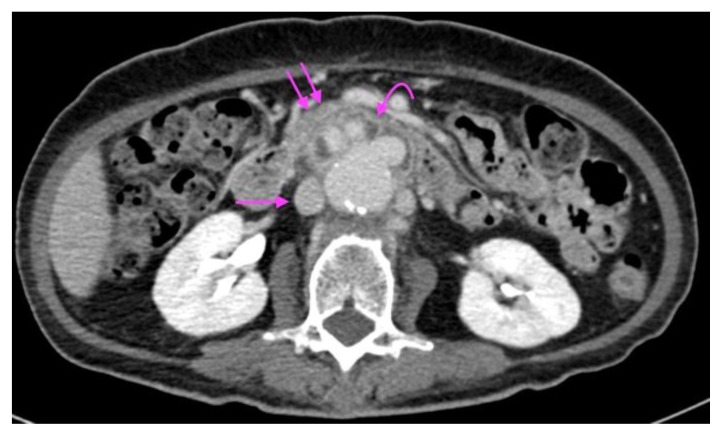
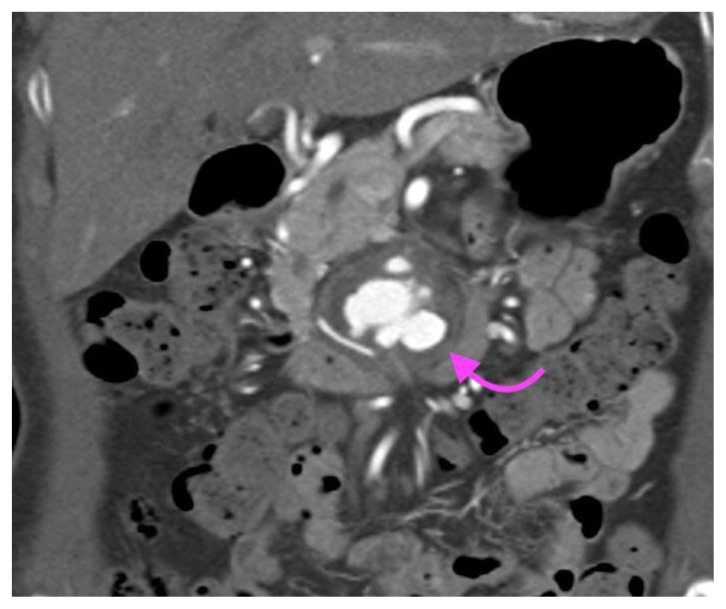
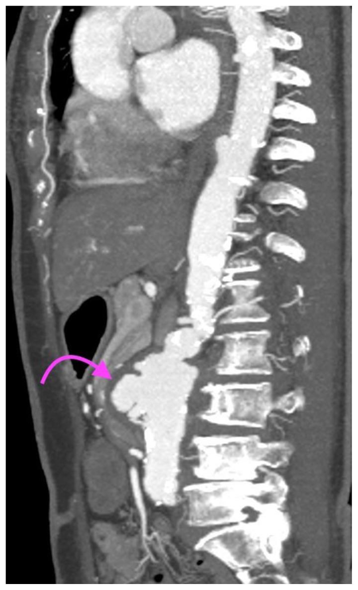
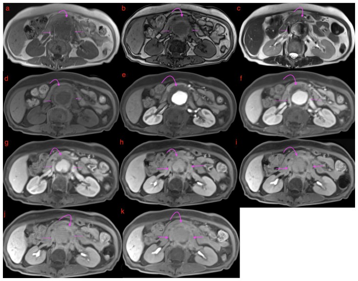
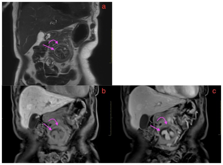
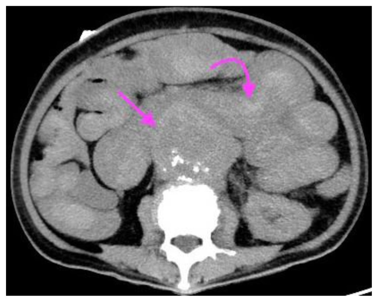
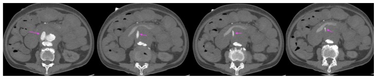
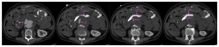
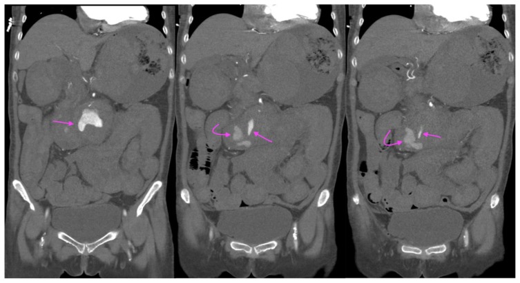
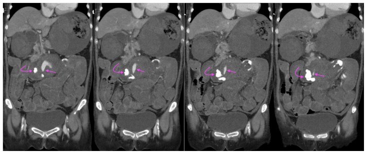
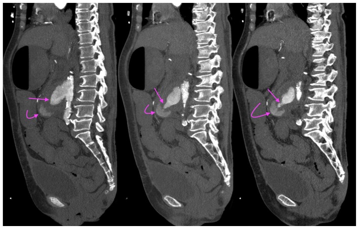
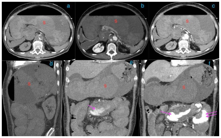
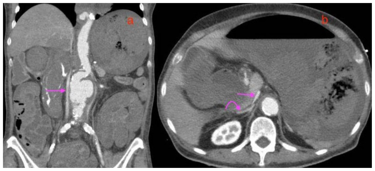
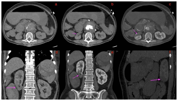
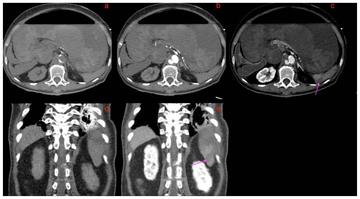
Primary aortoenteric fistulas are rare, with the annual incidence of such fistulas estimated to be 0.007 per million. The most common predisposing conditions for primary aortoenteric fistulas are atherosclerotic abdominal aortic aneurysms or penetrating atherosclerotic ulcers. We illustrate a rare case of an inflammatory aortic aneurysm causing a primary aortic fistula, with a direct fistulous jet from the aorta to the bowel with resultant catastrophic bleeding. In contrast to atherosclerotic aneurysms, most inflammatory aneurysms are symptomatic and show dense perianeurysmal fibrosis and periaortic wall thickening. A direct jet of contrast extravasation from the aorta into a bowel loop, while rarely seen, remains the most specific sign of a primary aorta-enteric fistula. A comprehensive literature review of the clinical presentation, imaging features, and differential diagnosis of a primary aortoenteric fistula are also discussed.

Keywords: Aortoenteric fistula; CT; MRI; abdomen; infectious aortitis; inflammatory abdominal aortic aneurysm; mycotic aneurysm; primary; radiotracer; secondary; ultrasound.


Figures

Figure 1
Figure 1
A 73-year-old lady with an inflammatory aortic aneurysm on initial ultrasound abdomen 7.5 months prior to presentation with an aortoenteric fistula and catastrophic hemorrhage. FINDINGS: 1(a) and 1(b) demonstrates a perianeurysmal cuff of hypoechoic soft tissue (curved arrow) around a fusiform abdominal aortic aneurysm (labelled A) with saccular outpouchings, measuring 3.9 × 4.0cm. 1(c) and 1(d) demonstrates turbulent flow within the abdominal aortic aneurysm with the “Pseudo Yin-Yang” sign. TECHNIQUE: Ultrasound of the abdominal aorta, low frequency 2–5 MHz curvilinear probe, 1(a) and 1(b) grey scale images, 1(c) and 1(d) color doppler images
Figure 2
Figure 2
A 73-year-old lady with an inflammatory aortic aneurysm on initial CT Aortogram 3.5 months from the initial ultrasound and 4 months prior to presentation with aortoenteric fistula and catastrophic hemorrhage. FINDINGS: Axial CT Aortogram in the arterial phase demonstrates a perianeurysmal cuff of fibrotic soft tissue (curved arrow) around a fusiform abdominal aortic aneurysm with saccular outpouchings and peripheral thrombi. A small bowel loop is seen tethered and draped over the fibrotic soft tissue (Double arrows). Incidental note of a dual inferior vena cava, one on each side of the posterior aspect of the abdominal aorta (Single arrows). TECHNIQUE: Axial CT Aortogram, arterial phase, 163 mAS, 100 KV, 3mm slice thickness, arterial phase, 75 ml Omnipaque 350
Figure 3
Figure 3
A 73-year-old lady with an inflammatory aortic aneurysm on initial CT Aortogram 4 months prior to presentation with aortoenteric fistula and catastrophic hemorrhage. FINDINGS: Axial CT Aortogram in the venous phase demonstrates a perianeurysmal cuff of fibrotic soft tissue (curved arrow) around a fusiform abdominal aortic aneurysm with saccular outpouchings and peripheral thrombi. A small bowel loop is seen tethered and draped over the fibrotic soft tissue (Double arrows). Incidental note of a dual inferior vena cava, one on each side of the posterior aspect of the abdominal aorta (single arrow). TECHNIQUE: Axial CT Aortogram, venous phase, 302 mAS, 100 KV, 3mm slice thickness, venous phase, 75 ml Omnipaque 350
Figure 4
Figure 4
A 73-year-old lady with an inflammatory aortic aneurysm on initial CT Aortogram 4 months prior to presentation with aortoenteric fistula and catastrophic hemorrhage. FINDINGS: Coronal slice of CT Aortogram in the arterial phase demonstrates a perianeurysmal cuff of fibrotic soft tissue (curved arrow) around a fusiform abdominal aortic aneurysm with saccular outpouchings. TECHNIQUE: CT Aortogram, coronal multiplanar reconstruction, 163 mAS, 100KV, 3mm slice thickness, arterial phase, 75 ml Omnipaque 350
Figure 5
Figure 5
A 73-year-old lady with an inflammatory aortic aneurysm on initial CT Aortogram 4 months prior to presentation with aortoenteric fistula and catastrophic hemorrhage. FINDINGS: Sagittal CT Aortogram in the arterial phase demonstrates a perianeurysmal cuff of fibrotic soft tissue (curved arrow) around a fusiform abdominal aortic aneurysm with saccular outpouchings. TECHNIQUE: CT Aortogram, sagittal MIP (maximum intensity projection), 10mm slice thickness, arterial phase, 163 mAS, 100 KV, 75 ml Omnipaque 350
Figure 6
Figure 6
A 73-year-old lady with an inflammatory aortic aneurysm depicted on MR liver (with primovist) performed for the purpose of further characterization for an incidental liver lesion picked up on CT aortogram performed earlier. FINDINGS: MRI liver with primovist shows aortic wall thickening and a cuff of perianeurysmal soft tissue (curved arrow) which shows intermediate T2 signal and progressive/persistent enhancement into the 20-minute phase with primovist. Double arrows point to dual inferior vena cava tethered to the inflammatory abdominal aortic aneurysm with a cleft like appearance. Note also elongation and draping of the duodenum over the inflammatory abdominal aortic aneurysm. TECHNIQUE: MRI liver with primovist, axial, 6(a)T1 Volumetric Interpolated Breath-hold Examination(VIBE) Dixon in phase (TR/TE 4.05/2.58, 3mm slice thickness), 6(b)T1 Volumetric Interpolated Breath-hold Examination(VIBE) Dixon oppose phase (TR/TE 4.05/1.35, 3mm slice thickness), 6(c)T2 Half-fourier-acquired single-shot turbo spin echo (HASTE) (TR/TE 1400/84, 5mm slice thickness), 6(d)T1 fat saturated pre contrast( TR/TE: 3.36/1.03, 3mm slice thickness), T1 dynavibe fat saturated with contrast at 6(e)1st, measurement(arterial phase), 6(f) 2nd measurement(venous phase), 6(g) 3rd measurement(equilibrium phase), 6(h) 5 minutes delayed, 6(i) 10 minutes delayed, 6(j) 15minutes delayed, 6(k) 20 minutes delayed ( TR/TE: 3.36/1.03, 3mm slice thickness, 9ml intravenous primovist).
Figure 7
Figure 7
A 73-year-old lady with an inflammatory aortic aneurysm depicted on coronal MRI liver (with primovist), performed for the purpose of further characterization for an incidental liver lesion picked up on CT aortogram performed earlier. FINDINGS: Coronal MRI liver with primovist shows a fusiform abdominal aortic aneurysm with saccular outpouchings, T2 hyperintense non enhancing peripheral thrombi(straight arrow), aortic wall thickening and a cuff of perianeurysmal soft tissue(curved arrow) which shows intermediate T2 signal and enhancement in the 7(b) 5 minutes delayed and 7(c) 20 minutes phase with primovist. TECHNIQUE: MR liver with primovist (a) Coronal oblique T2 HASTE (TR/TE 1800/101, slice thickness 3mm), Coronal T1 dynavibe fat saturated with contrast at 7(b) 5 minutes delayed and 7(c) 20 minutes delayed (TR/TE: 3.36/1.05, slice thickness 3mm, 9ml intravenous primovist)
Figure 8
Figure 8
A 73-year-old lady with an inflammatory abdominal aortic aneurysm causing a primary aortoenteric fistula with catastrophic hemorrhage. FINDINGS: Unenhanced axial CT mesenteric angiogram demonstrates hyperdense contents within the D3 segment of the duodenum compatible with acute blood products (curved arrow). Straight arrow points to the abdominal aortic aneurysm. TECHNIQUE: Axial CT mesenteric angiogram, plain, 138 mAS, 100 KV, 3mm slice thickness, nil contrast
Figure 9
Figure 9
A 73-year-old lady with an inflammatory abdominal aortic aneurysm causing a primary aortoenteric fistula with catastrophic hemorrhage. FINDINGS: axial CT mesenteric angiogram in the arterial phase demonstrates a jet of contrast extravasation from the abdominal aortic aneurysm to the D2/3 segment of the duodenum (straight arrow), with contrast filling of the duodenum. TECHNIQUE: Axial CT mesenteric angiogram, arterial phase,116 mAS, 100 KV, 3mm slice thickness, 75 ml of omnipaque 350
Figure 10
Figure 10
A 73-year-old lady with an inflammatory abdominal aortic aneurysm causing a primary aortoenteric fistula with catastrophic hemorrhage. FINDINGS: axial CT mesenteric angiogram in the venous phase demonstrates a jet of contrast extravasation from the abdominal aortic aneurysm to the D2/3 segment of the duodenum (straight arrow), with contrast accumulation within the duodenum (curved arrow). TECHNIQUE: Axial CT mesenteric angiogram, venous phase,116 mAS, 100 KV, 3mm slice thickness, 75 ml of omnipaque 350
Figure 11
Figure 11
A 73-year-old lady with an inflammatory abdominal aortic aneurysm causing a primary aortoenteric fistula with catastrophic hemorrhage. FINDINGS: CT mesenteric angiogram coronal multiplanar reconstruction in the arterial phase demonstrates a jet of contrast extravasation from the abdominal aortic aneurysm to the D2/3 segment of the duodenum (straight arrow), with contrast filling of the duodenum (curved arrow). TECHNIQUE: CT mesenteric angiogram, coronal multiplanar reconstruction, arterial phase, 116 mAS, 100 KV, 3mm slice thickness, 75 ml of omnipaque 350
Figure 12
Figure 12
A 73-year-old lady with an inflammatory abdominal aortic aneurysm causing a primary aortoenteric fistula. FINDINGS: CT mesenteric angiogram coronal multiplanar reconstruction in the venous phase demonstrates a jet of contrast extravasation from the abdominal aortic aneurysm to the D2/3 segment of the duodenum (straight arrow), with contrast accumulation within the duodenum (curved arrow). TECHNIQUE: CT mesenteric angiogram, coronal multiplanar reconstruction, venous phase, 116 mAS, 100 KV, 3mm slice thickness, 75 ml of omnipaque 350
Figure 13
Figure 13
A 73-year-old lady with an inflammatory abdominal aortic aneurysm causing a primary aortoenteric fistula with catastrophic hemorrhage. FINDINGS: CT mesenteric angiogram Sagittal maximum intensity projection (MIP) in the arterial phase demonstrates a jet of contrast extravasation from the abdominal aortic aneurysm to the D2/3 segment of the duodenum (straight arrow), with contrast accumulation within the duodenum (curved arrow). TECHNIQUE: CT mesenteric angiogram, sagittal, maximum intensity projection (MIP), arterial phase, 116 mAS, 100 KV, 3mm slice thickness, 75 ml of omnipaque 350
Figure 14
Figure 14
A 73-year-old lady with an inflammatory aortic aneurysm causing a primary aortoenteric fistula with catastrophic hemorrhage. FINDINGS: CT mesenteric angiogram in the axial 14(a)plain 14(b) arterial 14(c) venous phase and coronal multiplanar reconstruction in the 14(d) plain, 14(e) arterial, 14(d) venous phase. There is marked distension of the stomach (labelled S), with thin, poorly enhancing walls, suggestive of ischemia. Note hyperdense acute hematoma within the stomach in image 14(a) and 14(d), with contrast accumulation within the duodenum (single straight arrow) and jejunum (double straight arrows) in image 14(e) and 14(f). TECHNIQUE: CT mesenteric angiogram in the axial 14(a)plain 14(b) arterial 14(c) venous phase and coronal multiplanar reconstruction in the 14(d) plain, 14(e) arterial 14(d) venous phase, 116 mAS, 100 KV, 3mm slice thickness, 75 ml of omnipaque 350
Figure 15
Figure 15
A 73-year-old lady with an inflammatory abdominal aortic aneurysm causing a primary aortoenteric fistula with catastrophic hemorrhage, demonstrating the features of the CT hypoperfusion complex. FINDINGS: 15(a) Coronal multiplanar reconstruction CT mesenteric angiogram in the arterial phase and 15 (b) axial venous phase. Single straight arrows in 15(a) and 15(b) point to the collapsed slit like inferior vena cava and curved arrow in (b) points to the hyperenhancing right adrenal gland, features of the CT hypoperfusion complex. TECHNIQUE: CT mesenteric angiogram, 15(a) Coronal multiplanar reconstruction, arterial phase and 15(b) axial venous phase, 116 mAS, 100 KV, 3mm slice thickness, 75 ml of omnipaque 350
Figure 16
Figure 16
A 73-year-old lady with an inflammatory abdominal aortic aneurysm causing a primary aortoenteric fistula with catastrophic hemorrhage, demonstrating right lower renal pole infarct. FINDINGS: CT mesenteric angiogram in the axial 16(a) plain 16(b) arterial 16(c) venous phases, coronal multiplanar reconstruction in the 16(d) arterial and 16(b) venous phases, and sagittal maximum intensity projection in the 16(f)-arterial phase. Pink arrows in 16 b, c, d, e and f point to a wedge shaped right lower renal pole hypodensity representing an infarct. This is not well seen in the plain axial section of 16(a) TECHNIQUE: CT mesenteric angiogram in the axial 16(a)plain 16(b) arterial 16(c) venous phase, coronal multiplanar reconstruction in the 16(d) arterial 16(e) venous phase, sagittal maximum intensity projection in the 16(f) arterial phase, 116 mAS, 100 KV, 3mm slice thickness, 75 ml of omnipaque 350
Figure 17
Figure 17
A 73-year-old lady with an inflammatory abdominal aortic aneurysm causing a primary aortoenteric fistula with catastrophic hemorrhage, demonstrating a focal splenic infarct. FINDINGS: CT mesenteric angiogram in the axial 17(a) plain 17(b) arterial 17(c) venous phases, coronal multiplanar reconstruction in the 17(d) arterial and 17(e) venous phases. Pink arrows in 17(c) and 17(e) point to a wedge shaped hypodensity in the spleen representing an infarct. This is not well seen in the 17(a) axial plain phase, 17(b) axial arterial phase and 17(c) coronal arterial phase. TECHNIQUE: CT mesenteric angiogram in the axial 17(a)plain 17(b) arterial 17(c) venous phase, coronal multiplanar reconstruction in the 17(d) arterial 17(e) venous phase, 116 mAS, 100 KV, 3mm slice thickness, 75 ml of omnipaque 350

Similar articles

Cited by

References

    1. Restrepo CS, Ocazionez D, Suri R, Vargas D. Aortitis: imaging spectrum of the infectious and inflammatory conditions of the aorta. Radiographics. 2011 Mar-Apr;31(2):435–51. doi: 10.1148/rg.312105069.. - DOI - PubMed
    1. Tang T, Boyle JR, Dixon AK, Varty K. Inflammatory abdominal aortic aneurysms. Eur J Vasc Endovasc Surg. 2005 Apr;29(4):353–62. - PubMed
    1. Sintler M, Howell N, Mahmood A, Vohra RK. Ruptured inflammatory aortic aneurysm with aortoenteric fistula and infected with Streptococcus pneumoniae: a review of the literature. Indian J Surg. 2008 Jun;70(3):138–41. doi: 10.1007/s12262-008-0038-4. Epub 2008 Jul 24. - DOI - PMC - PubMed
    1. Rasmussen TE, Hallett JW., Jr Inflammatory aortic aneurysms. A clinical review with new perspectives in pathogenesis. Ann Surg. 1997 Feb;225(2):155–164. - PMC - PubMed
    1. Iijima M, Azuma R, Hieda T, Makino Y. A surgical case of inflammatory abdominal aortic aneurysm that responded remarkably to preoperative steroid therapy. J Surg Case Rep. 2018 Feb 20;2018(2):rjy020. doi: 10.1093/jscr/rjy020. eCollection 2018 Feb. - DOI - PMC - PubMed

MeSH terms

LinkOut - more resources