. 2020 Mar 6:8:e8408.
doi: 10.7717/peerj.8408. eCollection 2020.

Discrete analysis of camelid variable domains: sequences, structures, and in-silico structure prediction

Affiliations

Discrete analysis of camelid variable domains: sequences, structures, and in-silico structure prediction

Akhila Melarkode Vattekatte et al. PeerJ. .

Abstract

Antigen binding by antibodies requires precise orientation of the complementarity- determining region (CDR) loops in the variable domain to establish the correct contact surface. Members of the family Camelidae have a modified form of immunoglobulin gamma (IgG) with only heavy chains, called Heavy Chain only Antibodies (HCAb). Antigen binding in HCAbs is mediated by only three CDR loops from the single variable domain (VHH) at the N-terminus of each heavy chain. This feature of the VHH, along with their other important features, e.g., easy expression, small size, thermo-stability and hydrophilicity, made them promising candidates for therapeutics and diagnostics. Thus, to design better VHH domains, it is important to thoroughly understand their sequence and structure characteristics and relationship. In this study, sequence characteristics of VHH domains have been analysed in depth, along with their structural features using innovative approaches, namely a structural alphabet. An elaborate summary of various studies proposing structural models of VHH domains showed diversity in the algorithms used. Finally, a case study to elucidate the differences in structural models from single and multiple templates is presented. In this case study, along with the above-mentioned aspects of VHH, an exciting view of various factors in structure prediction of VHH, like template framework selection, is also discussed.

Keywords: Antibodies; Complementarity determining regions; Frameworks; Nanobodies; Secondary structure; Sequence structure relationship; Structural alphabet.


Conflict of interest statement

Frederic Cadet is associated with PEACCEL, Paris, France. Jean-Christophe Gelly and Alexandre G. de Brevern are associated with IBL, Paris, France. Jean-Philippe Meyneil is employed by ISoft, Paris, France. Alain Malpertuy is employed by Atragene, Paris, France. Nicolas K. Shinada is sponsored by Discngine, Paris, France and ANRT, France. All other authors declare no competing interests.

Figures

Figure 1
Figure 1. IgG and HCAb.
(A) IgG schematic representation with heavy chain (domains VH, CH1, CH2 and CH3) and light chain (domains VL and CL) and (B) Heavy Chain Only Antibodies (HCAbs), schematic representation with domains V HH, CH2 and CH3; (C) 2D representation of Immunoglobulin fold of V HH domain with demarcated Framework Regions (FRs) and Complementarity Determining Regions (CDRs), loop lengths are approximated for representation purposes. FR1 is composed of β-strands A1, A2 and B, FR2 is composed of β-strands C and C′, FR3 of 4 β-strands C″, D, E and F, FR4 end the V HH sequence by last β-strand, G. (D) 3D cartoon representation of VHH (PDB ID: 1BZQ chain K (Decanniere et al., 1999) and (E) 90° rotation of the same with the CDRs coloured.
Figure 2
Figure 2. Sequence conservation.
Sequence logo representation of multiple sequence alignment of complete dataset of VHH sequences. The relative frequency of amino acids at each position is shown here as sequence logo. The residues are colour coded according to their chemical properties. The residue positions are not in accordance with the numbering systems, sequence alignment creates a longer length than canonical VH.
Figure 3
Figure 3. Secondary structural profile of the 105 V HH domains.
The eight classes of DSSP elements are colour coded in the Figure legend. The symbols are E for extended conformation, (b-strand from b-sheet, T for hydrogen bond turn, S for bend, B for isolated b-bridge, G for 310-helix, H for a-helix, I for p-helix, and ‘-.’ for coils. The different β-strands that form the FRs are indicated above in red: from left to right are the β-strands A, A′, B, C, C′, C″, D, E, and F. The X-axis represents the numbering in the alignment.
Figure 4
Figure 4. Disulphide bridges of V HH domains with additional disulphide bonds.
(A) Conserved disulphide bond in type 1 V HH domains (B) Non conserved disulphide bonds in type 1 V HH domains, (C) Conserved disulphide bond in type 2 V HH domains and (D) non-conserved disulphide bonds in type 2 V HH domains. The disulphide bridges are indicated in yellow connecting any two cysteines.
Figure 5
Figure 5. Local conformational analysis of CDR clusters from PyIgClassify.
PB maps of CDR H1 region from V HH sequences from CDR H1 clusters (A) H1-13-1, (B) H1-13-3, and (C) H1-13-5, CDR H2 region from V HH sequences from (D) H2-9-1, (E) H2-10-1, and (F) H2-10-2 cluster, and Neq of (G) three CDR H1 clusters and (H) three CDR H2 clusters. The numbering of residues in each plot is according to the IMGT numbering system.
Figure 6
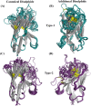
Figure 6. Analyses of structural template and best structural models.
First, the four templates Temp -m, Temp -l, Temp -a and Temp -h are superimposed. The colour coded regions in teal, pink, green and violet respectively are the CDRs. (A) Lateral view and (B) top view. Then, best models selected using DOPE score are superimposed. Four templates Temp-m, Temp-l, Temp-a, Temp-h and best structural model colour coded regions in teal, pink, green, violet and orange respectively are shown with different orientations. (A) Global view, (B) zoom on CDR1, FR1 and FR2, (C) on CDR2, FR2, and FR3, and (D) CDR3 with FR3 and FR4.
Figure 7
Figure 7. Positional PB entropy Neq and ΔPB.
(A) All five scenarios of modelling are represented in separate colours. X-axis represents residue positions and Y-axis represents Neq. (B) ΔPB between multi-template scenario and each mono template scenario.

Similar articles

Cited by

References

    1. Ablynx Ablynx 2016 Annual report 2016
    1. Adolf-Bryfogle J, Xu Q, North B, Lehmann A, Dunbrack Jr RL. PyIgClassify: a database of antibody CDR structural classifications. Nucleic Acids Research. 2015;43:D432–D438. doi: 10.1093/nar/gku1106. - DOI - PMC - PubMed
    1. Al-Lazikani B, Lesk AM, Chothia C. Standard conformations for the canonical structures of immunoglobulins 1 1Edited by I. A. Wilson. Journal of Molecular Biology. 1997;273:927–948. doi: 10.1006/jmbi.1997.1354. - DOI - PubMed
    1. Barnoud J, Santuz H, Craveur P, Joseph AP, Jallu V, de Brevern AG, Poulain P. PBxplore: a tool to analyze local protein structure and deformability with protein blocks. PeerJ. 2017;5:e4013. doi: 10.7717/peerj.4013. - DOI - PMC - PubMed
    1. Beard H, Cholleti A, Pearlman D, Sherman W, Loving KA. Applying physics-based scoring to calculate free energies of binding for single amino acid mutations in protein-protein complexes. PLOS ONE. 2013;8:e82849. doi: 10.1371/journal.pone.0082849. - DOI - PMC - PubMed

LinkOut - more resources