Albumin promotes proliferation of G1 arrested serum starved hepatocellular carcinoma cells
- PMID: 32185103
- PMCID: PMC7060934
- DOI: 10.7717/peerj.8568
Albumin promotes proliferation of G1 arrested serum starved hepatocellular carcinoma cells
Abstract
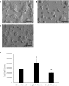
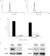
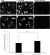
Albumin is the most abundant plasma protein and functions as a transport molecule that continuously interacts with various cell types. Because of these properties, albumin has been exploited by the pharmaceutical industry to improve drug delivery into target cells. The immediate effects of albumin on cells, however, require further understanding. The cell interacting properties and pharmaceutical applications of albumin incentivises continual research into the immediate effects of albumin on cells. The HepG2/C3A hepatocellular carcinoma cell line is used as a model for studying cancer pathology as well as liver biosynthesis and cellular responses to drugs. Here we investigated the direct effect of purified albumin on HepG2/C3A cell proliferation in the absence of serum, growth factors and other serum originating albumin bound molecules. We observed that the reduced cell counts in serum starved HepG2/C3A cultures were increased by the inclusion of albumin. Cell cycle analysis demonstrated that the percentage of cells in G1 phase during serum starvation was reduced from 86.4 ± 2.3% to 78.3 ± 3.2% by the inclusion of albumin whereas the percentage of cells in S phase was increased from 6.5 ± 1.5% to 14.3 ± 3.6%. A significant reduction in the cell cycle inhibitor protein, P21, accompanied the changes in the proportions of cell cycle phases upon treatment with albumin. We have also observed that the levels of dead cells determined by DNA fragmentation and membrane permeabilization caused by serum starvation (TUNEL: 16.6 ± 7.2%, ethidium bromide: 13.8 ± 4.8%) were not significantly altered by the inclusion of albumin (11.6 ± 10.2%, ethidium bromide: 16.9 ± 8.9%). Therefore, the increase in cell number was mainly caused by albumin promoting proliferation rather than protection against cell death. These primary findings demonstrate that albumin has immediate effects on HepG2/C3A hepatocellular carcinoma cells. These effects should be taken into consideration when studying the effects of albumin bound drugs or pathological ligands bound to albumin on HepG2/C3A cells.
Keywords: Albumin; Cell cycle; Cyclin d1; Ethidium bromide staining; HepG2/C3A cells; Hepatocellular carcinoma; Proliferation; TUNEL assay; Western blot; p21.
©2020 Ibrahim et al.
Conflict of interest statement
The authors declare there are no competing interests.
Figures


Similar articles
-
A novel all-trans retinoic acid derivative 4-amino‑2‑trifluoromethyl-phenyl retinate inhibits the proliferation of human hepatocellular carcinoma HepG2 cells by inducing G0/G1 cell cycle arrest and apoptosis via upregulation of p53 and ASPP1 and downregulation of iASPP.Oncol Rep. 2016 Jul;36(1):333-41. doi: 10.3892/or.2016.4795. Epub 2016 May 9. Oncol Rep. 2016. PMID: 27177208
-
Emodin-induced apoptosis through p53-dependent pathway in human hepatoma cells.Life Sci. 2004 Mar 19;74(18):2279-90. doi: 10.1016/j.lfs.2003.09.060. Life Sci. 2004. PMID: 14987952
-
Involvement of down-regulation of Cdk2 activity in hepatocyte growth factor-induced cell cycle arrest at G1 in the human hepatocellular carcinoma cell line HepG2.J Biochem. 2004 Nov;136(5):701-9. doi: 10.1093/jb/mvh177. J Biochem. 2004. PMID: 15632311
-
Cyproheptadine, an antihistaminic drug, inhibits proliferation of hepatocellular carcinoma cells by blocking cell cycle progression through the activation of P38 MAP kinase.BMC Cancer. 2015 Mar 17;15:134. doi: 10.1186/s12885-015-1137-9. BMC Cancer. 2015. PMID: 25886177 Free PMC article.
-
SC-III3, a novel scopoletin derivative, induces cytotoxicity in hepatocellular cancer cells through oxidative DNA damage and ataxia telangiectasia-mutated nuclear protein kinase activation.BMC Cancer. 2014 Dec 19;14:987. doi: 10.1186/1471-2407-14-987. BMC Cancer. 2014. PMID: 25527123 Free PMC article.
Cited by
-
Prognostic value of preoperative systemic immune-inflammation index/albumin for patients with hepatocellular carcinoma undergoing curative resection.World J Gastroenterol. 2024 Dec 28;30(48):5130-5151. doi: 10.3748/wjg.v30.i48.5130. World J Gastroenterol. 2024. PMID: 39735268 Free PMC article.
-
Albumin promotes the progression of fibroblasts through late G1 into S-phase in the absence of growth factors.Cell Cycle. 2020 Sep;19(17):2158-2167. doi: 10.1080/15384101.2020.1795999. Epub 2020 Jul 26. Cell Cycle. 2020. PMID: 32715871 Free PMC article.
-
Inhibition of vitamin D analog eldecalcitol on hepatoma in vitro and in vivo.Open Med (Wars). 2020 Jul 13;15(1):663-671. doi: 10.1515/med-2020-0137. eCollection 2020. Open Med (Wars). 2020. PMID: 33336024 Free PMC article.
References
-
- Chen RF. Removal of fatty acids from serum albumin by charcoal treatment. The Journal of Biological Chemistry. 1967;242:173–181. - PubMed
LinkOut - more resources
Full Text Sources
Research Materials

