

Fixation Methods for Mandibular Advancement and Their Effects on Temporomandibular Joint: A Finite Element Analysis Study
- PMID: 32185199
- PMCID: PMC7060428
- DOI: 10.1155/2020/2810763
Fixation Methods for Mandibular Advancement and Their Effects on Temporomandibular Joint: A Finite Element Analysis Study
Abstract
Objectives: Bilateral sagittal split osteotomy (BSSO) is a common surgical procedure to correct dentofacial deformities that involve the mandible. Usually bicortical bone fixation screw or miniplates with monocortical bone fixation screw were used to gain stability after BSSO. On the other hand, the use of resorbable screw materials had been reported. In this study, our aim is to determine first stress distribution values at the temporomandibular joint (TMJ) and second displacement amounts of each mandibular bone segment.
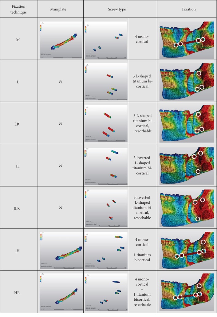
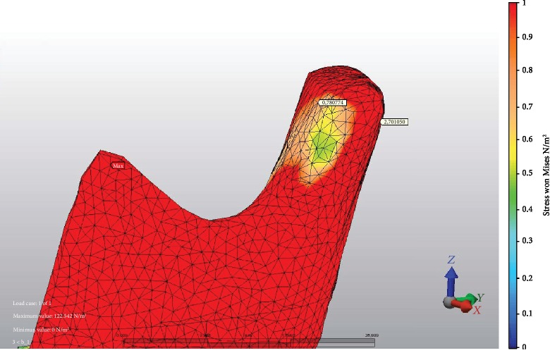
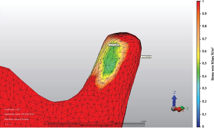
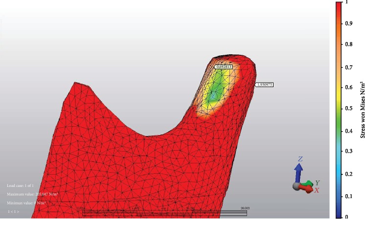
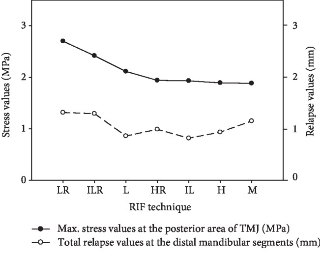
Methods: A three-dimensional virtual mesh model of the mandible was constructed. Then, BSSO with 9 mm advancement was simulated using the finite element model (FEM). Fixation between each mandibular segment was also virtually performed using seven different combinations of fixation materials, as follows: miniplate only (M), miniplate and a titanium bicortical bone fixation screw (H), miniplate and a resorbable bicortical bone fixation screw (HR), 3 L-shaped titanium bicortical bone fixation screws (L), 3 L-shaped resorbable bicortical bone fixation screws (LR), 3 inverted L-shaped titanium bicortical bone fixation screws (IL), and 3 inverted L-shaped resorbable bicortical bone fixation screws (ILR).
Results: At 9 mm advancement, the biggest stress values at the anterior area TMJ was seen at M fixation and LR fixation at posterior TMJ. The minimum stress values on anterior TMJ were seen at L fixation and M fixation at posterior TMJ. Minimum displacement was seen in IL method. It was followed by L, H, HR, M, ILR, and LR, respectively.
Conclusion: According to our results, bicortical screw fixation was associated with more stress on the condyle. In terms of total stress value, especially LR and ILR lead to higher amounts.
Copyright © 2020 Sabit Demircan et al.
Conflict of interest statement
The authors report no financial or other conflict of interest.
Figures
References
-
- Sato F. R., Asprino L., Noritomi P. Y., da Silva J. V., de Moraes M. Comparison of five different fixation techniques of sagittal split ramus osteotomy using three-dimensional finite elements analysis. International Journal of Oral and Maxillofacial Surgery. 2012;41(8):934–941. doi: 10.1016/j.ijom.2012.03.018. - DOI - PubMed
-
- Verweij J. P., Houppermans P. N., Mensink G., van Merkesteyn J. Removal of bicortical screws and other osteosynthesis material that caused symptoms after bilateral sagittal split osteotomy: a retrospective study of 251 patients, and review of published papers. The British Journal of Oral & Maxillofacial Surgery. 2014;52(8):756–760. doi: 10.1016/j.bjoms.2014.05.017. - DOI - PubMed
MeSH terms
Substances
LinkOut - more resources
Full Text Sources
