Review
. 2020 Jun;23(2):207-215.
doi: 10.1007/s40477-020-00447-w. Epub 2020 Mar 17.

S-Detect characterization of focal breast lesions according to the US BI RADS lexicon: a pictorial essay

Affiliations
Review

S-Detect characterization of focal breast lesions according to the US BI RADS lexicon: a pictorial essay

Tommaso Vincenzo Bartolotta et al. J Ultrasound. 2020 Jun.

Abstract

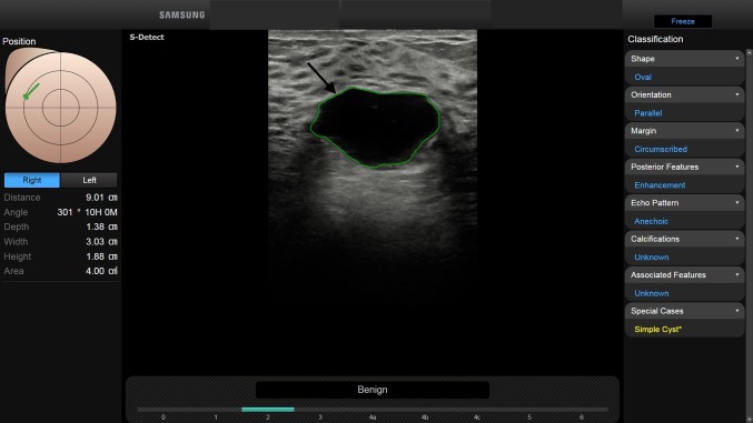
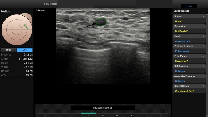
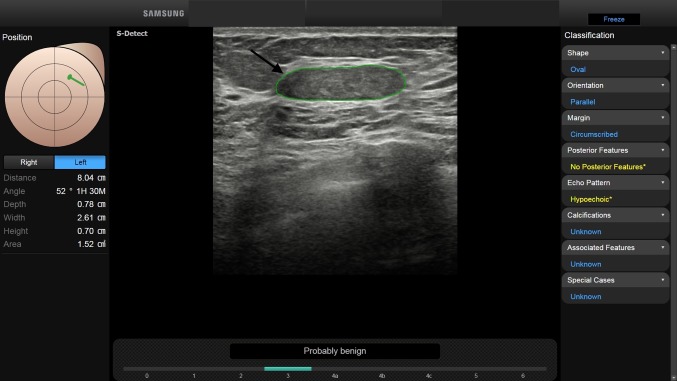
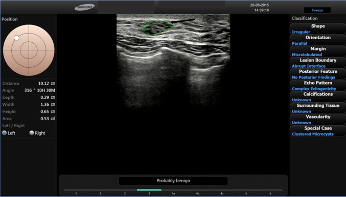
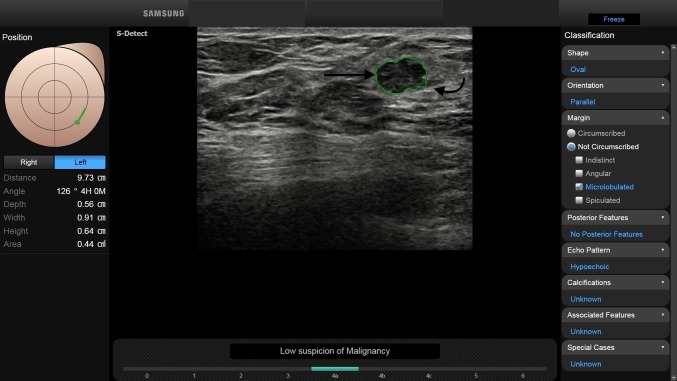
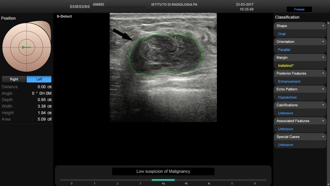
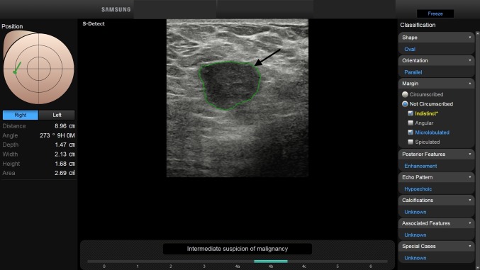
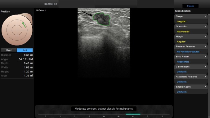
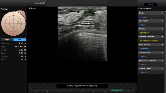
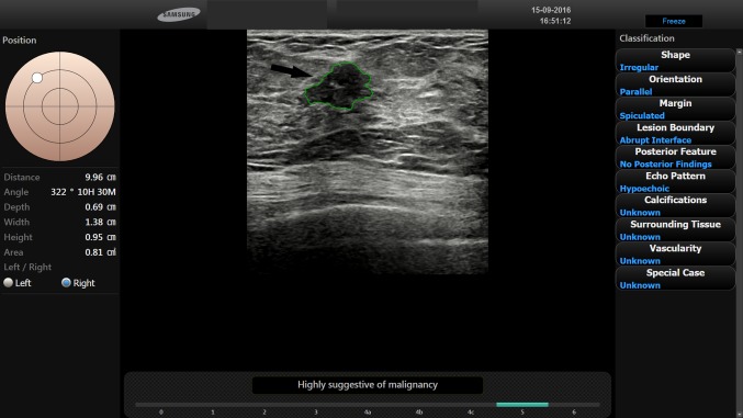
High-resolution ultrasonography (US) is a valuable tool in breast imaging. Nevertheless, US is an operator-dependent technique: to overcome this issue, the American College of Radiology (ACR) has developed the breast imaging-reporting and data system (BI-RADS) US lexicon. Despite this effort, the variability in the assessment of focal breast lesions (FBLs) with the use of BI-RADS US lexicon is still an issue. Within this framework, evidence shows that computer-aided image analysis may be effective in improving the radiologist's assessment of FBLs. In particular, S-Detect is a newly developed image-analytic computer program that provides assistance in morphologic analysis of FBLs seen on US according to the BI-RADS US lexicon. This pictorial essay describes state-of-the-art of sonographic characterization of FBLs by using S-Detect.

Keywords: BI-RADS; Breast neoplasms; Computer-assisted diagnosis; Decision-making; Problem-solving; Ultrasonography.


Conflict of interest statement

Tommaso Vincenzo Bartolotta is lecturer and scientific advisor for Samsung. Other authors declare that they have no conflict of interest.

Figures

Fig. 1
Fig. 1
BI-RADS 2. In a 28-year-old-woman, with palpable right breast lump, B-mode US depicts a 3 cm FBL. According to BI-RADS lexicon, with S-Detect (green line contour) this FBL was classified as oval-shaped, with parallel orientation and circumscribed margins (arrow). The echo pattern was judged anechoic, with enhancement as posterior feature. Final diagnosis: simple cyst
Fig. 2
Fig. 2
BI-RADS 3. In a 35-year-old-woman, B-mode US shows a 6 mm FBL. According to BI-RADS lexicon, with S-Detect (green line contour) this FBL was assessed as round-shaped, with not parallel orientation and circumscribed margins (arrow). The echo pattern was assessed as hypoechoic, with enhancement as posterior feature. Final diagnosis: complicated cyst
Fig. 3
Fig. 3
BI-RADS 3. In a 32-year-old-woman, with palpable right breast lump, B-mode US depicted a 2.6 cm FBL. According to BI-RADS lexicon, with S-Detect (green line contour) this FBL was classified as oval-shaped, with parallel orientation and circumscribed margins (arrow). The echo pattern was assessed as hypoechoic, with no posterior features. Final diagnosis: fibroadenoma
Fig. 4
Fig. 4
BI-RADS 3. In a 38-year-old-woman, B-mode US showed a 1.3 cm FBL in her left breast. According to BI-RADS lexicon, with S-Detect (green line contour) this FBL was classified as irregular-shaped, with parallel orientation and microlobulated margins. The echo pattern was assessed as complex echogenicity, with no posterior features. Final diagnosis: clustered microcysts
Fig. 5
Fig. 5
BI-RADS 4a. In a 40-year-old-woman, with a history of multiple bilateral fibroadenomas and a new palpable left breast lump, B-mode US showed a 9 mm FBL. According to BI-RADS lexicon, with S-Detect (green line contour) this FBL was evaluated as an oval-shaped hypoechoic mass with parallel orientation (arrow). Margins were partially circumscribed (< 75%) and partially microlobulated (curved arrow). Core-needle biopsy proved the mass to be a fibroadenoma
Fig. 6
Fig. 6
BI-RADS 4a. In a 47-year-old-woman, with a palpable left breast lump, B-mode US showed a 3.4 cm FBL. According to BI-RADS, with S-Detect (green line contour) this FBL was described as an oval-shaped hypoechoic mass with parallel orientation and enhancement as posterior feature (arrow). Margins were partially circumscribed (< 75%) and partially indistinct. Core-needle biopsy proved the mass to be a mucinous carcinoma
Fig. 7
Fig. 7
BI-RADS 4b. In a 43-year-old-woman, with a palpable left breast lump, B-mode US depicted a 2 cm FBL. According to BI-RADS lexicon, with S-Detect (green line contour) this FBL was assessed as an oval-shaped hypoechoic mass with parallel orientation and enhancement as posterior feature (arrow). Margins were not circumscribed, indistinct and microlobulated. Core-needle biopsy proved the mass to be a medullary breast carcinoma
Fig. 8
Fig. 8
BI-RADS 4c. In a 50-year-old-woman, with a palpable left breast lump, B-mode US depicted a 1.6 cm FBL. According to BI-RADS lexicon, with S-Detect (green line contour) this FBL was judged as an irregular-shaped hypoechoic mass with angular margins, not parallel orientation and no posterior features (arrow). Core-needle biopsy proved the mass to be an invasive ductal breast carcinoma
Fig. 9
Fig. 9
BI-RADS 5. In a 41-year-old-woman, with a palpable left breast lump, B-mode US showed a 1.3 cm FBL. According to BI-RADS lexicon, with S-Detect (green line contour) this FBL was classified as an irregular-shaped hypoechoic mass with spiculated margins, not parallel orientation and no posterior features (arrow). Core-needle biopsy proved the mass to be an invasive ductal breast carcinoma.
Fig. 10
Fig. 10
BI-RADS 5. In a 55-year-old-woman, with a palpable left breast lump, B-mode US depicted a 1.4 cm FBL. According to BI-RADS lexicon, with S-Detect (green line contour) this FBL was judged as an irregular-shaped hypoechoic mass with spiculated margins, parallel orientation and no posterior features (arrow). Core-needle biopsy proved the mass to be an invasive ductal breast carcinoma

References

    1. Hooley RJ, Scoutt LM, Philpotts LE. Breast ultrasonography: state of the art. Radiology. 2013;268(3):642–659. doi: 10.1148/radiol.13121606. - DOI - PubMed
    1. Bartolotta TV, Ienzi R, Cirino A, Genova C, Ienzi F, Pitarresi D, Safina E, Midiri M. Characterisation of indeterminate focal breast lesions on grey-scale ultrasound: role of ultrasound elastography. Radiol Med. 2011;116(7):1027–1038. doi: 10.1007/s11547-011-0648-y. - DOI - PubMed
    1. Mendelson EB, Böhm-Vélez M, Berg WA, et al. ACR BI-RADS® atlas, breast imaging reporting and data system. Reston, VA: American College of Radiology; 2013. ACR BI-RADS® urltrasound.
    1. Hong AS, Rosen EL, Soo MS, Baker JA. BI-RADS for sonography: positive and negative predictive values of sonographic features. Am J Roentgenol. 2005;184(4):1260–1265. doi: 10.2214/ajr.184.4.01841260. - DOI - PubMed
    1. Raza S, Goldkamp AL, Chikarmane SA, Birdwell RL. US of breast masses categorized as BI-RADS 3, 4, and 5: pictorial review of factors influencing clinical management. Radiographics. 2010;30(5):1199–1213. doi: 10.1148/rg.305095144. - DOI - PubMed

MeSH terms