Multicenter Study
. 2020 Oct;20(10):2768-2780.
doi: 10.1111/ajt.15863. Epub 2020 Apr 26.

Discovery of non-HLA antibodies associated with cardiac allograft rejection and development and validation of a non-HLA antigen multiplex panel: From bench to bedside

Affiliations
Multicenter Study

Discovery of non-HLA antibodies associated with cardiac allograft rejection and development and validation of a non-HLA antigen multiplex panel: From bench to bedside

Carrie L Butler et al. Am J Transplant. 2020 Oct.

Abstract

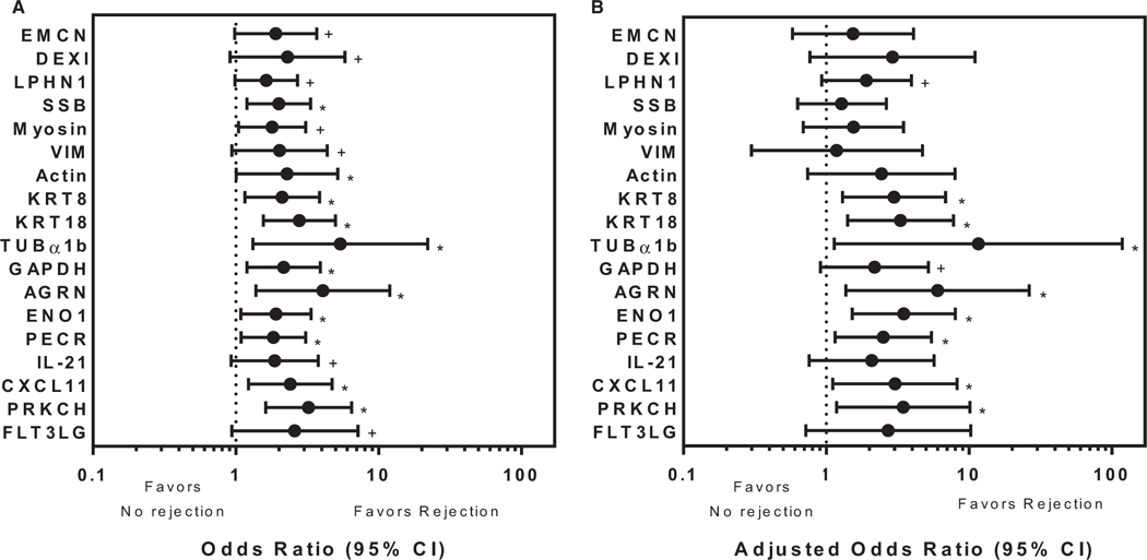
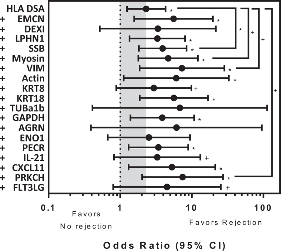
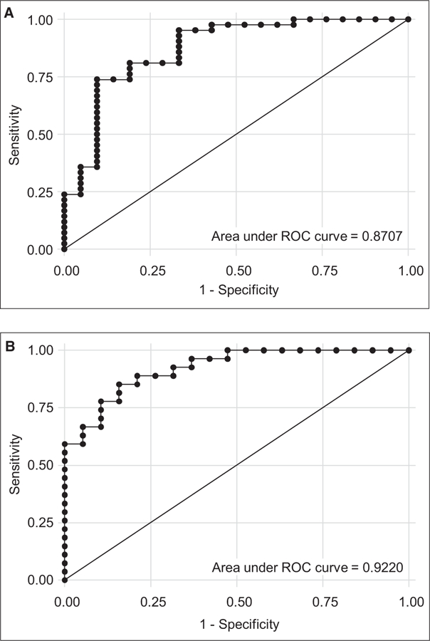
We analyzed humoral immune responses to nonhuman leukocyte antigen (HLA) after cardiac transplantation to identify antibodies associated with allograft rejection. Protein microarray identified 366 non-HLA antibodies (>1.5 fold, P < .5) from a discovery cohort of HLA antibody-negative, endothelial cell crossmatch-positive sera obtained from 12 cardiac allograft recipients at the time of biopsy-proven rejection. From these, 19 plasma membrane proteins and 10 autoantigens identified from gene ontology analysis were combined with 48 proteins identified through literature search to generate a multiplex bead array. Longitudinal sera from a multicenter cohort of adult cardiac allograft recipients (samples: n = 477 no rejection; n = 69 rejection) identified 18 non-HLA antibodies associated with rejection (P < .1) including 4 newly identified non-HLA antigenic targets (DEXI, EMCN, LPHN1, and SSB). CART analysis showed 5/18 non-HLA antibodies distinguished rejection vs nonrejection. Antibodies to 4/18 non-HLA antigens synergize with HLA donor-specific antibodies and significantly increase the odds of rejection (P < .1). The non-HLA panel was validated using an independent adult cardiac transplant cohort (n = 21 no rejection; n = 42 rejection, >1R) with an area under the curve of 0.87 (P < .05) with 92.86% sensitivity and 66.67% specificity. We conclude that multiplex bead array assessment of non-HLA antibodies identifies cardiac transplant recipients at risk of rejection.

Keywords: autoantibody; autoantigen; clinical research/practice; heart transplantation/cardiology; histocompatibility; immunogenetics; microarray/protein array; organ transplantation in general; rejection; translational research/science.


Conflict of interest statement

DISCLOSURE

The authors of this manuscript have conflicts of interest to disclose as described by the American Journal of Transplantation.

Figures

FIGURE 1
FIGURE 1
Non-HLA antibody discovery and generation of a high throughput multiplex bead set. Non-HLA antibodies associated with cardiac allograft rejection were identified by protein microarray analysis (9000 full length proteins) using sera from 12 heart transplant patients with biopsy proven rejection in the absence of HLA donor specific antibodies (DSA), but with positive endothelial cell crossmatch (ECXM) (n = 12). 366 proteins binding non-HLA antigens were identified (>1.5 fold increase, P < .05) compared to control sera from rejection negative, DSA negative, and ECXM negative heart transplant patients (n = 6). Of the 366, gene ontology analysis identified 22 plasma membrane proteins and 10 autoantigens. Nineteen of these, with an additional 48 proteins identified by literature search as associated with solid organ rejection, were conjugated to polystyrene beads to generate a multiplex panel of non-HLA proteins for downstream high throughput testing of non-HLA antibodies
FIGURE 2
FIGURE 2
Non-HLA antibodies significantly associated with cardiac allograft rejection. High throughput multiplex bead analysis was used to identify non-HLA antibodies in sera from a multicenter adult cardiac transplant cohort. A, Antibodies to 18 non-HLA antigens (y-axis) were identified to be significantly associated with rejection with an odds ratio >1 (x-axis; n = 69 rejection sera acute-cellular rejection [ACR] ≥ 1b, n = 477 nonrejection sera). Four of these, endomucin, dexamethasone-induced transcript, latrophilin 1 and Sjogren syndrome antigen B, are newly described in relationship to transplant rejection. Bars represent the 95% confidence interval (CI). The odds ratios for antibody binding to the remaining non-HLA antigens on the multiplex panel that were not significantly different from 1 are not shown. +Denotes P < .1, *Denotes P < .05. B, Odds ratios (x-axis) of the 18 non-HLA antibodies identified in A were adjusted to remove the effects of HLA donor specific antibodies using a random effects logistic regression (n = 26 rejection sera ACR ≥ 1b, n = 106 nonrejection sera). Antibodies to 10/18 non-HLA antigens are significantly associated with rejection with odds ratio >1. Bars represent the 95% CI. The odds ratios for antibody binding to the remaining 49 non-HLA antigens not significantly different from 1 are not shown. +Denotes P < .1, *Denotes P < .05
FIGURE 3
FIGURE 3
Heart allograft rejection is associated with an increased number of non-HLA antibodies. The number of non-HLA antibodies identified by multiplex analysis is significantly increased (P < .001) in sera obtained at the time of rejection in comparison to nonrejection. The median number of non-HLA antibodies identified from rejection samples (n = 69) is 3 (interquartile range: 1–5) in comparison to 1 (interquartile range: 1–3) in nonrejection samples (n = 477). Horizontal bar indicates interquartile range. Black circle indicates median
FIGURE 4
FIGURE 4
Correlation matrix analysis showing hierarchical clustering of non-HLA antibodies in cardiac allograft rejection sera. The matrix describes correlation of non-HLA antibodies found in sera of rejection patients. Non-HLA antibodies associated with cardiac allograft rejection (n = 69/546) selectively cluster into 9 groups (red boxes). The coefficient of correlation (scale bar at right) is indicated by the dot color and size and describes the tendency that the non-HLA antibodies will be identified independently or together in the sera of rejection patients. Dark blue and large dots along the diagonal represent a marker’s perfect correlation with itself. Correlation to other non-HLA antibodies is evaluated off the diagonal and indicated by the dot color and size (smaller dot size and lighter color indicates lower correlation; blue color indicates positive correlation). Three of the 4 non-HLA antibodies that are newly identified in this study (Sjogren syndrome antigen B, endomucin [EMCN], and latrophilin 1) are independently associated with rejection and do not group with other non-HLA antibodies. In comparison, non-HLA antibodies to 11 other antibodies cluster together and are not considered independently informative. At left, solid boxes indicate non-HLA antigens newly identified in this study; antigens identified in the CART analysis (shown in Figure 5) are indicated by dotted line boxes. EMCN is boxed in both a solid and dotted line designating it as newly identified, and identified in the CART analysis
FIGURE 5
FIGURE 5
Classification algorithm identifying non-HLA antibodies that predict cardiac allograft rejection. Classification and regression tree (CART) analysis showing a binary decision tree that assesses the classification of rejection based on non-HLA median fluorescence intensity (MFI) in sera isolated from cardiac allograft transplant patients (n = 546 sera). The algorithm selects MFI values which optimize the differentiation of the sera into rejection and nonrejection categories at each node in the tree. For display, selected MFI’s were rounded to the nearest 100. The root node, KRT18, that includes all 546 sera, 13% of which are rejection samples (69/546 = 0.13) splits at an MFI > 1500 into child nodes. As the algorithm progresses to terminal nodes, 86% (n = 467) of nonrejection samples are correctly identified (far left). Sera samples with KRT18 MFI > 1500 strongly correlate to cardiac allograft rejection (far right). KRT18 appears in the model twice as nodes are split recursively in decision tree construction. Scale bar indicates association with rejection with lighter terminal node boxes correlating to nonrejection
FIGURE 6
FIGURE 6
Synergy of non-HLA antibodies and HLA donor specific antibodies (DSA) in cardiac allograft rejection. Logistic regression models (using 546 samples) showing the odds ratio (x-axis) of HLA DSA alone or HLA DSA plus each non-HLA antibody (y-axis) in comparison to no antibodies. The gray shaded box highlights the significant association of cardiac allograft rejection when HLA DSA alone is present (HLA DSA, odds ratio = 2.31, P < .05). The odds ratio of 14 of the 18 non-HLA antibodies initially identified as significantly associated with cardiac allograft rejection retain significance in this model (*P < .05, +P < .1). When conditions are compared, the odds ratio of HLA DSA with non-HLA antibody raised against Sjogren syndrome antigen B, Myosin, vimentin and PRKCH with cardiac allograft rejection are significantly increased in comparison to HLA DSA alone suggesting synergistic interactions between the 2 types of antibodies (brackets, *P < .05, +P < .1). Bars represent the 95% confidence interval (CI)
FIGURE 7
FIGURE 7
Validation of the panel of 18 non-HLA antibodies for the prediction of cardiac allograft rejection using an independent single-center adult cardiac transplant cohort. A, ROC analysis identified an area under the curve (AUC) of 0.87 and sensitivity and specificity of 92.86% and 66.67%, respectively (P < .05) in an independent cohort of cardiac transplant recipients (n = 63 patients, 21 with no rejection and 42 with rejection). B, After removing those patients with HLA donor-specific antibodies (DSAs), ROC analysis identified an AUC of 0.92 and sensitivity and specificity of 88.89% and 68.42%, respectively (P < .05) in the independent cohort of cardiac transplant recipients (n = 46 patients without HLA DSAs, 19 with no rejection and 27 with rejection)
FIGURE 8
FIGURE 8
Non-HLA antigenic targets are expressed in human tissues and endothelial cells. A, mRNA expression of the 18 non-HLA antibody antigens associated with cardiac allograft rejection from publically available data of the 18 non-HLA antibodies in cardiac, kidney, lung, and liver tissue as well as HUVEC. B, Expression of Sjogren syndrome antigen B (SSB) and endomucin (EMCN) on cardiac biopsies. Immunohistochemistry of biopsies taken at the time of rejection shows expression of SSB and EMCN proteins on cardiac endothelium. Micrographs are representative of biopsies tested from 3 cardiac transplant recipients from the multicenter cohort. SSB is a nuclear protein expressed in endothelial cell (EC) microvasculature. EMCN is expressed in the membrane and cytoplasm of some EC in the microvasculature and in all cardio myocytes

Comment in

Similar articles

Cited by

References

    1. Tonsho M, Michel S, Ahmed Z, Alessandrini A, Madsen JC. Heart transplantation: challenges facing the field. Cold Spring Harb Perspect Med. 2014;4(5):pii:a015636. - PMC - PubMed
    1. Stehlik J, Edwards LB, Kucheryavaya AY, et al. The Registry of the International Society for Heart and Lung Transplantation: 29th official adult heart transplant report–2012. J Heart Lung Transplant. 2012;31(10):1052–1064. - PubMed
    1. Colvin-Adams M, Smith JM, Heubner BM, et al. OPTN/SRTR 2013 annual data report: heart. Am J Transplant. 2015;15(Suppl 2):1–28. - PubMed
    1. Tible M, Loupy A, Vernerey D, et al. Pathologic classification of antibody-mediated rejection correlates with donor-specific antibodies and endothelial cell activation. J Heart Lung Transplant. 2013;32(8):769–776. - PubMed
    1. Tait BD, Süsal C, Gebel HM, et al. Consensus guidelines on the testing and clinical management issues associated with HLA and non-HLA antibodies in transplantation. Transplantation. 2013;95(1):19–47. - PubMed

Publication types