. 2020 Apr;22(4):e13177.
doi: 10.1111/cmi.13177.

Tracking the cargo of extracellular symbionts into host tissues with correlated electron microscopy and nanoscale secondary ion mass spectrometry imaging

Affiliations

Tracking the cargo of extracellular symbionts into host tissues with correlated electron microscopy and nanoscale secondary ion mass spectrometry imaging

Stephanie K Cohen et al. Cell Microbiol. 2020 Apr.

Abstract

Extracellular bacterial symbionts communicate biochemically with their hosts to establish niches that foster the partnership. Using quantitative ion microprobe isotopic imaging (nanoscale secondary ion mass spectrometry [NanoSIMS]), we surveyed localization of 15 N-labelled molecules produced by the bacterium Vibrio fischeri within the cells of the symbiotic organ of its host, the Hawaiian bobtail squid, and compared that with either labelled non-specific species or amino acids. In all cases, two areas of the organ's epithelia were significantly more 15 N enriched: (a) surface ciliated cells, where environmental symbionts are recruited, and (b) the organ's crypts, where the symbiont population resides in the host. Label enrichment in all cases was strongest inside host cell nuclei, preferentially in the euchromatin regions and the nucleoli. This permissiveness demonstrated that uptake of biomolecules is a general mechanism of the epithelia, but the specific responses to V. fischeri cells recruited to the organ's surface are due to some property exclusive to this species. Similarly, in the organ's deeper crypts, the host responds to common bacterial products that only the specific symbiont can present in that location. The application of NanoSIMS allows the discovery of such distinct modes of downstream signalling dependent on location within the host and provides a unique opportunity to study the microbiogeographical patterns of symbiotic dialogue.

Keywords: 15N-labeled bacteria; host-microbe communication; squid-vibrio.


Conflict of interest statement

CONFLICT OF INTEREST

The authors declare no conflict of interest.

Figures

Figure 1
Figure 1
Relevant morphology, anatomy, and ultrastructure of the host, Euprymna scolopes. (A) A diagram of a ventral dissection revealing the mantle cavity with relevant tissues and organs that were examined in this study. br, brain; dg, digestive gland; lo, light organ; mc, mantle cavity; st, statolith; w, white body. (B) A diagram of the bilateral light organ. Relevant surface features (left half) include the juvenile-specific ciliated epithelium with two appendages, anterior (aa) and posterior (pa), and the field of cilia surrounding three pores. Relevant interior features include the three independent regions of symbiont migration and residence. ante, antechamber diverticulum; bs, blood sinus; cr, crypt; du, duct; endo, endothelial cells lining the blood sinus; hc, hemocytes; po, pores. (C) A TEM with symbiont-containing crypt region of the light organ, false colored to correspond to colored regions in (B). cr 1–3, crypts 1–3; ce, crypt epithelium; ct, connective tissue; is, ink sac; ref, reflector.
Figure 2
Figure 2
Uptake of isotope-labeled V. fischeri products into light-organ epithelial cells of a juvenile host. (A) 15N-enrichment (expressed as δ15N) in the light organ of a newly-hatched animal at 2 h post-exposure to 106 intact 15N-labeled V. fischeri cells/ml of seawater; the data were obtained from all nuclei in the mosaic, independent of enrichment level. A composite TEM micrograph and the corresponding mosaic of 65 individual, high-resolution NanoSIMS images. Labeling was abundant and appeared as hotspots in the nucleoli of all light-organ epithelia (e.g., yellow arrows) with which V. fischeri cells interface, i.e., the surface ciliated field, here showing the epithelia of the anterior appendage (ap), the pore (po), the duct/antechamber (du/ante) and crypts (crypt epithelia, ce 1–3); ct, connective tissue; ref, reflector. Inset, upper left, the average level of 15N-enrichment in cell nuclei in different epithelial regions. Data were derived from the mosaic (Ap, n=56; Po, n=58; Du, n=36; Cr, n=142). Lower case letters indicate statistically significant differences between the light-organ compartments (Kruskal-Wallis test with Nemenyi post hoc, P<0.0001). Error bars represent 1 standard deviation. (B) 15N-labeled V. fischeri cells migrating through the ducts. (C, C’) TEM (left) and NanoSIMS (right) images of two cells of the crypt epithelium, defining the region scanned (start – end) for 15N/14N ratio within the nucleus, showing strong enrichment in the nucleoli (green arrows) and euchromatin (eu); ht, heterochromatin. C’, NanoSIMS at two sensitivities (main, lower and inset higher sensitivity, respectively) to highlight less intense labeling of the chromatin. (D) A representative image of the quantification of 15N enrichment through the region scanned in (C). Points are averages of NanoSIMS scans (n=10) +/− one standard deviation.
Figure 3
Figure 3
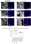
The 15N-enrichments in tissues not directly associating with the symbiont. Nuclei and nucleoli (hotspots) in light organ non-epithelial tissues (A-C) and in epithelial tissues in other squid organs (D-F) after 3.5 h inoculation with 15N-labelled V. fischeri cells. Left panels show tissue structure (NanoSIMS 12C14N image). Right panels show corresponding, quantified NanoSIMS 15N/14N ratio image. (G) Representative 15N-enrichments in nuclei of a single animal (excluding the nucleolus when visible) in different tissues across the body. Non-epithelial tissue inside the light organ (l.o.; n=72): connective tissue (n=59), and tissues outside of the light organ: gill epithelia (n=34), blood vessel endothelial cells (n=48), gut epithelia (n=100), digestive gland cells (n=31), brain (n=16); retina (r; n=11), tentacles (n=29), white body (n=27), and epithelium supporting statolith (n=27). Only enriched tissues were considered in the statistical analysis. Lower case letters indicate statistically significant differences between the different tissues (Kruskal-Wallis test with Nemenyi post hoc, P<0.0001). Points are averages +/− one standard deviation. Dashed line, limit of detection, or no difference between the control value for the natural distribution of 15N over 14N in the sample.
Figure 4
Figure 4
15N-enrichment (δ15N) in juvenile tissues with 15N-labeled non-symbiotic Vibrionaceae cells. Animals were analyzed at 3.5 h post inoculation. (A/B) Left panels, tissue structure (NanoSIMS 12C14N image); right panels, corresponding quantified image of NanoSIMS 15N/14N ratio; nucleoli, green arrows. Ap, anterior appendage of the light organ. (C) Average 15N enrichment in the nucleus (n = 236, V. campbellii; n=196, P. leiognathi) and nucleolus (n = 73, V. campbellii; n=57, P. leiognathi) of the light-organ surface epithelium from 5–7 individual animals. Points are average +/− one standard deviation. Lower case letters indicate statistically significant differences between the light-organ compartments (Kruskal-Wallis test with Nemenyi post hoc, P<0.0001).
Figure 5
Figure 5
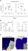
15N enrichment of light organ regions upon exposure to outer membrane vesicles (OMVs). (A) Relative Labeling Index (RLI) for 15N-enrichment in light organs exposed for different time periods. Left, to intact V. fischeri cells for 2 h (n=3) or 3.5 h (n=6); 6–10 nucleoli/region for each animal; right, to purified OMVs for 1 h (n=5) or 3 h (n=4). Ap, anterior appendage (Cells: n=37, 2h; n=90, 3.5 h; OMVs: n=44, 1h; n=48, 3 h); Po, pore (Cells: n=44, 2 h; n=54, 3.5h; OMVS: n=55, 1h; n=42, 3 h); Cr, crypt (cells: n=80, 2 h; n=73, 3.5 h; OMVs: n=65, 1h; n=44, 3 h). Points are averages +/− one standard deviation. Lower case letters indicate statistically significant differences between the light-organ compartments (Two-way ANOVA with Tukey post hoc, P<0.0001). (B) Quantification of the distribution of 15N-enrichment in subcellular regions of the anterior appendage and crypt in animals exposed to the OMVs of V. fischeri (n=3), V. campbellii (n=5), and E. coli (n=3). For each animal, cytoplasm n =31–64; nucleus n= 101–373; nucleolus n=829–868. Points are averages +/− one standard deviation. Lower case letters indicate significant differences between the cell organelles (Kruskal-Wallis test with Nemenyi post hoc, P<0.0001); asterisks indicate significant differences between bacteria species (Kruskal-Wallis test, P<0.0001). (C) A comparison of 15N enrichment in epithelium of the superficial anterior appendage of the light organ between animals exposed to labeled OMVs from V. fischeri or V. campbellii. Images of NanoSIMS 15N/14N ratios; inset, corresponding mosaic TEM micrographs; nucleoli, green arrows.

Similar articles

Cited by

References

    1. Altura MA, Heath-Heckman EA, Gillette A, Kremer N, Krachler AM, Brennan C, … McFall-Ngai MJ (2013). The first engagement of partners in the Euprymna scolopes-Vibrio fischeri symbiosis is a two-step process initiated by a few environmental symbiont cells. Environmental Microbiology, 15, 2937–2950. doi: 10.1111/1462-2920.12179. - DOI - PMC - PubMed
    1. Aschtgen MS, Lynch JB, Koch E, Schwartzman J, McFall-Ngai M, & Ruby E (2016a). Rotation of Vibrio fischeri flagella produces outer membrane vesicles that induce host development. Journal of Bacteriology, 198, 2156–2165. doi: 10.1128/JB.00101-16. - DOI - PMC - PubMed
    1. Aschtgen MS, Wetzel K, Goldman W, McFall-Ngai M, & Ruby E (2016b). Vibrio fischeri- derived outer membrane vesicles trigger host development. Cellular Microbiology, 18, 488–499. doi: 10.1111/cmi.12525. - DOI - PMC - PubMed
    1. Berleman J, & Auer M (2013). The role of bacterial outer membrane vesicles for intra- and interspecies delivery. Environmental Microbiology, 15, 347–354. doi: 10.1111/1462-2920.12048. - DOI - PubMed
    1. Berry D, & Loy A (2018). Stable-isotope probing of human and animal microbiome function. Trends in Microbiology, 26, 999–1007. doi: 10.1016/j.tim.2018.06.004. - DOI - PMC - PubMed

Publication types