. 2020 Jun;45(6):1851-1863.
doi: 10.3892/ijmm.2020.4545. Epub 2020 Mar 17.

Long non‑coding RNA EBLN3P promotes the recovery of the function of impaired spiral ganglion neurons by competitively binding to miR‑204‑5p and regulating TMPRSS3 expression

Affiliations

Long non‑coding RNA EBLN3P promotes the recovery of the function of impaired spiral ganglion neurons by competitively binding to miR‑204‑5p and regulating TMPRSS3 expression

Wenqi Jiang et al. Int J Mol Med. 2020 Jun.

Abstract

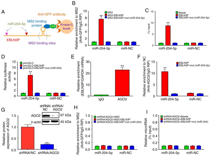
Sensorineural hearing loss (SNHL) is one of the major leading causes of hearing impairment, and is typically characterized by the degeneration of spiral ganglion neurons (SGNs). In previous studies by the authors, it was demonstrated that microRNA (miRNA or miR)‑204‑5p decreased the viability of SGNs by inhibiting the expression of transmembrane protease, serine 3 (TMPRSS3), which was closely associated with the development of SGNs. However, the upstream regulatory mechanism of miR‑204‑5p was not fully elucidated. The present study found that an important upstream regulatory factor of miR‑204‑5p, long non‑coding RNA (lncRNA) EBLN3P, was expressed at low levels in impaired SGNs, whereas it was expressed at high levels in normal SGNs. Mechanistic analyses demonstrated that lncRNA EBLN3P functioned as a competing endogenous RNA (ceRNA) when regulating miR‑204‑5p in normal SGNs. In addition, lncRNA EBLN3P regulated TMPRSS3 expression via the regulation of miR‑204‑5p in normal SGNs. In vitro functional analysis revealed that lncRNA EBLN3P promoted the recovery of the viability of normal SGNs and inhibited the apoptosis of normal SGNs. Finally, the results revealed a recovery‑promoting effect of lncRNA EBLN3P on the structure and function of impaired SGNs in models of deafness. On the whole, the findings of the present study demonstrate that lncRNA EBLN3P promotes the recovery of the function of impaired SGNs by competitively binding to miR‑204‑5p and regulating TMPRSS3 expression. This suggests that lncRNA EBLN3P may be a potential therapeutic target for diseases involving SNHL.


Figures

Figure 1
Figure 1
Expression of lncRNA EBLN3P and miR-204-5p in models of deafness. (A) Schematic representation of the predicted binding sites of miR-204-5p in the lncRNA EBLN3P transcript according to online prediction software (www.biostars.org). (B) Schematic representation of models of deafness. (C) Estimation of acoustic sensibility in mice. The green and red dots represent the mean of threshold shift to the 8-kHz stimuli before and after the injection of kanamycin and saline at days 0, 3, 7, 10 and 14. *P<0.05 (Student's t-test), vs. control saline. (D) SGNs were dissected from P4-P6 mouse cochlea by trypsinization. (E and F) Fluorescence in situ hybridization was performed to detect the expression of lncRNA EBLN3P and miR-204-5p in primary SGNs in a model of deafness. (G and H) RT-qPCR was performed to detect the expression of lncRNA EBLN3P and miR-204-5p in primary SGNs from the mice in the model of deafness. lncRNA, long non-coding RNA; miR, microRNA; SGN, spiral ganglion neuron. **P<0.01 (Student's t-test), vs. controls.
Figure 2
Figure 2
lncRNA EBLN3P functions as a competing endogenous RNA by regulating miR-204-5p in normal SGNs. (A and B) MS2-RIP assay followed by RT-qPCR to detect endogenous miRNAs associated tightly with lncRNA EBLN3P. (C) Cell lysates of normal SGNs were incubated with biotin-labeled EBLN3P. After pull-down, the miRNAs were separated and assayed by RT-qPCR. (D) Luciferase activities in normal SGNs cells co-transfected with miR-204-5p and empty luciferase reporters, lncRNA EBLN3P wild transcript or lncRNA EBLN3P mutant transcript. Data are presented as the relative ratio of Firefly luciferase activity to Renilla luciferase activity. (E) RIP assay of the binding between lncRNA EBLN3P and AGO2 using control IgG and AGO2 special antibody. lncRNA EBLN3P and GAPDH expression was quantified using RT-qPCR, and analyzed as enrichment in RNA-binding protein RIP in contrast to control IgG RIP. (F) Anti-AGO2 RIP was performed in SGNs that were transiently transfected with miR-204-5p mimics, followed by RT-qPCR to detect lncRNA EBLN3P or lncRNA EBLN3P-mut (miR-204-5p) associated with AGO2. (G) After silencing AGO2 in normal SGNs for 48 h, the protein expression level of AGO2 was assessed by western blotting. (H) After silencing the AGO2 gene in normal SGNs for 48 h, MS2-RIP analysis followed by RT-qPCR was performed to detect miR-204-5p endogenously associated with lncRNA EBLN3P. (I) After silencing AGO2 in normal SGNs for 48 h, the cells were lysed, and the cell lysates were incubated with biotin-labeled lncRNA EBLN3P. After pull-down, the whole miRNAs were extracted, and miR-204-5p was measured by RT-qPCR. **P<0.01 (ANOVA with Tukey's post hot-test). lncRNA, long non-coding RNA; miR, microRNA; miRNA, microRNA; SGN, spiral ganglion neuron; RIP, RNA immunoprecipitation; RIP, RNA immunoprecipitation; AGO2, argonaute 2.
Figure 3
Figure 3
lncRNA EBLN3P regulates the expression of TMPRSS3 through miR-204-5p in normal SGNs. (A) RT-qPCR was performed to detect the effects of lncRNA EBLN3P knockdown on the expression of EBLN3P in primary normal SGNs. (B) Luciferase activity was evaluated to observe the effect of lncRNA EBLN3P knockdown on the psiCHECK-2/TMPRSS3 wild-type 3′-UTR, psiCHECK-2/TMPRSS3 mutated 3′-UTR and empty 3′-UTR vectors (control). (C) RT-qPCR was performed to detect the effects of lncRNA EBLN3P knockdown on the mRNA expression of TMPRSS3 in primary normal SGNs. (D) Western blot analysis was performed to detect the effects of lncRNA EBLN3P knockdown on the protein expression of TMPRSS3 in primary normal SGNs. (E) RT-qPCR was performed to detect the effects of lncRNA EBLN3P overexpression on the expression of EBLN3P in primary normal SGNs. (F) Normal SGNs were treated with over-NC, over-lncRNA EBLN3P or anti-miR-204-5p + over-lncRNA EBLN3P, and the luciferase activity was evaluated. (G-I) Normal SGNs were treated with control over-NC, over-lncRNA EBLN3P or anti-miR-204-5p + over-lncRNA EBLN3P. RT-qPCR and western blot analysis was performed to detect the mRNA and protein expression of TMPRSS3 in primary normal SGNs. **P<0.01 (Student's t-test) vs. NCs in panels A, B, C, D and E. **P<0.01 (ANOVA with Tukey's post hoc test) in panels F, G and I. lncRNA, long non-coding RNA; SGN, spiral ganglion neuron; TMPRSS3, transmembrane protease, serine 3; UTR, untranslated region; NC, negative control; miR, microRNA.
Figure 4
Figure 4
lncRNA EBLN3P promotes the viability and inhibits the apoptosis of normal SGNs. (A) Normal SGNs were treated with lncRNA EBLN3P shRNA and negative control shRNA via transfection with Lipofectamine 3000. SGN cell viability was determined by MTT assay. (B) Cell survival of SGNs was measured by Trypan blue staining in SGNs treated with lncRNA EBLN3P shRNA and negative control shRNA. (C) Necrosis of SGNs was determined by detection of lactate dehydrogenase release to the medium in SGNs treated with lncRNA EBLN3P shRNA and negative control shRNA. (D) Flow cytometry was performed to observe the effect of lncRNA EBLN3P knockdown on the apoptosis of normal SGNs cells. (E and F) Western blot analysis and quantitative analysis were performed to observe the effect of lncRNA EBLN3P knockdown on the levels of cleaved caspase-3 in normal SGNs. *P<0.05 and **P<0.01 (Student's t-test) vs. negative controls. lncRNA, long non-coding RNA; SGN, spiral ganglion neuron; shRNA, small hairpin RNA.
Figure 5
Figure 5
Recovery-promoting effect of Ad-EBLN3P on the structure and function of impaired organ of Corti and spiral ganglion. (A) Auditory brainstem response test was performed to evaluate the function of the organ of Corti and spiral ganglion in deafness models. Colored dots show the mean of the threshold shift to the 8-kHz stimuli before and after injection of Ad-EBLN3P in deafness models. (B) Hematoxylin and eosin staining was applied to observe the recovering effect of long non-coding RNA EBLN3P on the structure of impaired organ of Corti and spiral ganglions. (C and D) Immunohistochemistry and quantitative analysis were performed to observe the levels of β-tubulin in cochlear spiral ganglion neurons. β-tubulin was used as an early marker for neurons. **P<0.01 (Student's t-test) vs. model.
Figure 6
Figure 6
Expression of lncRNA EBLN3P, TMPRSS3 and NT-3 in the cochlear SGNs of deafness models. (A) Fluorescence in situ hybridization was performed to observe the expression of lncRNA EBLN3P in the cochlear SGNs of deafness models. (B and C) IHC and quantitative analysis were performed to observe the expression of TMPRSS3 in the cochlear SGNs of deafness models. (D and E) IHC and quantitative analysis were performed to observe the expression of NT-3, an important growth factor that helps stimulate and control neurogenesis, in the cochlear SGNs models of deafness. **P<0.01 (Student's t-test) vs. model. lncRNA, long non-coding RNA; SGN, spiral ganglion neuron; TMPRSS3, transmembrane protease, serine 3; NT-3, neurotrophin-3; IHC, immunohistochemistry.
Figure 7
Figure 7
Schematic model of lncRNA EBLN3P functioning as a competing endogenous RNA for miR-204-5p and regulating the expression of TMPRSS3. miR-204-5p could bind to the 3′-untranslated region of TMPRSS3 to inhibit the expression of TMPRSS3. lncRNA EBLN3P could bind to miR-204-5p at two sites, '314-322' and '973-981', in the 1,200-nt gene fragment at the 5′-end of EBLN3P, and block the regulatory function of miR-204-5p on TMPRSS3 expression. lncRNA, long non-coding RNA; miR, microRNA; TMPRSS3, transmembrane protease, serine 3.

Similar articles

Cited by

References

    1. Charizopoulou N, Lelli A, Schraders M, Ray K, Hildebrand MS, Ramesh A, Srisailapathy CR, Oostrik J, Admiraal RJ, Neely HR, et al. Gipc3 mutations associated with audiogenic seizures and sensorineural hearing loss in mouse and human. Nat Commun. 2011;2:201. doi: 10.1038/ncomms1200. - DOI - PMC - PubMed
    1. Clark GM. Cochlear Implants: Fundamentals and applications Graeme Clark. Springer; New York: 2003. - DOI
    1. Feghali JG, Lefebvre PP, Staecker H, Kopke R, Frenz DA, Malgrange B, Liu W, Moonen G, Ruben RJ, Van de Water TR. Mammalian auditory hair cell regeneration/repair and protection: A review and future directions. Ear Nose. Throat J. 1998;77:276–280. 282–285. doi: 10.1177/014556139807700409. - DOI - PubMed
    1. Hardie NA, Shepherd RK. Sensorineural hearing loss during development: Morphological and physiological response of the cochlea and auditory brainstem. Hear Res. 1999;128:147–165. doi: 10.1016/S0378-5955(98)00209-3. - DOI - PubMed
    1. Dodson HC, Mohuiddin A. Response of spiral ganglion neurones to cochlear hair cell destruction in the guinea pig. J Neurocytol. 2000;29:525–537. doi: 10.1023/A:1007201913730. - DOI - PubMed