. 2020 Apr 24;83(4):1249-1257.
doi: 10.1021/acs.jnatprod.0c00038. Epub 2020 Mar 18.

Boholamide A, an APD-Class, Hypoxia-Selective Cyclodepsipeptide

Affiliations

Boholamide A, an APD-Class, Hypoxia-Selective Cyclodepsipeptide

Joshua P Torres et al. J Nat Prod. .

Abstract

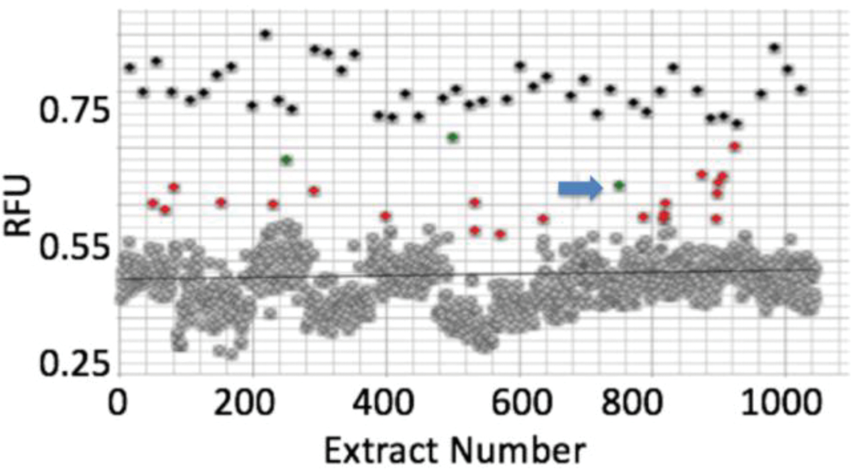
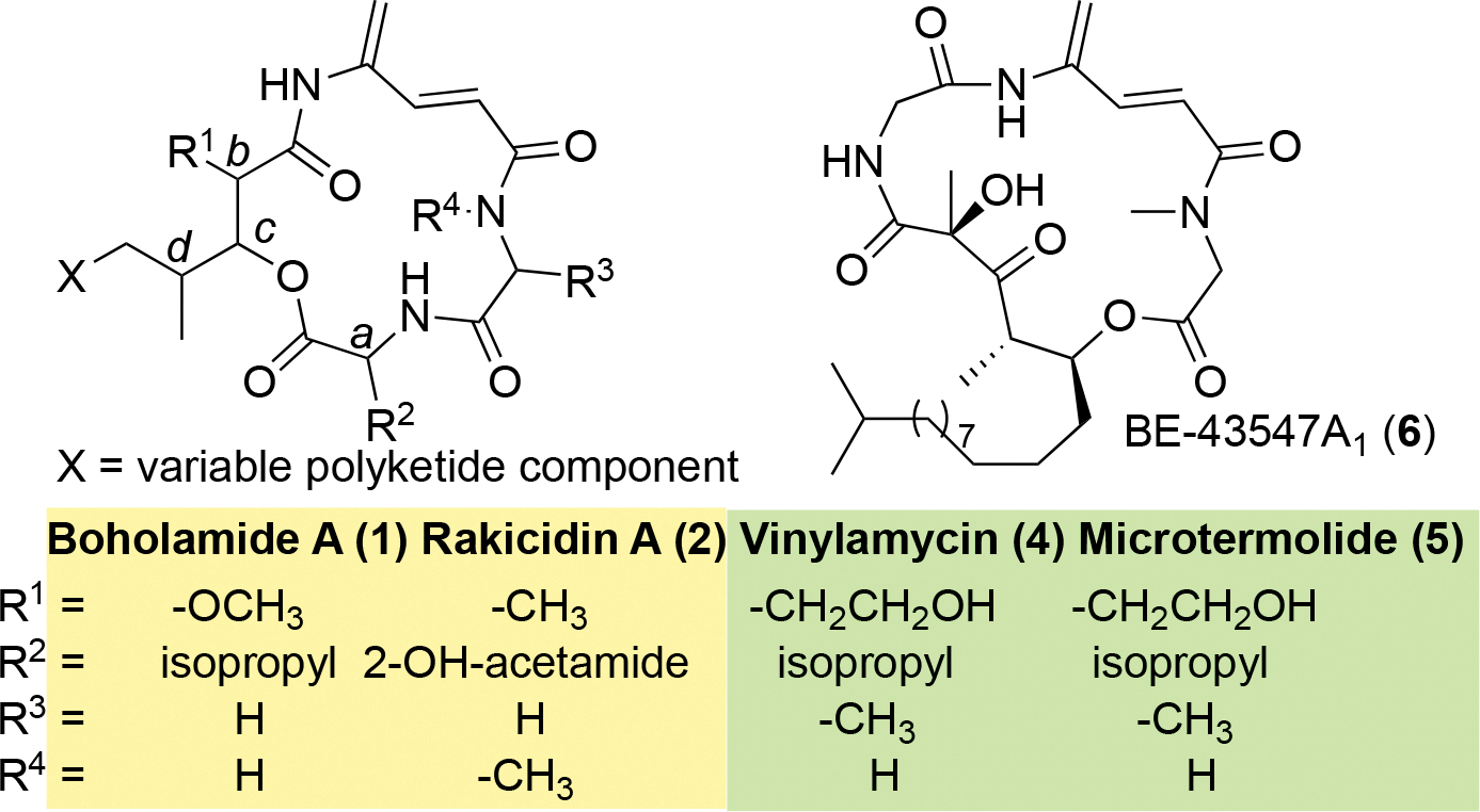
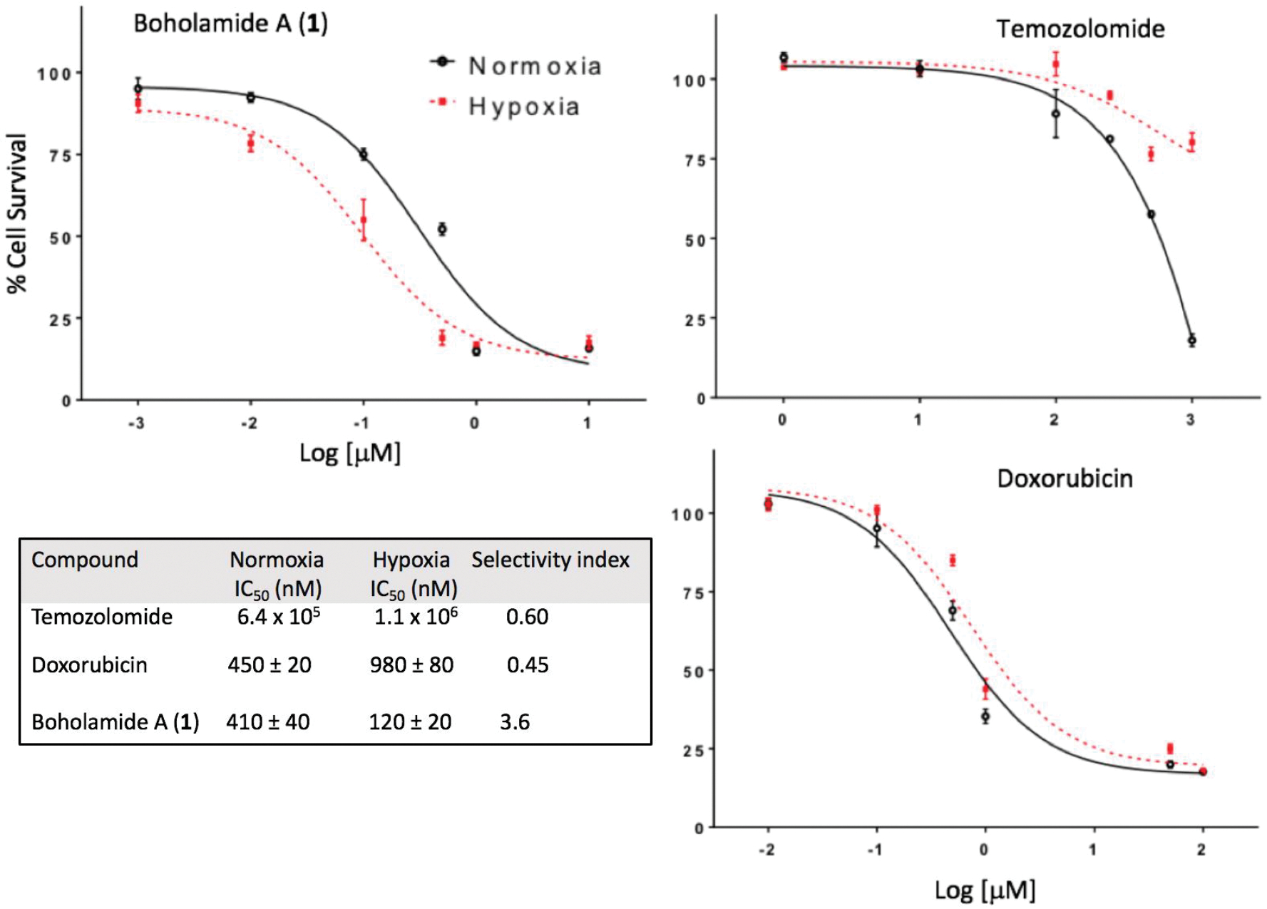
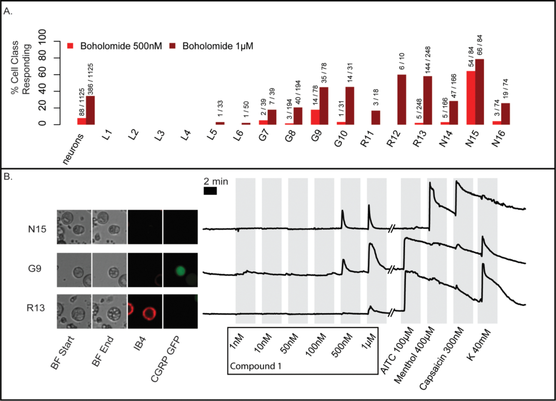
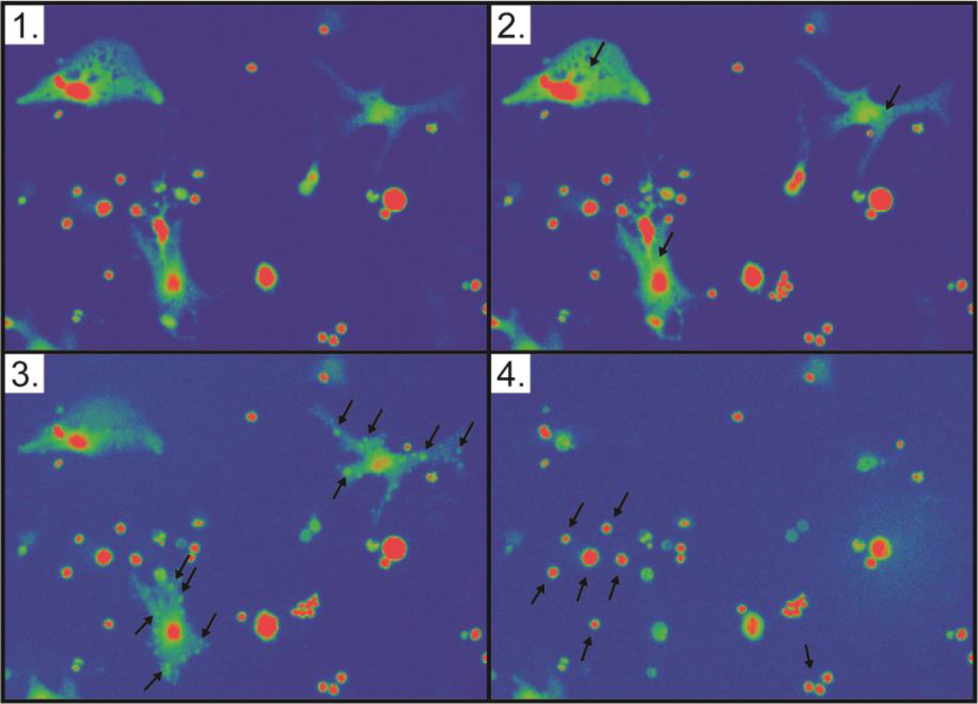
Calcium homeostasis is implicated in some cancers, leading to the possibility that selective control of calcium might lead to new cancer drugs. On the basis of this idea, we designed an assay using a glioblastoma cell line and screened a collection of 1000 unique bacterial extracts. Isolation of the active compound from a hit extract led to the identification of boholamide A (1), a 4-amido-2,4-pentadieneoate (APD)-class peptide. Boholamide A (1) applied in the nanomolar range induces an immediate influx of Ca2+ in glioblastoma and neuronal cells. APD-class natural products are hypoxia-selective cytotoxins that primarily target mitochondria. Like other APD-containing compounds, 1 is hypoxia selective. Since APD natural products have received significant interest as potential chemotherapeutic agents, 1 provides a novel APD scaffold for the development of new anticancer compounds.


Figures

Figure 1.
Figure 1.
Discovery of boholamide A (1) as a Ca2+-perturbing agent. Ca2+ imaging assay results for a subset of ~1,000 bacterial extracts using glioblastoma U87MG cells. Relative fluorescence units (RFU) were measured for each extract using a Ca2+-selective dye. Black points are from positive controls (detergent lysis of cells), while gray points are inactive extracts that cluster around a black bar indicating mean activity. Red and green dots are active extracts, with extract 3158H.R.1.a.03 indicated by a blue arrow.
Figure 2.
Figure 2.
Key HMBC, ROESY and COSY correlations of 1.
Figure 3.
Figure 3.
Key ROESY correlations of the HDMN in compounds 3a/3b.
Figure 4.
Figure 4.
Comparative structures of APD compounds. The yellow box indicates compounds with the configuration abcd = SSRS, while the green box indicates the enantiomeric abcd = RRSR.
Figure 6.
Figure 6.
Hypoxia-selective cytotoxicity of boholamide A (1) against U87MG glioblastoma cells. 1 is compared with standard glioblastoma treatments doxorubicin and temozolomide, showing that 1 is hypoxia-selective, whereas the standards are less cytotoxic to hypoxic cells.
Figure 7.
Figure 7.
DRG experiments using a 7-second pulse of boholamide A (1). (A) Summary of neurons affected in an experiment containing 1,125 individual cells. In the y-axis, the % of cells responding to boholamide (1) at 0.5 and 1 μM is shown, while the x-axis indicates the individual cell types affected. (B) The raw data underlying the experiment are shown. Three individual representative cells out of 1,125 total were selected. Cells are shown using bright-field (BF) microscopy at the start and end of the experiment. Small spheres within G9 and R13 are indicative of vesiculation. Fluorescent microscopy shows cells that are stained using IB4 antibodies or that express CGRP-GFP, differentiating cell types. To the right of the micrographs, a series of traces are shown representing fluorescence from Ca2+ entry into the cytoplasm. The y-axis represents relative fluorescence. On the x-axis, the individual treatments with reagents is shown. K = potassium chloride 40 mM.
Figure 8:
Figure 8:
Boholamide A (1) causes lysis of mouse brain cortex cells. Each panel is a still shot taken from Movie S1. (1) Cells prior to application of compound 1. (2) Upon application of 1, Ca2+ immediately floods cells, as shown by increasing fluorescence of cells as indicated with arrows. (3) Within seconds, puncta form on the surface of cells, indicated with arrows. (4) Shortly thereafter, the cytoplasm is lost from the cell, leaving a ghost membrane. Arrows indicate cells that were not affected by the compound.

References

    1. Ostrom QT; Gittleman H; Farah P; Ondracek A; Chen Y; Wolinsky Y; Stroup NE; Kruchko C; Barnholtz-Sloan JS Neuro. Oncol. 2013, 15 Suppl 2, ii1–56. - PMC - PubMed
    1. Taylor OG; Brzozowski JS; Skelding KA Front. Oncol. 2019, 9, 963. - PMC - PubMed
    1. Haar CP; Hebbar P; Wallace GCT; Das A; Vandergrift WA; Smith JA; Giglio P; Patel SJ; Ray SK; Banik NL Neurochem. Res. 2012, 37, 1192–1200. - PMC - PubMed
    1. Maklad A; Sharma A; Azimi I Cancers 2019, 11. - PMC - PubMed
    1. Monteith GR; Davis FM; Roberts-Thomson SJ J. Biol. Chem. 2012, 287, 31666–31673. - PMC - PubMed

Publication types

MeSH terms