Review
. 2020 Jun 20:89:309-332.
doi: 10.1146/annurev-biochem-011420-111224. Epub 2020 Mar 18.

Anti-CRISPRs: Protein Inhibitors of CRISPR-Cas Systems

Affiliations
Review

Anti-CRISPRs: Protein Inhibitors of CRISPR-Cas Systems

Alan R Davidson et al. Annu Rev Biochem. .

Abstract

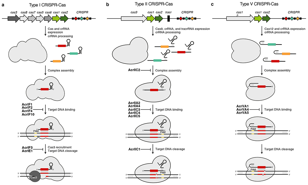
Clustered regularly interspaced short palindromic repeats (CRISPR) together with their accompanying cas (CRISPR-associated) genes are found frequently in bacteria and archaea, serving to defend against invading foreign DNA, such as viral genomes. CRISPR-Cas systems provide a uniquely powerful defense because they can adapt to newly encountered genomes. The adaptive ability of these systems has been exploited, leading to their development as highly effective tools for genome editing. The widespread use of CRISPR-Cas systems has driven a need for methods to control their activity. This review focuses on anti-CRISPRs (Acrs), proteins produced by viruses and other mobile genetic elements that can potently inhibit CRISPR-Cas systems. Discovered in 2013, there are now 54 distinct families of these proteins described, and the functional mechanisms of more than a dozen have been characterized in molecular detail. The investigation of Acrs is leading to a variety of practical applications and is providing exciting new insight into the biology of CRISPR-Cas systems.

Keywords: Acr; CRISPR-Cas; anti-CRISPR; bacteriophage; genome editing; mobile genetic element.


Figures

Figure 1
Figure 1
Anti-CRISPR proteins inhibit CRISPR-Cas systems at distinct stages. (a) A generalized type I CRISPR-Cas locus is depicted. Cas genes and the CRISPR array are expressed. The array transcript is subsequently processed into mature crRNA. Cas proteins assemble around the mature crRNA to form the a CRISPR-Cas complex, which identifies foreign DNA targets through complementary base pairing with its crRNA and an appropriate PAM sequence. AcrIF1, AcrIF2, AcrIF4, and AcrIF10 prevent Cascade from interacting with DNA. Annealing of the crRNA triggers R-loop formation, the recruitment of the Cas3 helicase/nuclease, and the destruction of the DNA target. AcrIF3 and AcrIE1 disable Cas3 to prevent target cleavage. (b) A generalized type II CRISPR-Cas locus is depicted. Cas9 and processed crRNA and tracrRNA assemble in a complex that can recognize PAM flanked sequences with complementarity to its crRNA. AcrIIC2 inhibits crRNA loading into Cas9 preventing proper complex assembly. AcrIIA2, AcrIIA4, AcrIIC3, AcrIIC4, and AcrIIC5 prevent the complex from recognizing target DNA. Following target recognition, Cas9 creates a double stranded DNA break target, leading to its destruction. AcrIIC1 inhibits the nuclease activity of Cas9 to prevent target cleavage. (c) A generalized type V CRISPR-Cas locus is depicted. Cas12 and the crRNA are expressed, processed, and assembled into a surveillance complex. The complex binds target DNA with a crRNA complementary, PAM flanked target. AcrVA1, AcrVA4, and AcrVA5 prevent DNA recognition. Target binding triggers the nuclease activity of Cas12, generating a staggered double-stranded DNA break to destroy the target. Cas genes = coloured arrows. CRISPR array: repeats = black boxes; spacers = coloured diamonds. PAM = yellow. cas1 and cas2 encode proteins involved in CRISPR adaptation
Figure 2
Figure 2
The commonly used approaches for acr gene discovery. (a) Representative acr regions are shown from the Pae phages in which these genes were first identified. Gray arrows depict predicted open reading frames flanking the acr regions. Dark green arrows correspond to genes encoding the highly conserved anti-CRISPR-associated gene 1 (aca1) predicted to encode a protein with a helix-turn-helix (HTH) motif. The remaining colored arrows represent the acr genes. (b) The “guilt-by-association” method is a homology-based computational search approach that uses the proteins encoded by the acr and aca genes as queries. For example, a search with AcrIF6 (pink arrow) of Pae identified a homologue in Oceanimonas smirnovii. The gene encoding this protein was found adjacent to aca2, which is a distinct HTH protein. Searches with Aca2, resulted in homologues being detected in the genomes of Vibrio parahaemolyticus and Brackiella oedipodis, which led to the discovery of acrIF9 (purple arrow) and acrIIC1 (yellow arrow) positioned upstream of aca2 in these bacteria. Finally, homology-based searches using the protein encoded by acrIIC1 resulted in the discovery of aca3 (green arrow). This back and forth approach has facilitated the discovery of many families of Acrs. (c) The self-targeting approach is also commonly used for acr gene discovery. If a prokaryotic strain has a functional CRISPR-Cas system (e.g., type II-A depicted on the left) and contains a spacer (checkered box in the CRISPR-array) that matches a protospacer (checkered box within the dark gray arrow on the right) located adjacent to a known PAM (red box within the dark gray arrow on the right) within an MGE, then that organism must also have an acr gene—otherwise, its CRISPR-Cas system would degrade its own genome. Black diamonds represent the palindromic repeats in the CRISPR-array. Candidate acr genes are unique genes (patterned blue arrows) present in the targeted MGE (patterned blue arrows; top) and absent from a related MGE that elicits no Acr activity (patterned blue arrows; bottom).
Figure 3
Figure 3
All of the solved structures of Acrs are shown. Some of these were solved on their own and some were solved bound to CRISPR-Cas complexes. No significant structural changes have been observed between the bound and unbound forms of Acrs when both have been solved. The PDB identification codes for each structure are shown in parentheses.
Figure 4
Figure 4
Structures of type I-F Acrs bound to the Pae Csy complex determined by cryoEM. (a) The cryo-EM structure of AcrIF1 bound to the Csy complex in the presence of a crRNA (yellow) is shown. Two copies of AcrIF1 (cyan) bind to the Cas7f backbone (gray) of the complex. Cas8f and Cas5f are not shown here for simplification. On the right is shown a close-up where residues 8-15 and 33-35 of AcrIF1 (blue spheres) reach into the DNA binding groove to sterically clash and prevent target DNA hybridization. These residues are indicated with red arrows. (b) The left image shows AcrIF2 (red) bound to the Cas8f hook domain (cyan) of the Csy complex. The middle image shows the structure of AcrIF10 (magenta) bound to the Cas8f hook domain (cyan). The right image shows that the two Acrs bind in different locations and cause the hook to move in different directions. (c) The left image shows the complete Csy complex. The helical bundle of Cas8f that binds to Cas3 is in cyan in the center of the structure. In the middle image, this bundle is overlaid with AcrIF3. The two helices that overlay well and are in bolder colors are both critical for binding to Cas3. On the right is the structure of Cas3 bound to AcrIF3.
Figure 5
Figure 5
Structures of Cas9 bound to Acrs. (a) The structure on the left shows SpyCas9 bound to AcrIIA2 (cyan) primarily interacting with the PAM-interacting domain (PID) of Cas9 (pale green), as well as the HNH (light orange) and REC domains (grey). The structure on the right shows SpyCas9 bound to AcrIIA4 (violet) primarily interacting with the PID (pale green), as well as the RuvC domain (pale yellow). Both Acrs bind in the same region, which is also where DNA binds. (b) AcrIIC1 (salmon) and AcrIIC3 (bluepurple) interact with different surfaces of the HNH domain (light orange) from NmeCas9.
Figure 6
Figure 6
AcrVA5 acetylates K635 and prevents MbCas12a from recognizing the PAM. On the left is a superposition of PAM-bound LbCas12a and MbCas12a acetylated at K635. In the close-up on the right, it can be seen that the acetylated K635 (red) sits within the PID and can sterically block recognition of the PAM. Thus, MbCas12a is unable to interact with target DNA when K635 is methylated.

Similar articles

Cited by

References

    1. Doron S, Melamed S, Ofir G, Leavitt A, Lopatina A, et al. 2018. Systematic discovery of antiphage defense systems in the microbial pangenome. Science 359: 1008–18 - PMC - PubMed
    1. Dy RL, Richter C, Salmond GP, Fineran PC. 2014. Remarkable Mechanisms in Microbes to Resist Phage Infections. Annu Rev Virol 1: 307–31 - PubMed
    1. Makarova KS, Wolf YI, Alkhnbashi OS, Costa F, Shah SA, et al. 2015. An updated evolutionary classification of CRISPR-Cas systems. Nat Rev Microbiol 13: 722–36 - PMC - PubMed
    1. Amitai G, Sorek R. 2016. CRISPR-Cas adaptation: insights into the mechanism of action. Nat Rev Microbiol 14: 67–76 - PubMed
    1. Sorek R, Lawrence CM, Wiedenheft B. 2013. CRISPR-mediated adaptive immune systems in bacteria and archaea. Annu Rev Biochem 82: 237–66 - PubMed

Publication types

MeSH terms

LinkOut - more resources