Review
. 2020 Mar 17;52(3):464-474.
doi: 10.1016/j.immuni.2020.02.017.

Neuro-immune Interactions in the Tissues

Affiliations
Review

Neuro-immune Interactions in the Tissues

Coco Chu et al. Immunity. .

Abstract

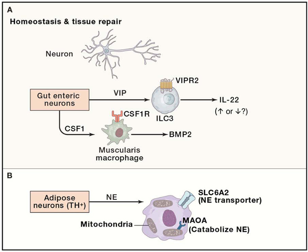
The ability of the nervous system to sense environmental stimuli and to relay these signals to immune cells via neurotransmitters and neuropeptides is indispensable for effective immunity and tissue homeostasis. Depending on the tissue microenvironment and distinct drivers of a certain immune response, the same neuronal populations and neuro-mediators can exert opposing effects, promoting or inhibiting tissue immunity. Here, we review the current understanding of the mechanisms that underlie the complex interactions between the immune and the nervous systems in different tissues and contexts. We outline current gaps in knowledge and argue for the importance of considering infectious and inflammatory disease within a conceptual framework that integrates neuro-immune circuits both local and systemic, so as to better understand effective immunity to develop improved approaches to treat inflammation and disease.

Keywords: ▪▪▪.


Conflict of interest statement

Declaration of Interests D.A. has contributed to scientific advisory boards at FARE, Genentech, KRF, Pfizer, and Takeda in the last twelve months. I.M.C. receives sponsored research support from GSK and Allergan Pharmaceuticals and is a member of scientific advisory boards for GSK and Kintai pharmaceuticals.

Figures

Figure 1.
Figure 1.. Neuro-immune interactions in host defense.
(A) Skin, lung and gut-innervating Nav1.8+ and/or TRPV1+ nociceptor neurons release the neuropeptide calcitonin gene-related peptide (CGRP) from their nerve terminals during infection, which acts via the calcitonin receptor-like receptor/receptor activity-modifying protein 1 (CALCRL/RAMP1) receptor complex on immune cells. CGRP inhibits TNF-α expression and production by macrophages. It also modulates the ability of neutrophils to survey and kill bacterial pathogens. In the intestine, CGRP regulates Peyer’s patch M cells to reduce bacterial invasion. (B) Enteric neurons secrete the cytokine IL-18, which acts on goblet cells to promote the production of antimicrobial peptides and bacterial killing. (C) Gut-innervating tyrosine hydroxylase (TH+) sympathetic neurons secrete norepinephrine (NE), which acts via the beta-2 adrenergic receptor (β2AR) on muscularis macrophages to polarize a M2 phenotype, which feedback to neurons to prevent cell loss in bacterial infection. Gut-innervating sympathetic neurons also act on ILC2 via β2AR to inhibit type 2 cytokine production and helminth expulsion. (D) Lung and gut-innervating choline acetyltransferase positive (ChAT+) neurons release neuropeptide neuromedin U (NMU), which acts via the neuromedin U receptor 1 (NMUR1) on ILC2s to promote type 2 cytokine production and helminth expulsion.
Figure 2.
Figure 2.. Neuro-immune interactions in inflammation.
(A) The cholinergic “anti-inflammatory reflex” circuit. The vagus nerve signals to TH+ splenic nerves, which secrete NE to activate ChAT+ T cells to produce acetylcholine (ACh), which in turn signal to macrophages via the α7 nictonic acetylcholine receptor to reduce pro-inflammatory cytokine production. The vagus nerve also directly activates ChAT+ myenteric nerves to drive its ACh production to reduce pro-inflammatory cytokines from macrophages in the intestine. (B) Skin-innervating nociceptors release the neuropeptides Substance P (SP) and CGRP. SP acts via the Mas-related G-protein coupled receptor member B2 (MRGPRB2) on mast cells to promote the release of inflammatory mediators. CGRP acts via the CALCRL-RAMP1 receptor complex on DCs to induce IL-23 from DCs which in turn leads to γδT cell and Th17 cell activation and pro-inflammatory cytokine and type 17 cytokine production. (C) Enteric ChAT+ neurons and lung-innervating sensory neurons release neuropeptide CGRP, inhibiting ILC2 proliferation and IL-13 production. Lung sensory neurons also release neuropeptide vasoactive intestinal peptide (VIP), which acts via the vasoactive intestinal peptide receptor 2 (VIPR2) on ILC2 and Th2 cells, increasing the production of type 2 cytokines.
Figure 3.
Figure 3.. Neuro-immune interactions in tissue repair and homeostasis.
(A) Enteric neurons release neuropeptide VIP, which acts on VIPR2 expressed by ILC3s to modulate IL-22 production (increase or decrease). Enteric neurons also secrete macrophage colony-stimulating factor 1 (CSF1), which binds to its receptor CSF1R on muscularis macrophages to regulate the production of bone morphogenetic protein 2 (BMP2) from muscularis macrophages and gastrointestinal motility. (B) Adipose tissue TH+ neuron-derived NE can be taken up and catabolized by tissue resident macrophages via the sodium-dependent noradrenaline transporter SLC6A2 and (monoamine oxidase A) MAOA, respectively.

References

    1. Abraira VE, and Ginty DD (2013). The sensory neurons of touch. Neuron 79, 618–639. - PMC - PubMed
    1. Angus DC, and van der Poll T. (2013). Severe sepsis and septic shock. N Engl J Med 369, 840–851. - PubMed
    1. Baral P, Udit S, and Chiu IM (2019). Pain and immunity: implications for host defence. Nat Rev Immunol 19, 433–447. - PMC - PubMed
    1. Baral P, Umans BD, Li L, Wallrapp A, Bist M, Kirschbaum T, Wei Y, Zhou Y, Kuchroo VK, Burkett PR, et al. (2018). Nociceptor sensory neurons suppress neutrophil and gammadelta T cell responses in bacterial lung infections and lethal pneumonia. Nat Med 24, 417–426. - PMC - PubMed
    1. Basbaum AI, Bautista DM, Scherrer G, and Julius D. (2009). Cellular and molecular mechanisms of pain. Cell 139, 267–284. - PMC - PubMed

Publication types