

Haruspex: A Neural Network for the Automatic Identification of Oligonucleotides and Protein Secondary Structure in Cryo-Electron Microscopy Maps
- PMID: 32187813
- PMCID: PMC7497202
- DOI: 10.1002/anie.202000421
Haruspex: A Neural Network for the Automatic Identification of Oligonucleotides and Protein Secondary Structure in Cryo-Electron Microscopy Maps
Abstract
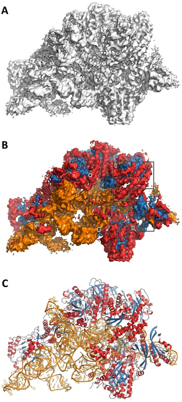
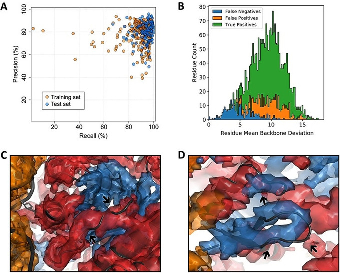
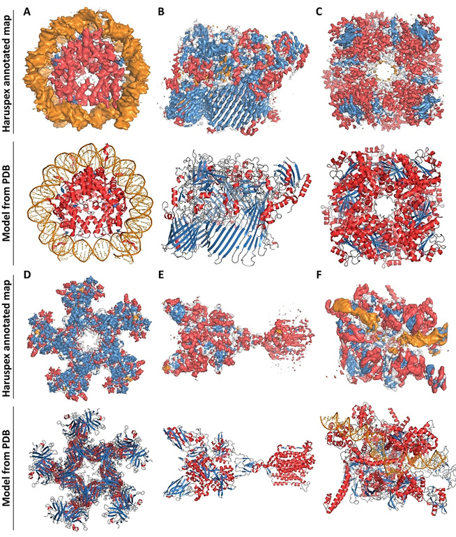
In recent years, three-dimensional density maps reconstructed from single particle images obtained by electron cryo-microscopy (cryo-EM) have reached unprecedented resolution. However, map interpretation can be challenging, in particular if the constituting structures require de-novo model building or are very mobile. Herein, we demonstrate the potential of convolutional neural networks for the annotation of cryo-EM maps: our network Haruspex has been trained on a carefully curated set of 293 experimentally derived reconstruction maps to automatically annotate RNA/DNA as well as protein secondary structure elements. It can be straightforwardly applied to newly reconstructed maps in order to support domain placement or as a starting point for main-chain placement. Due to its high recall and precision rates of 95.1 % and 80.3 %, respectively, on an independent test set of 122 maps, it can also be used for validation during model building. The trained network will be available as part of the CCP-EM suite.
Keywords: DNA structures; RNA structures; electron microscopy; neural networks; protein structures.
© 2020 The Authors. Published by Wiley-VCH Verlag GmbH & Co. KGaA.
Conflict of interest statement
The authors declare no conflict of interest.
Figures
Similar articles
-
Protein secondary structure detection in intermediate-resolution cryo-EM maps using deep learning.Nat Methods. 2019 Sep;16(9):911-917. doi: 10.1038/s41592-019-0500-1. Epub 2019 Jul 29. Nat Methods. 2019. PMID: 31358979 Free PMC article.
-
Automated detection and de novo structure modeling of nucleic acids from cryo-EM maps.Nat Commun. 2024 Oct 30;15(1):9367. doi: 10.1038/s41467-024-53721-4. Nat Commun. 2024. PMID: 39477926 Free PMC article.
-
Protein Secondary Structure and DNA/RNA Detection for Cryo-EM and Cryo-ET Using Emap2sec and Emap2sec.Methods Mol Biol. 2025;2867:105-120. doi: 10.1007/978-1-0716-4196-5_6. Methods Mol Biol. 2025. PMID: 39576577
-
Refinement of Atomic Structures Against cryo-EM Maps.Methods Enzymol. 2016;579:277-305. doi: 10.1016/bs.mie.2016.05.033. Epub 2016 Jun 24. Methods Enzymol. 2016. PMID: 27572731 Review.
-
Finding the right fit: chiseling structures out of cryo-electron microscopy maps.Curr Opin Struct Biol. 2014 Apr;25:118-25. doi: 10.1016/j.sbi.2014.04.001. Epub 2014 May 9. Curr Opin Struct Biol. 2014. PMID: 24814094 Review.
Cited by
-
The structural basis for HIV-1 Vif antagonism of human APOBEC3G.Nature. 2023 Mar;615(7953):728-733. doi: 10.1038/s41586-023-05779-1. Epub 2023 Feb 8. Nature. 2023. PMID: 36754086 Free PMC article.
-
Cryo2StructData: A Large Labeled Cryo-EM Density Map Dataset for AI-based Modeling of Protein Structures.bioRxiv [Preprint]. 2024 Jan 2:2023.06.14.545024. doi: 10.1101/2023.06.14.545024. bioRxiv. 2024. Update in: Sci Data. 2024 May 6;11(1):458. doi: 10.1038/s41597-024-03299-9. PMID: 37398020 Free PMC article. Updated. Preprint.
-
Deep learning for reconstructing protein structures from cryo-EM density maps: Recent advances and future directions.Curr Opin Struct Biol. 2023 Apr;79:102536. doi: 10.1016/j.sbi.2023.102536. Epub 2023 Feb 9. Curr Opin Struct Biol. 2023. PMID: 36773336 Free PMC article. Review.
-
Using cryo-EM to uncover mechanisms of bacterial transcriptional regulation.Biochem Soc Trans. 2021 Dec 17;49(6):2711-2726. doi: 10.1042/BST20210674. Biochem Soc Trans. 2021. PMID: 34854920 Free PMC article. Review.
-
Fast and automated protein-DNA/RNA macromolecular complex modeling from cryo-EM maps.Brief Bioinform. 2023 Mar 19;24(2):bbac632. doi: 10.1093/bib/bbac632. Brief Bioinform. 2023. PMID: 36682003 Free PMC article.
References
-
- Frank J., Agrawal R. K., Nature 2000, 406, 318–322. - PubMed
Publication types
MeSH terms
Substances
LinkOut - more resources
Full Text Sources
