

Porphyrins as Colorimetric and Photometric Biosensors in Modern Bioanalytical Systems
- PMID: 32187831
- PMCID: PMC7383976
- DOI: 10.1002/cbic.202000067
Porphyrins as Colorimetric and Photometric Biosensors in Modern Bioanalytical Systems
Abstract
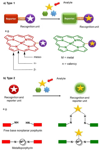
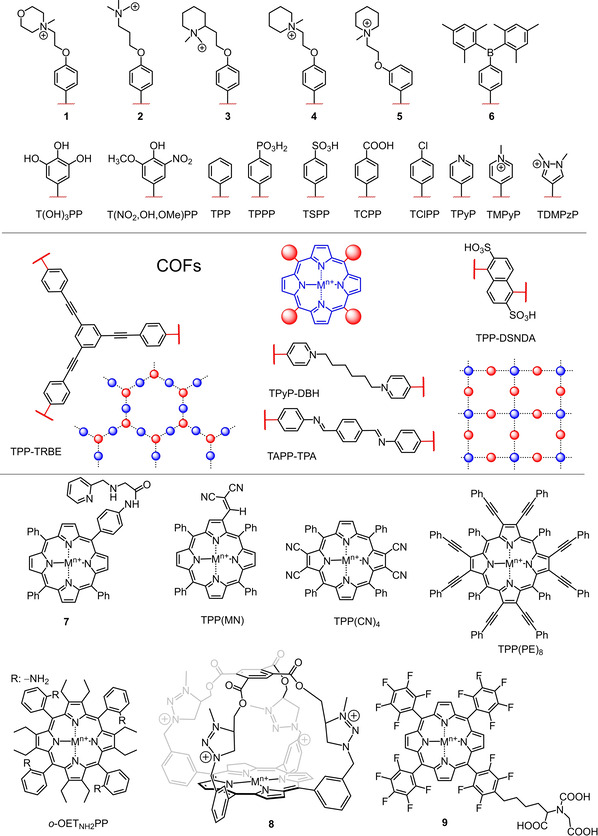
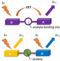
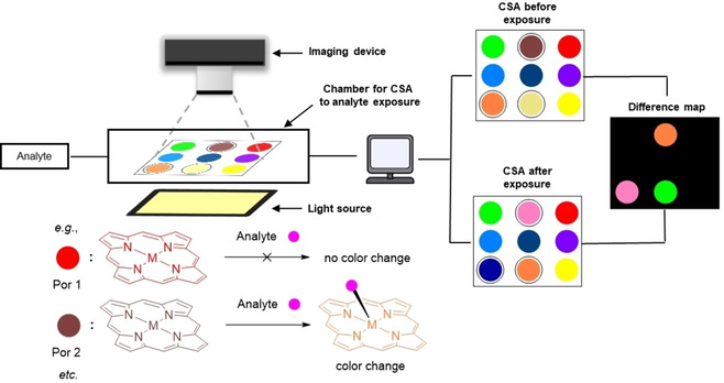
Advances in porphyrin chemistry have provided novel materials and exciting technologies for bioanalysis such as colorimetric sensor array (CSA), photo-electrochemical (PEC) biosensing, and nanocomposites as peroxidase mimetics for glucose detection. This review highlights selected recent advances in the construction of supramolecular assemblies based on the porphyrin macrocycle that provide recognition of various biologically important entities through the unique porphyrin properties associated with colorimetry, spectrophotometry, and photo-electrochemistry.
Keywords: Biorecognition; biosensing; colorimetry; detection; porphyrin.
© 2020 The Authors. Published by Wiley-VCH Verlag GmbH & Co. KGaA.
Conflict of interest statement
The authors declare no conflict of interest.
Figures
Similar articles
-
One-pot synthesis of porphyrin functionalized γ-Fe2O3 nanocomposites as peroxidase mimics for H2O2 and glucose detection.Mater Sci Eng C Mater Biol Appl. 2015 Oct;55:193-200. doi: 10.1016/j.msec.2015.05.028. Epub 2015 May 9. Mater Sci Eng C Mater Biol Appl. 2015. PMID: 26117755
-
Glucose-sensitive colorimetric sensor based on peroxidase mimics activity of porphyrin-Fe3O4 nanocomposites.Mater Sci Eng C Mater Biol Appl. 2014 Aug 1;41:142-51. doi: 10.1016/j.msec.2014.04.038. Epub 2014 Apr 26. Mater Sci Eng C Mater Biol Appl. 2014. PMID: 24907747
-
Enhanced Mimetic Enzyme Activity of Phosphorylated Porphyrin Nanocomposite Induced by Localized Surface Plasmon Resonance for Colorimetric Assay.Anal Sci. 2019 Jun 10;35(6):691-699. doi: 10.2116/analsci.19P004. Epub 2019 Mar 8. Anal Sci. 2019. PMID: 30853695
-
Porphyrin colorimetric indicators in molecular and nano-architectures.J Nanosci Nanotechnol. 2007 Sep;7(9):2969-93. doi: 10.1166/jnn.2007.910. J Nanosci Nanotechnol. 2007. PMID: 18019127 Review.
-
Recent progress of heterostructure-based photoelectrodes in photoelectrochemical biosensing: a mini review.Analyst. 2020 Feb 17;145(4):1121-1128. doi: 10.1039/c9an02448d. Analyst. 2020. PMID: 31984380 Review.
Cited by
-
Could the Length of the Alkyl Chain Affect the Photodynamic Activity of 5,10,15,20-Tetrakis(1-alkylpyridinium-4-yl)porphyrins?Molecules. 2024 Mar 14;29(6):1285. doi: 10.3390/molecules29061285. Molecules. 2024. PMID: 38542921 Free PMC article.
-
Metalloprotein enabled redox signal transduction in microbes.Curr Opin Chem Biol. 2023 Oct;76:102331. doi: 10.1016/j.cbpa.2023.102331. Epub 2023 Jun 11. Curr Opin Chem Biol. 2023. PMID: 37311385 Free PMC article. Review.
-
Advancements in Biosensors Based on the Assembles of Small Organic Molecules and Peptides.Biosensors (Basel). 2023 Jul 29;13(8):773. doi: 10.3390/bios13080773. Biosensors (Basel). 2023. PMID: 37622859 Free PMC article. Review.
-
A novel TiO2-modified THPP-BCP composite optical waveguide sensor for the determination of ethylenediamine at ppb level.Anal Sci. 2024 Feb;40(2):291-300. doi: 10.1007/s44211-023-00458-7. Epub 2023 Nov 17. Anal Sci. 2024. PMID: 37976016
-
Weak Interactions and Conformational Changes in Core-Protonated A2- and Ax-Type Porphyrin Dications.Molecules. 2020 Jul 13;25(14):3195. doi: 10.3390/molecules25143195. Molecules. 2020. PMID: 32668713 Free PMC article.
References
Publication types
MeSH terms
Substances
LinkOut - more resources
Full Text Sources
