

Drug Delivery to the Posterior Segment of the Eye: Biopharmaceutic and Pharmacokinetic Considerations
- PMID: 32188045
- PMCID: PMC7151081
- DOI: 10.3390/pharmaceutics12030269
Drug Delivery to the Posterior Segment of the Eye: Biopharmaceutic and Pharmacokinetic Considerations
Abstract
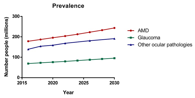
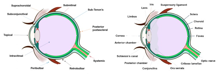
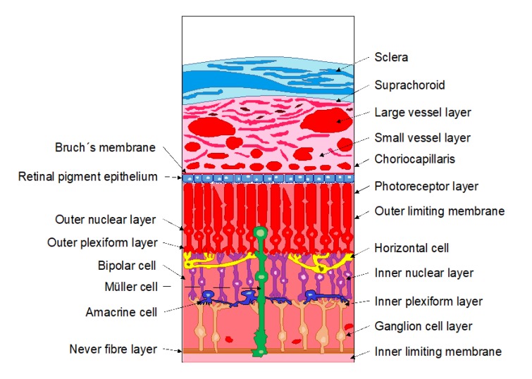
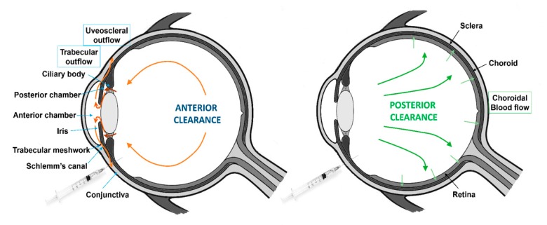
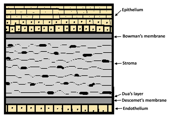
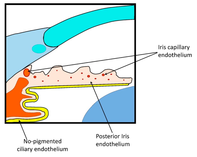
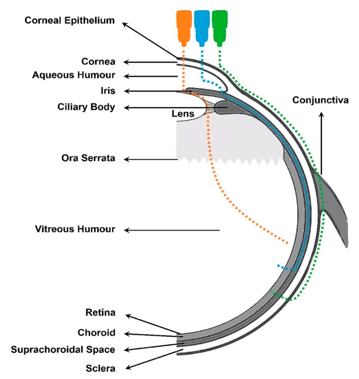
The treatment of the posterior-segment ocular diseases, such as age-related eye diseases (AMD) or diabetic retinopathy (DR), present a challenge for ophthalmologists due to the complex anatomy and physiology of the eye. This specialized organ is composed of various static and dynamic barriers that restrict drug delivery into the target site of action. Despite numerous efforts, effective intraocular drug delivery remains unresolved and, therefore, it is highly desirable to improve the current treatments of diseases affecting the posterior cavity. This review article gives an overview of pharmacokinetic and biopharmaceutics aspects for the most commonly-used ocular administration routes (intravitreal, topical, systemic, and periocular), including information of the absorption, distribution, and elimination, as well as the benefits and limitations of each one. This article also encompasses different conventional and novel drug delivery systems designed and developed to improve drug pharmacokinetics intended for the posterior ocular segment treatment.
Keywords: intravitreal administration; ocular drug delivery systems; ocular pharmacokinetics; ocular routes of drug administration; topical administration.
Conflict of interest statement
The authors have declared no potential conflicts of interest, financial or otherwise.
Figures
References
-
- WHO Team . World Report on Vision. WHO; Geneva, Switzerland: 2019. p. 180.
Publication types
Grants and funding
LinkOut - more resources
Full Text Sources
Other Literature Sources
