. 2020 Mar 16;9(3):728.
doi: 10.3390/cells9030728.

SIRT1 Alleviates LPS-Induced IL-1β Production by Suppressing NLRP3 Inflammasome Activation and ROS Production in Trophoblasts

Affiliations

SIRT1 Alleviates LPS-Induced IL-1β Production by Suppressing NLRP3 Inflammasome Activation and ROS Production in Trophoblasts

Sumi Park et al. Cells. .

Abstract

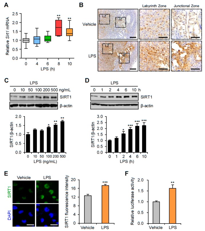
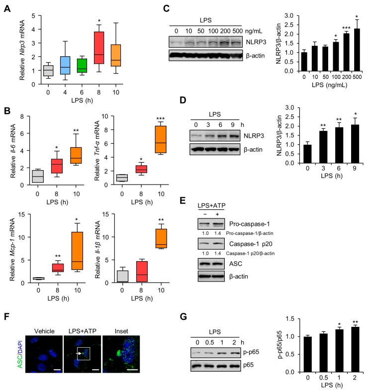
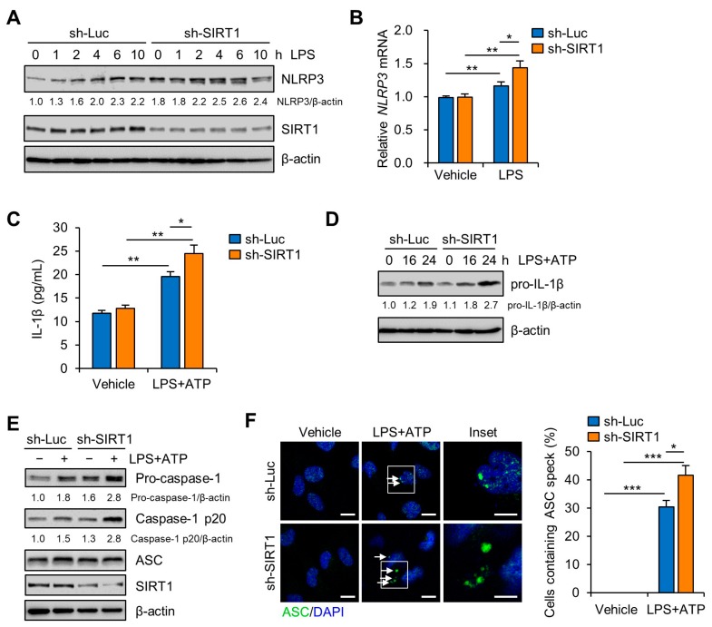
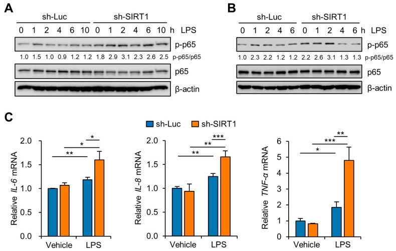
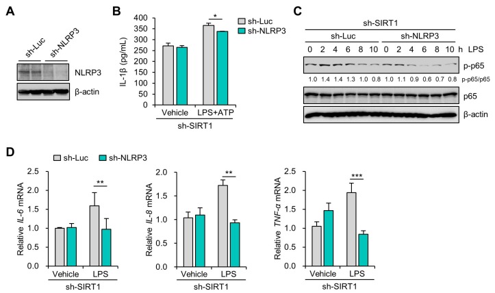
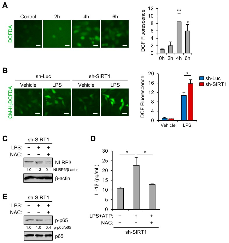
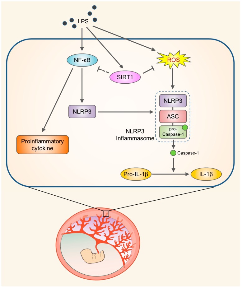
Emerging evidence indicates that aberrant maternal inflammation is associated with several pregnancy-related disorders such as preeclampsia, preterm birth, and intrauterine growth restriction. Sirtuin1 (SIRT1), a class III histone deacetylase, is involved in the regulation of various physiopathological processes including cellular inflammation and metabolism. However, the effect of SIRT1 on the placental proinflammatory environment remains to be elucidated. In this study, we investigated the effect of SIRT1 on lipopolysaccharide (LPS)-induced NLRP3 inflammasome activation and its underlying mechanisms in human first-trimester trophoblasts (Sw.71 and HTR-8/SVneo cells). Treatment with LPS elevated SIRT1 expression and induced NLRP3 inflammasome activation in mouse placental tissues and human trophoblasts. Knockdown of SIRT1 enhanced LPS-induced NLRP3 inflammasome activation, inflammatory signaling, and subsequent interleukin (IL)-1β secretion. Furthermore, knockdown of NLRP3 considerably attenuated the increase of IL-1β secretion in SIRT1-knockdown cells treated with LPS. Moreover, SIRT1 inhibited LPS-induced NLRP3 inflammasome activation by reducing oxidative stress. This study revealed a novel mechanism via which SIRT1 exerts anti-inflammatory effects, suggesting that SIRT1 is a potential therapeutic target for the prevention of inflammation-associated pregnancy-related complications.

Keywords: NLRP3 inflammasome; SIRT1; human trophoblasts; maternal inflammation; oxidative stress; placenta.


Conflict of interest statement

The authors declare no conflicts of interest.

Figures

Figure 1
Figure 1
Expression of SIRT1 in placenta of mice and in human trophoblasts exposed to lipopolysaccharide (LPS). (A) C57BL/6 pregnant mice were injected intraperitoneally with LPS (100 μg/kg) on gestation day 17.5. After the indicated number of hours, placentas were harvested and analyzed by real-time PCR for expression of SIRT1 (n = 5–11). (B) Immunohistochemistry analysis of SIRT1 in placental tissues from mice exposed to LPS or vehicle for 8 h. Nuclei were stained with hematoxylin. Boxed areas are magnified in middle panels. LZ and JZ represent the labyrinth zone and the junctional zone respectively. Scale bar, 200 μm; inset, 100 μm. (C) Sw.71 cells were treated with the indicated concentration of LPS for 4 h. Cell lysates were immunoblotted with an anti-SIRT1 antibody. β-Actin served as a loading control. Band intensities were quantified and normalized over the β-actin values. (D) Sw.71 cells were treated with 200 ng/mL LPS for the indicated time periods. Cell lysates were immunoblotted with an anti-SIRT1 antibody. β-Actin served as a loading control. Band intensities were quantified and normalized over the β-actin values. (E) Immunofluorescence staining of SIRT1 (green) in Sw.71 cells treated with LPS (200 ng/mL) or vehicle for 4 h. Nuclei were stained with DAPI (blue). Scale bars, 40 μm. (F) Sw.71 cells that were transfected with firefly luciferase constructs containing an SIRT1 promoter sequence. At 48 h after transfection, cells were treated with LPS (100 ng/mL) for 4 h. Firefly luciferase activity was measured and normalized to the Renilla luciferase activity. Data are shown as mean ± SEM. Results are representative of at least three independent experiments. * p < 0.05; ** p < 0.01; *** p < 0.001 (Student’s t-test).
Figure 2
Figure 2
NLRP3 inflammasome activation in placentas of mice and in human trophoblasts exposed to LPS. (A,B) C57BL/6 pregnant mice were injected intraperitoneally with LPS (100 μg/kg) on gestation day 17.5. After the indicated number of hours, placentas were harvested and analyzed using real-time PCR for the expression of NLRP3, IL-6, TNF-α, MCP-1, and IL-1β (n = 4–7). (C) Sw.71 cells were treated with the indicated concentration of LPS for 4 h. Cell lysates were immunoblotted with an anti-NLRP3 antibody. (D,G) Sw.71 cells were treated with 200 ng/mL LPS for the indicated time periods. Cell lysates were immunoblotted with anti-NLRP3 (D) and anti-p-p65 NF-κB, and anti-p65 NF-κB (G) antibodies. (E) Sw.71 cells were treated with LPS (100 ng/mL) for 24 h and then followed by ATP (5 mM) treatment for 45 min, or were treated with vehicle. Cell lysates were immunoblotted with anti-caspase-1 and anti-ASC (apoptosis-associated speck-like protein) antibodies. β-Actin or p65 served as a loading control. Band intensities were quantified and normalized over the β-actin or p65 values. (F) Immunofluorescence staining of ASC (green) in Sw.71 cells treated with LPS (100 ng/mL) for 24 h and then followed by ATP (5 mM) treatment for 45 min, or treated with vehicle. The white arrow represents ASC specks. Nuclei were stained with DAPI (blue). Scale bars, 20 μm; inset, 10 μm. Data are shown as mean ± SEM. Results are representative of at least three independent experiments. * p < 0.05; ** p < 0.01; *** p < 0.001 (Student’s t-test).
Figure 3
Figure 3
SIRT1 knockdown enhances LPS-induced NLRP3 inflammasome activation in trophoblasts. (A) Sw.71 cells were infected with lentiviruses expressing shRNAs targeting luciferase (sh-Luc) or SIRT1 (sh-SIRT1) and then treated with LPS (200 ng/mL) for the indicated time. Cell lysates were immunoblotted with anti-NLRP3 and anti-SIRT1 antibodies. (B) Relative mRNA expression of NLRP3 in Sw.71 cells infected with sh-Luc or sh-SIRT1 lentivirus and treated LPS (200 ng/mL) for 12 h. (C) Conditioned media were collected from Sw.71 cells infected with sh-Luc or sh-SIRT1 lentivirus and then treated or not (vehicle) with LPS (200 ng/mL) for 24 h, followed by ATP (5 mM) treatment for 45 min. Levels of IL-1β in the conditioned media were quantified by ELISA. (D) Sw.71 cells were infected with sh-Luc or sh-SIRT1 lentivirus and then treated or not (vehicle) with LPS (200 ng/mL) for the indicated time, followed by ATP (5 mM) treatment for 45 min. Cell lysates were immunoblotted with anti-IL-1β antibody. (E,F) Sw.71 cells were treated with LPS (100 ng/mL) for 24 h and then followed by ATP (5 mM) treatment for 45 min, or were treated with vehicle. (E) Cell lysates were immunoblotted with anti-caspase-1, anti-ASC, and anti-SIRT1 antibodies. (F) Immunofluorescence staining of ASC (green). The white arrow represents ASC specks. Nuclei were stained with DAPI (blue). Scale bars, 20 μm; inset, 10 μm. Endogenous ASC specks were quantified. β-Actin served as a loading control. Numbers below the immunoblot bands indicate the fold changes normalized to the control bands. Data are shown as mean ± S.E.M. Results are representative of at least three independent experiments. * p < 0.05; ** p < 0.01; *** p < 0.001 (Student’s t-test).
Figure 4
Figure 4
SIRT1 knockdown enhances LPS-induced inflammatory response in trophoblasts. (A,B) Sw.71 cells (A) and HTR-8/SVneo cells (B) were infected with lentiviruses expressing shRNAs targeting luciferase (sh-Luc) or SIRT1 (sh-SIRT1) and were then treated with 200 ng/mL LPS for the indicated time. Cell lysates were immunoblotted with anti-p-p65 and anti-p65 antibodies. β-Actin served as a loading control. Numbers below the immunoblot bands indicate the fold changes normalized to the p65 bands. (C) Relative mRNA expression of IL-6, IL-8, and TNF-α in Sw.71 cells infected with sh-Luc or sh-SIRT1 lentivirus and treated with 200 ng/mL LPS for 6 h. Data are shown as mean ± SEM. Results are representative of at least three independent experiments. * p < 0.05; ** p < 0.01; *** p < 0.001 (Student’s t-test).
Figure 5
Figure 5
SIRT1 knockdown increases LPS-induced IL-1β secretion in an NLRP3-dependent manner. (A) Sw.71 cells were infected with lentiviruses expressing shRNAs targeting luciferase (sh-Luc) or NLRP3 (sh-NLRP3). Cell lysates were immunoblotted with anti-NLRP3 antibody. β-Actin served as a loading control. (B) Conditioned media were collected from Sw.71 cells infected with double sh-SIRT1/sh-Luc or sh-SIRT1/sh-NLRP3 lentivirus and then treated with 200 ng/mL LPS or vehicle for 48 h, with the addition of 5 mM ATP for the last 45 min. Levels of IL-1β in the conditioned media were quantified by ELISA. (C) Sw.71 cells were infected with double sh-SIRT1/sh-Luc or sh-SIRT1/sh-NLRP3 lentivirus and then treated with 200 ng/mL LPS for the indicated time. Cell lysates were immunoblotted with anti-p-p65 and anti-p65 antibodies. β-Actin served as a loading control. Numbers below the immunoblot bands indicate the fold changes normalized to the p65 bands. (D) Relative mRNA expression of IL-6, IL-8, and TNF-α in Sw.71 cells infected with double sh-SIRT1/sh-Luc or sh-SIRT1/sh-NLRP3 lentivirus and then treated with 200 ng/mL LPS for 6 h. Data are shown as mean ± SEM. Results are representative of at least three independent experiments. * p < 0.5; ** p < 0.01; *** p < 0.001 (Student’s t-test).
Figure 6
Figure 6
SIRT1 knockdown enhances LPS-induced NLRP3 inflammasome activation by reducing oxidative stress. (A) Sw.71 cells were treated with 200 ng/mL LPS for the indicated time. Reactive oxygen species (ROS) levels were analyzed using CM-H2DCFDA. Scale bars, 20 μm. (B) Sw.71 cells were infected with lentiviruses expressing shRNAs targeting luciferase (sh-Luc) or SIRT1 (sh-SIRT1) and then treated with 200 ng/mL LPS for 4 h. ROS levels were analyzed using CM-H2DCFDA. Scale bars, 40 μm. (C,E) Sw.71 cells were infected with sh-SIRT1 lentivirus and then treated with 200 ng/mL LPS or vehicle, each in the presence or absence of 5 mM N-acetylcysteine (NAC). Cell lysates were immunoblotted with anti-NLRP3, anti-p-p65, and anti-p65 antibodies. β-Actin or p65 served as the loading control. Numbers below the immunoblot bands indicate fold changes normalized to the control bands. (D) Conditioned media were collected from Sw.71 cells infected with sh-SIRT1 lentivirus and then treated with 200 ng/mL LPS alone or with 5 mM NAC for 34 h, with the addition of 5 mM ATP for the last 45 min. Levels of IL-1β in the conditioned media were quantified using ELISA. Data are indicated as mean ± SEM. Results are representative of at least three independent experiments. * p < 0.05; ** p < 0.01 (Student’s t-test).
Figure 7
Figure 7
Schematic diagram illustrating the role of SIRT1 in trophoblasts upon exposure to LPS. SIRT1 not only attenuates LPS-induced NLRP3 inflammasome activation but also inhibits inflammatory response and IL-1β secretion by reducing oxidative stress.

References

    1. Mor G., Cardenas I., Abrahams V., Guller S. Inflammation and Pregnancy: The Role of the Immune System at the Implantation Site. Ann. N. Y. Acad. Sci. 2011;1221:80–87. doi: 10.1111/j.1749-6632.2010.05938.x. - DOI - PMC - PubMed
    1. He N., van Iperen L., de Jong D., Szuhai K., Helmerhorst F.M., van der Westerlaken L.A., Chuva de Sousa Lopes S.M. Human Extravillous Trophoblasts Penetrate Decidual Veins and Lymphatics before Remodeling Spiral Arteries During Early Pregnancy. PLoS ONE. 2017;12:e0169849. doi: 10.1371/journal.pone.0169849. - DOI - PMC - PubMed
    1. Graham C.H., Lala P.K. Mechanisms of Placental Invasion of the Uterus and Their Control. Biochem. Cell Biol. 1992;70:867–874. doi: 10.1139/o92-135. - DOI - PubMed
    1. Benyo D.F., Smarason A., Redman C.W., Sims C., Conrad K.P. Expression of Inflammatory Cytokines in Placentas from Women with Preeclampsia. J. Clin. Endocrinol. Metab. 2001;86:2505–2512. doi: 10.1210/jc.86.6.2505. - DOI - PubMed
    1. Boutsikou T., Mastorakos G., Kyriakakou M., Margeli A., Hassiakos D., Papassotiriou I., Kanaka-Gantenbein C., Malamitsi-Puchner A. Circulating Levels of Inflammatory Markers in Intrauterine Growth Restriction. Mediat. Inflamm. 2010;2010:790605. doi: 10.1155/2010/790605. - DOI - PMC - PubMed

Publication types

MeSH terms

LinkOut - more resources