Review
. 2020 Mar 16;21(6):2024.
doi: 10.3390/ijms21062024.

Pituitary Hyperplasia, Hormonal Changes and Prolactinoma Development in Males Exposed to Estrogens-An Insight From Translational Studies

Affiliations
Review

Pituitary Hyperplasia, Hormonal Changes and Prolactinoma Development in Males Exposed to Estrogens-An Insight From Translational Studies

Branka Šošić-Jurjević et al. Int J Mol Sci. .

Abstract

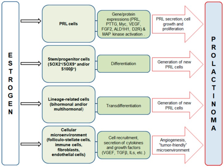
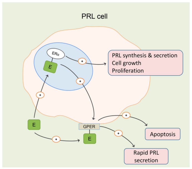
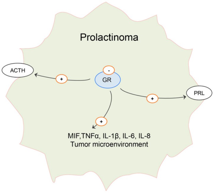
Estrogen signaling plays an important role in pituitary development and function. In sensitive rat or mice strains of both sexes, estrogen treatments promote lactotropic cell proliferation and induce the formation of pituitary adenomas (dominantly prolactin or growth-hormone-secreting ones). In male patients receiving estrogen, treatment does not necessarily result in pituitary hyperplasia, hyperprolactinemia or adenoma development. In this review, we comprehensively analyze the mechanisms of estrogen action upon their application in male animal models comparing it with available data in human subjects. Sex-specific molecular targets of estrogen action in lactotropic (PRL) cells are highlighted in the context of their proliferative and secretory activity. In addition, putative effects of estradiol on the cellular/tumor microenvironment and the contribution of postnatal pituitary progenitor/stem cells and transdifferentiation processes to prolactinoma development have been analyzed. Finally, estrogen-induced morphological and hormone-secreting changes in pituitary thyrotropic (TSH) and adrenocorticotropic (ACTH) cells are discussed, as well as the putative role of the thyroid and/or glucocorticoid hormones in prolactinoma development, based on the current scarce literature.

Keywords: adrenocorticotropic hormone; estrogen; folliculo-stellate cells; men; microenvironment; pituitary gland; prolactin; prolactinoma; rat; thyroid-stimulating hormone.


Conflict of interest statement

The authors declare no conflict of interest.

Figures

Figure 1
Figure 1
Proposed cellular and molecular targets of estrogen treatment action that contribute to pituitary lactotropic (PRL) cell hyperplasia, hyperprolactinemia and prolactinoma development. Estrogen may affect already-differentiated PRL cells, stem/progenitor cells, lineage-related cells and cells comprising the microenvironment. Transcriptional regulation of the following genes is mainly associated with tumorigenic action of estrogen in the pituitary: pituitary tumor transforming gene (PTTG); Myc; aldehyde dehydrogenase 1A1 (ALDH1A1); D2R, dopamine D2 receptor; mitogen-activated protein kinases (MAPK); vascular endothelial growth factor (VEGF); fibroblast growth factor 2 (FGF2); transforming growth factor β (TGFβ) and other growth factors and cytokines, such as ILs, interleukines [15,16,40,41,42,43,44,45,46,47,48,49,50,51,52,53].
Figure 2
Figure 2
Representative micrographs of immunohistochemically (IHC) or histochemically stained anterior pituitary sections of orchidectomized (Orx) and estradiol-treated orchidectomized (Orx+E) middle-aged rats. Hypertrophy and hyperplasia of prolactin-immunopositive cells (PRL, red arrows; adopted from our previous publication [55] and reprinted by permission of the Licensors - publishers Elsevier (Licence number 4774000297301)), stronger vascular endothelial growth factor A (VEGFA) immunopositivity, more abundant blood vessels (red arrows, Novelli histochemical staining) and lower nuclear estrogen receptor α immunopositivity (red arrows, ERα), respectively, are observable after estradiol treatment. Micrographs were obtained according to the same procedures described in our earlier papers [55]. Briefly, for IHC characterization of anterior pituitary tissue, the primary rabbit antisera directed against PRL (Abcam, Cambridge, UK; 1:200), VEGFA (Abcam, Cambridge, UK; 1:100) or ERα (1:100; Santa Cruz Biotechnology), were applied overnight at 4 °C. Swine anti-rabbit IgG- horseradish peroxidase (HRP; Dakopatts, Glostrup, Denmark; 1:100) was applied as a secondary antiserum for 1 h, while visualization was performed using diaminobenzidine tetrahydrochloride (DAB; Dakopatts, Glostrup, Denmark) at concentrations suggested by the manufacturer. Sections were counter-stained with hematoxylin and mounted in DPX medium (Sigma-Aldrich, Barcelona, Spain). For Novelli histochemical staining, sections were incubated in hot 1 N HCl (60 °C, 3 min), followed by staining in 1% acid fuchsin (Fluka Chemie AG, Buchs, Switzerland; 30 s) and 1% light green (Sigma-Aldrich, St. Louis, MO, USA; 3 min), respectively. In between, the slides were washed in PBS (for Nowelly, distilled water), and after the last step, mounted in DPX (Sigma-Aldrich, Barcelona, Spain). Scale bar is shown in the right corner.
Figure 3
Figure 3
Physiological and pathophysiological mechanisms of estrogen (E) action on proliferation, growth and secretion of lactotropic (PRL) cells are probably largely mediated by nuclear estrogen receptor (ER) α. Membrane G-protein-coupled estrogen receptor 1 (GPER)-mediated estrogen signaling seems to be more involved in antiproliferative and apoptotic actions in the pituitary and may contribute to rapid secretion of PRL under physiological conditions. Its expression is under ERα-mediated nuclear signaling [15,59,68,69,70].
Figure 4
Figure 4
Representative micrographs of immunofluorescently (IFC) stained anterior pituitary sections of orchidectomized (Orx) and estradiol-treated orchidectomized (Orx+E) middle-aged rats. Weaker immunofluorescent signal of S100β, stronger growth hormone (GH; adopted from our previous publication [99] and reprinted by permission of the Licensors - publishers Springer (Licence number 4774320912101)) and adrenocorticotropic hormone (ACTH) immunofluorescent signal, and unchanged immunofluorescent signal of subunit β of thyroid-stimulating hormone (TSHβ; adopted from our previous publication [55] and reprinted by permission of the Licensors - publishers Elsevier (Licence number 4774000297301)), respectively, are observable after estradiol treatment. All figures were obtained according to the same procedure described in our earlier papers [55,99]. Briefly, for IFC staining, the following primary antibodies were applied overnight at 4 °C: mouse monoclonal anti-S100β antibody (Abcam, Cambridge, UK; 1:100), polyclonal rabbit anti–rat TSHβ, GH and ACTH (donation from Dr. A. F. Parlow, National Institute of Health, Bethesda, MD, USA; dilutions 1:500, 1:200, 1:200, respectively). Alexa Fluor 488 donkey anti-rabbit IgG (Invitrogen Life technologies, CA, USA; 1:300) was applied as secondary antiserum for 1 h. The sections were mounted with Mowiol 4–88 (Sigma-Aldrich, St. Louis, MO, USA). Scale bar is shown in the right corner.
Figure 5
Figure 5
Hypothetic role of attenuated glucocorticoid hormone signaling in prolactinoma. Expression of glucocorticoid receptor (GR) is decreased in estrogen-induced prolactinoma and may contribute to increased adrenocorticotropic hormone (ACTH) and prolactin (PRL) secretion, as well as to changes in local secretion of growth factors and interleukins such as mitogen inhibiting factor (MIF), tumor necrosis factor α (TNFα), interleukins (IL) 1β, 6 and 8, thus promoting “tumor-friendly“ microenvironment [110,118,119,120].
Figure 6
Figure 6
Hypothetic role of enhanced thyroid hormone signaling in prolactinoma. Deiodinase type 2 (Dio 2) expression in folliculo-stellate cells is increased in prolactinomas, which may lead to increased production of l-triiodothyronine (T3) and 3,5-T2 (T2) from l-thyroxine (T4) in the pituitary. T3- thyroid receptor (TR) β complex prevents proteolisis of estrogen receptor (ER) α in the cell nucleus, while mutated thyroid receptor (mTRβ) binds to pituitary tumor transforming gene (PTTG), thus preventing its proteolysis. Both T4 and T3 may bind to its membrane integrin αvβ3 receptor and activate mitogen-activated protein kinase (MAPK) cascade and phosphorylation processes, including ERα phosphorylation (dashed arrow). Increased concentration of T2, demonstrated in pituitary adenomas, may contribute to progression of prolactinoma through stimulation of energy metabolism in mitochondria [132,133,134,135,136,137,138,139,140].

Similar articles

Cited by

References

    1. Ogasawara K., Nogami H., Tsuda M.C., Gustafsson J.A., Korach K.S., Ogawa S., Harigaya T., Hisano S. Hormonal regulation of prolactin cell development in the fetal pituitary gland of the mouse. Endocrinology. 2009;150:1061–1068. doi: 10.1210/en.2008-1151. - DOI - PubMed
    1. Eckstrum K.S., Weis K.E., Baur N.G., Yoshihara Y., Raetzman L.T. Icam5 Expression Exhibits Sex Differences in the Neonatal Pituitary and Is Regulated by Estradiol and Bisphenol, A. Endocrinology. 2016;157:1408–1420. doi: 10.1210/en.2015-1521. - DOI - PMC - PubMed
    1. Mitsui T., Ishida M., Izawa M., Arita J. Differences between rat strains in the development of PRL-secreting pituitary tumors with long-term estrogen treatment: In vitro insulin-like growth factor-1-induced lactotroph proliferation and gene expression are affected in Wistar-Kyoto rats with low estrogen-susceptibility. Endocr. J. 2013;60:1251–1259. - PubMed
    1. Bisson J.R., Chan K.J., Safer J.D. Prolactin levels do not rise among transgender women treated with estradiol and spironolactone. Endocr. Pract. 2018;24:646–651. doi: 10.4158/EP-2018-0101. - DOI - PubMed
    1. Vroonen L., Daly A.F., Beckers A. Epidemiology and Management Challenges in Prolactinomas. Neuroendocrinology. 2019;109:20–27. doi: 10.1159/000497746. - DOI - PubMed

MeSH terms