

Perivascular Adipose Tissue Regulates Vascular Function by Targeting Vascular Smooth Muscle Cells
- PMID: 32188271
- PMCID: PMC7441816
- DOI: 10.1161/ATVBAHA.120.312464
Perivascular Adipose Tissue Regulates Vascular Function by Targeting Vascular Smooth Muscle Cells
Abstract
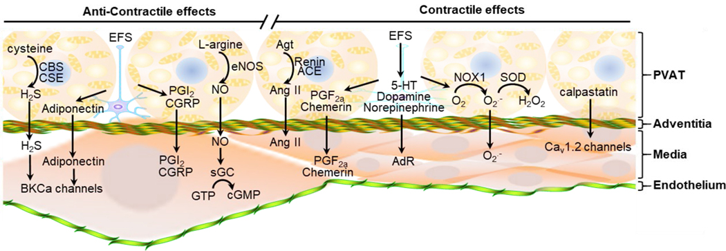
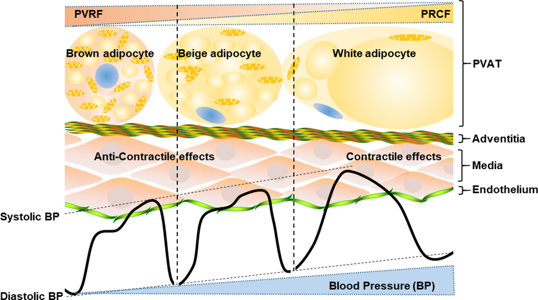
Adipose tissues are present at multiple locations in the body. Most blood vessels are surrounded with adipose tissue which is referred to as perivascular adipose tissue (PVAT). Similarly to adipose tissues at other locations, PVAT harbors many types of cells which produce and secrete adipokines and other undetermined factors which locally modulate PVAT metabolism and vascular function. Uncoupling protein-1, which is considered as a brown fat marker, is also expressed in PVAT of rodents and humans. Thus, compared with other adipose tissues in the visceral area, PVAT displays brown-like characteristics. PVAT shows a distinct function in the cardiovascular system compared with adipose tissues in other depots which are not adjacent to the vascular tree. Growing and extensive studies have demonstrated that presence of normal PVAT is required to maintain the vasculature in a functional status. However, excessive accumulation of dysfunctional PVAT leads to vascular disorders, partially through alteration of its secretome which, in turn, affects vascular smooth muscle cells and endothelial cells. In this review, we highlight the cross talk between PVAT and vascular smooth muscle cells and its roles in vascular remodeling and blood pressure regulation.
Keywords: brown adipose tissue; cardiovascular diseases; endothelial cells; hypertension; vascular smooth muscle cells.
Figures

References
-
- Pond CM. Paracrine interactions of mammalian adipose tissue. J Exp Zool A Comp Exp Biol. 2003;295:99–110. - PubMed
-
- Wu J, Bostrom P, Sparks LM, Ye L, Choi JH, Giang AH, Khandekar M, Virtanen KA, Nuutila P, Schaart G, Huang K, Tu H, van Marken Lichtenbelt WD, Hoeks J, Enerback S, Schrauwen P and Spiegelman BM. Beige adipocytes are a distinct type of thermogenic fat cell in mouse and human. Cell. 2012;150:366–376. - PMC - PubMed
-
- Efremova A, Senzacqua M, Venema W, Isakov E, Di Vincenzo A, Zingaretti MC, Protasoni M, Thomski M, Giordano A and Cinti S. A large proportion of mediastinal and perirenal visceral fat of Siberian adult people is formed by UCP1 immunoreactive multilocular and paucilocular adipocytes. J Physiol Biochem. 2019; December 18[Online ahead of print]. - PubMed
-
- Chang L, Villacorta L, Li R, Hamblin M, Xu W, Dou C, Zhang J, Wu J, Zeng R and Chen YE. Loss of perivascular adipose tissue on peroxisome proliferator-activated receptor-gamma deletion in smooth muscle cells impairs intravascular thermoregulation and enhances atherosclerosis. Circulation. 2012;126:1067–1078. - PMC - PubMed
Publication types
MeSH terms
Grants and funding
LinkOut - more resources
Full Text Sources
Medical
Research Materials
