Review
. 2020 Mar 18;20(1):102.
doi: 10.1186/s12883-020-01682-8.

A ruptured posterior communicating artery aneurysm presenting as tentorial and spinal isolated subdural hemorrhage: a case report and literature review

Affiliations
Review

A ruptured posterior communicating artery aneurysm presenting as tentorial and spinal isolated subdural hemorrhage: a case report and literature review

Abdulrahman Hamad Al-Abdulwahhab et al. BMC Neurol. .

Abstract

Background: Ruptured intracranial aneurysms are often associated with subarachnoid or intraparenchymal hemorrhage. However, the prevalence of subdural hemorrhage post aneurysmal rupture is low and rarely reported in scientific studies. Here, we report an unusual case of a ruptured posterior communicating artery aneurysm resulting in an isolated subdural hematoma located in the tentorial and spinal canal without subarachnoid or intraparenchymal hemorrhage.

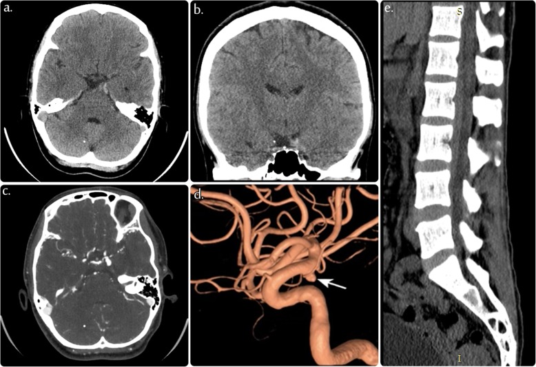
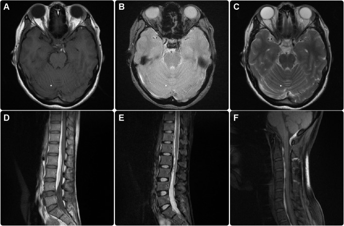
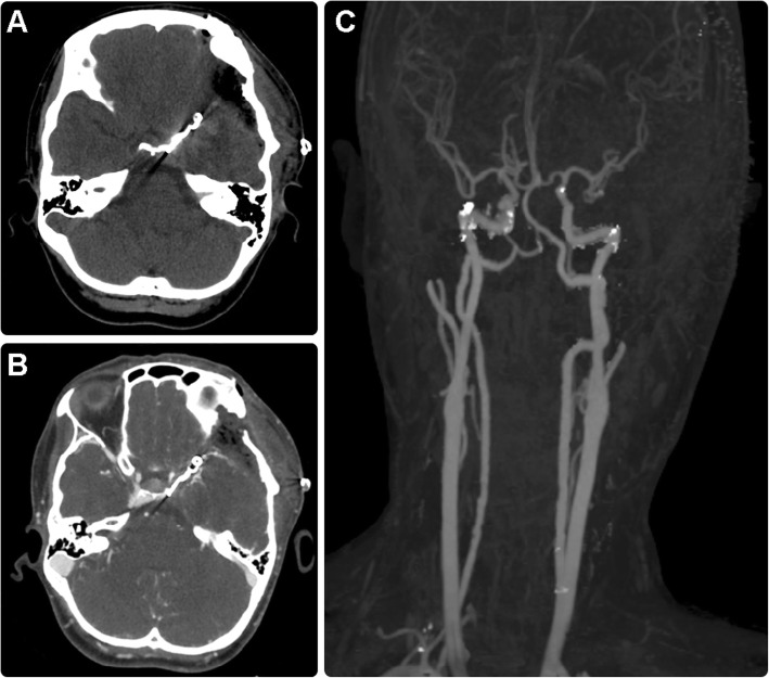
Case presentation: In this case, a 34-year-old woman with no history of trauma or coagulopathy was diagnosed with a subdural hematoma in the tentorium cerebellum tracing to the subdural space of the spinal column. Computed tomography angiography was used to identify the source of the bleeding, which revealed a ruptured left-sided posterior communicating artery saccular aneurysm. The aneurysm was clipped, and the hematoma was evacuated. The patient recovered without any neurological complications.

Conclusions: Our results suggest that a diagnosis of ruptured intracranial aneurysm should be considered in patients with nontraumatic subdural hematoma. Prompt diagnostic imaging and interventional diagnostic procedures are required to ensure proper management of these patients and to avoid unnecessary complications.

Keywords: Clipping; Posterior fossa; Ruptured aneurysm; Saccular aneurysm; Subdural hemorrhage.


Conflict of interest statement

The authors declare that they have no conflict of interest.

Figures

Fig. 1
Fig. 1
a, b, c Selective images of axial and coronal unenhanced computed tomography (CT) of the brain showing a small subdural hematoma at the anterior edge of the left tentorium, medial to the left uncus. d Selective 3D image of the left internal carotid artery (ICA) demonstrating unilocular posterior communicating artery (PCOM) aneurysm. (e) Selective image of the sagittal CT of the lumbar spine showing a subdural hematoma exerting a mass effect on the spinal canal
Fig. 2
Fig. 2
Multi-sequential magnetic resonance imaging (MRI) of the brain: axial T1-weighted (a), gradient (b), and T2-weighted (c) images showing a small area of extra-axial hyperintensity in the T1 image, and iso- to low-signal intensity in the T2 image, and a susceptibility artifact located at the anterior left tentorial edge, medial to the uncus, representing a subacute to chronic subdural hemorrhage in the gradient image. Multi-sequential MRI of the spine: sagittal T1-weighted (d), short tau inversion recovery (STAIR) of the lumbar spine (e), and T1-weighted fat saturation (f) images of the cervicothoracic spine showing massive extradural (subdural) bleeding presenting as predominantly high signal intensity on T1 (d) and predominantly low signal intensity on T2 STAIR (e) images, which show blood collection along the spinal canal extending from the posterior foramen magnum down to the lumbar spine, thus representing subdural hemorrhage
Fig. 3
Fig. 3
Magnetic resonance angiogram (time-of-fight) showing a large aneurysm in the left posterior communicating artery
Fig. 4
Fig. 4
Axial unenhanced computed tomography (CT) scan of the brain (a), axial CT angiogram (b), and maximum intensity projection image (c) showing clipped matter in situ with no evidence of aneurysmal sac recanalization or recurrence. Left frontotemporal craniotomy is shown with the expected postoperative changes

Similar articles

Cited by

References

    1. Gong J, Sun H, Shi XY, Liu WX, Shen Z. Pure subdural haematoma caused by rupture of middle cerebral artery aneurysm: case report and literature review. J Int Med Res. 2014;42:870–878. doi: 10.1177/0300060514524929. - DOI - PubMed
    1. Mrfka M, Pistracher K, Augustin M, Kurschel-Lackner S, Mokry M. Acute subdural hematoma without subarachnoid hemorrhage or intraparenchymal hematoma caused by rupture of a posterior communicating artery aneurysm: case report and review of the literature. J Emerg Med. 2013;44:e369–e373. doi: 10.1016/j.jemermed.2012.11.073. - DOI - PubMed
    1. Heit JJ, Iv M, Wintermark M. Imaging of intracranial hemorrhage. J Stroke. 2017;19:11–27. doi: 10.5853/jos.2016.00563. - DOI - PMC - PubMed
    1. De Blasi R, Salvati A, Renna M, Chiumarulo L. Pure subdural hematoma due to cerebral aneurysmal rupture: an often delayed diagnosis. Cardiovasc Intervent Radiol. 2010;33:870–873. doi: 10.1007/s00270-009-9772-7. - DOI - PubMed
    1. Thapa A, Bidur KC, Shakya B. Pure acute-on-chronic subdural hematoma due to ruptured posterior communicating artery aneurysm: unsuspecting entity. World Neurosurg. 2018;114:335–338. doi: 10.1016/j.wneu.2018.03.211. - DOI - PubMed

MeSH terms