

Early progression of pulmonary hypertension in the monocrotaline model in males is associated with increased lung permeability
- PMID: 32188512
- PMCID: PMC7079376
- DOI: 10.1186/s13293-020-00289-5
Early progression of pulmonary hypertension in the monocrotaline model in males is associated with increased lung permeability
Abstract
Background: The mechanisms involved in pulmonary hypertension (PH) development in patients and pre-clinical models are poorly understood. PH has a well-established sex dimorphism in patients with increased frequency of PH in females, and more severe disease with poor survival prognosis in males. Previously, we found that heme signaling plays an essential role in the development phase of the Sugen/Hypoxia (SU/Hx) model. This study is focused on the elucidation of sex differences in mechanisms of PH development related to heme action at the early stage of the monocrotaline (MCT) PH model.
Methods: Rats received MCT injection (60 mg/kg, i.p.) and followed for 14 days to investigate early disease changes. Hemodynamic parameters were recorded at the end of the study; plasma, lung homogenates, and nuclear fractions were used for the evaluation of protein levels.
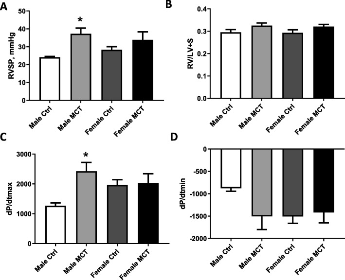
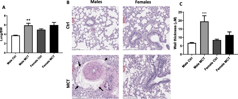
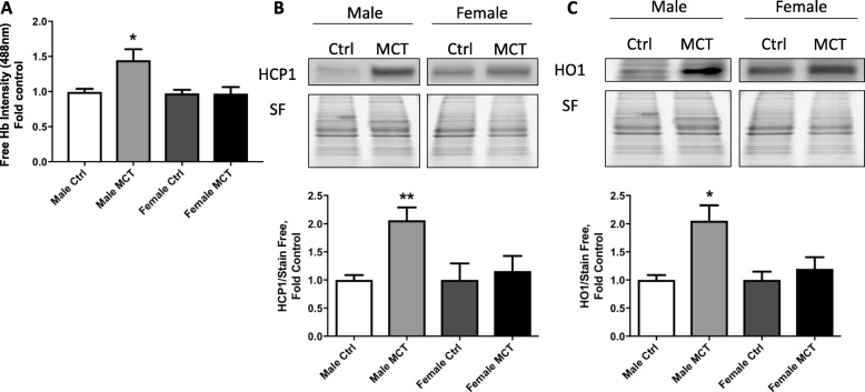
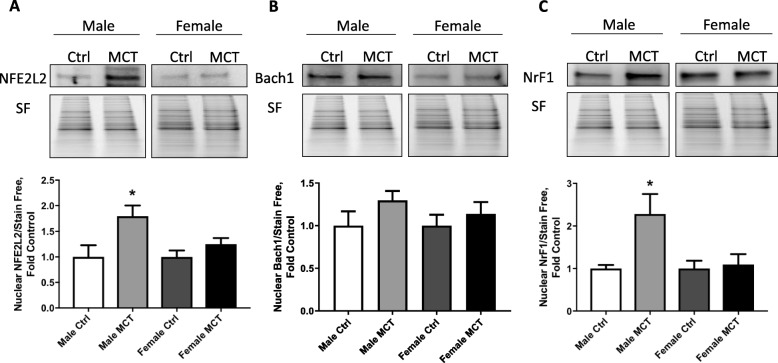
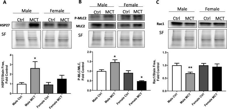
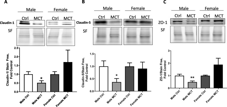
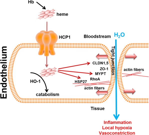
Results: Our data indicate that on day 14, rats did not show any significant increase in the Fulton index due to the early disease phase. However, the right ventricular systolic pressure was significantly increased in male rats, while female rats showed only a trend. Interestingly, only males demonstrated an increased lung-to-bodyweight ratio that indicated lung edema. Indeed, lung histology confirmed severe perivascular edema in males. Previously, we have reported that the increased perivascular edema in SU/Hx model correlated with intravascular hemolysis and activated heme signaling. Here, we found that elevated free hemoglobin levels and perivascular edema were increased, specifically in males showing more rapid progress of PH. A high level of heme carrier protein 1 (HCP-1), which is involved in heme uptake from the bloodstream into the cells, was also found elevated in the lungs of males. The upregulation of heme oxygenase in males indicated increased intracellular heme catabolism. Increased heme signaling resulted in the activation of heme-mediated barrier-disruptive mechanisms. Thus, hemolysis in males can be responsible for increased permeability of the lungs and early disease development.
Conclusions: Our study indicates the importance of barrier-disruptive mechanisms as an earlier event in the induction of pulmonary hypertension. Importantly, males are more susceptible to hemolysis and develop PH earlier than females.
Keywords: Endothelial barrier function; Heme signaling; Lung permeability; Pulmonary hypertension; Sex difference.
Conflict of interest statement
The authors declare that they have no competing interests.
Figures
Similar articles
-
Hemolysis-induced Lung Vascular Leakage Contributes to the Development of Pulmonary Hypertension.Am J Respir Cell Mol Biol. 2018 Sep;59(3):334-345. doi: 10.1165/rcmb.2017-0308OC. Am J Respir Cell Mol Biol. 2018. PMID: 29652520 Free PMC article.
-
Increased pulmonary heme oxygenase-1 and delta-aminolevulinate synthase expression in monocrotaline-induced pulmonary hypertension.Curr Neurovasc Res. 2005 Apr;2(2):133-9. doi: 10.2174/1567202053586794. Curr Neurovasc Res. 2005. PMID: 16181105
-
2-Methoxyestradiol mediates the protective effects of estradiol in monocrotaline-induced pulmonary hypertension.Vascul Pharmacol. 2006 Dec;45(6):358-67. doi: 10.1016/j.vph.2006.05.007. Epub 2006 Jun 8. Vascul Pharmacol. 2006. PMID: 16872912
-
Heme on Pulmonary Malaria: Friend or Foe?Front Immunol. 2020 Aug 25;11:1835. doi: 10.3389/fimmu.2020.01835. eCollection 2020. Front Immunol. 2020. PMID: 32983096 Free PMC article. Review.
-
The role of macrophages in pulmonary hypertension: Pathogenesis and targeting.Int Immunopharmacol. 2020 Nov;88:106934. doi: 10.1016/j.intimp.2020.106934. Epub 2020 Sep 2. Int Immunopharmacol. 2020. PMID: 32889242 Review.
Cited by
-
Glucose-6-phosphate dehydrogenase deficiency contributes to metabolic abnormality and pulmonary hypertension.Am J Physiol Lung Cell Mol Physiol. 2021 Apr 1;320(4):L508-L521. doi: 10.1152/ajplung.00165.2020. Epub 2021 Jan 27. Am J Physiol Lung Cell Mol Physiol. 2021. PMID: 33502933 Free PMC article.
-
Experimental animal models of pulmonary hypertension: Development and challenges.Animal Model Exp Med. 2022 Sep;5(3):207-216. doi: 10.1002/ame2.12220. Epub 2022 Mar 25. Animal Model Exp Med. 2022. PMID: 35333455 Free PMC article.
-
Low Thyroid Hormones Level Attenuates Mitochondrial Dysfunction and Right Ventricular Failure in Pulmonary Hypertensive Rats.Cardiovasc Drugs Ther. 2024 Aug 31. doi: 10.1007/s10557-024-07618-5. Online ahead of print. Cardiovasc Drugs Ther. 2024. PMID: 39215901
-
Circulating free heme induces cytokine storm and pulmonary hypertension through the MKK3/p38 axis.Am J Physiol Lung Cell Mol Physiol. 2024 Oct 1;327(4):L574-L586. doi: 10.1152/ajplung.00422.2022. Epub 2024 Aug 28. Am J Physiol Lung Cell Mol Physiol. 2024. PMID: 39197168 Free PMC article.
-
Deficiency of cold-inducible RNA-binding protein exacerbated monocrotaline-induced pulmonary artery hypertension through Caveolin1 and CAVIN1.J Cell Mol Med. 2021 May;25(10):4732-4743. doi: 10.1111/jcmm.16437. Epub 2021 Mar 23. J Cell Mol Med. 2021. PMID: 33755319 Free PMC article.
References
-
- Patel M, Predescu D, Tandon R, Bardita C, Pogoriler J, Bhorade S, Wang M, Comhair S, Hemnes AR, Chen J, et al. A novel p38 mitogen-activated protein kinase/Elk-1 transcription factor-dependent molecular mechanism underlying abnormal endothelial cell proliferation in plexogenic pulmonary arterial hypertension. J Biol Chem. 2013;288(36):25701–25716. - PMC - PubMed
Publication types
MeSH terms
Substances
Grants and funding
LinkOut - more resources
Full Text Sources
Medical
Research Materials
Miscellaneous
